Cited by 113Protein Kinase Regulation and GTPase SignalingCellular transport and secretionProtein Tyrosine Phosphatases

The DOI matches its Crossref record; 68 of the 69 references it cites were found in a registry or an index.

Requirement for Ras Guanine Nucleotide Releasing Protein 3 in Coupling Phospholipase C-γ2 to Ras in B Cell Receptor Signaling

Open access
Oh-hora, Masatsugu, Johmura, Sachiko, Hashimoto, Ari, Hikida, Masaki, Kurosaki, Tomohiro·2003·The Journal of Experimental Medicine

Abstract

Two important Ras guanine nucleotide exchange factors, Son of sevenless (Sos) and Ras guanine nucleotide releasing protein (RasGRP), have been implicated in controlling Ras activation when cell surface receptors are stimulated. To address the specificity or redundancy of these exchange factors, we have generated Sos1/Sos2 double- or RasGRP3-deficient B cell lines and determined their ability to mediate Ras activation upon B cell receptor (BCR) stimulation. The BCR requires RasGRP3; in contrast, epidermal growth factor receptor is dependent on Sos1 and Sos2. Furthermore, we show that BCR-induced recruitment of RasGRP3 to the membrane and the subsequent Ras activation are significantly attenuated in phospholipase C-γ2–deficient B cells. This defective Ras activation is suppressed by the expression of RasGRP3 as a membrane-attached form, suggesting that phospholipase C-γ2 regulates RasGRP3 localization and thereby Ras activation.

Overview

The claims and statistics of this paper haven’t been checked yet.

Run the check — free, no account needed. It reads the open-access text and checks the sources it cites.

A few free checks a day · papers already verified open instantly · results are public

Integrity signals

Registry

Crossref · 10.1084/jem.20031547

Cites

68 of 68 references verified0 citation errors · 0 not checkable

Statistics

Figures

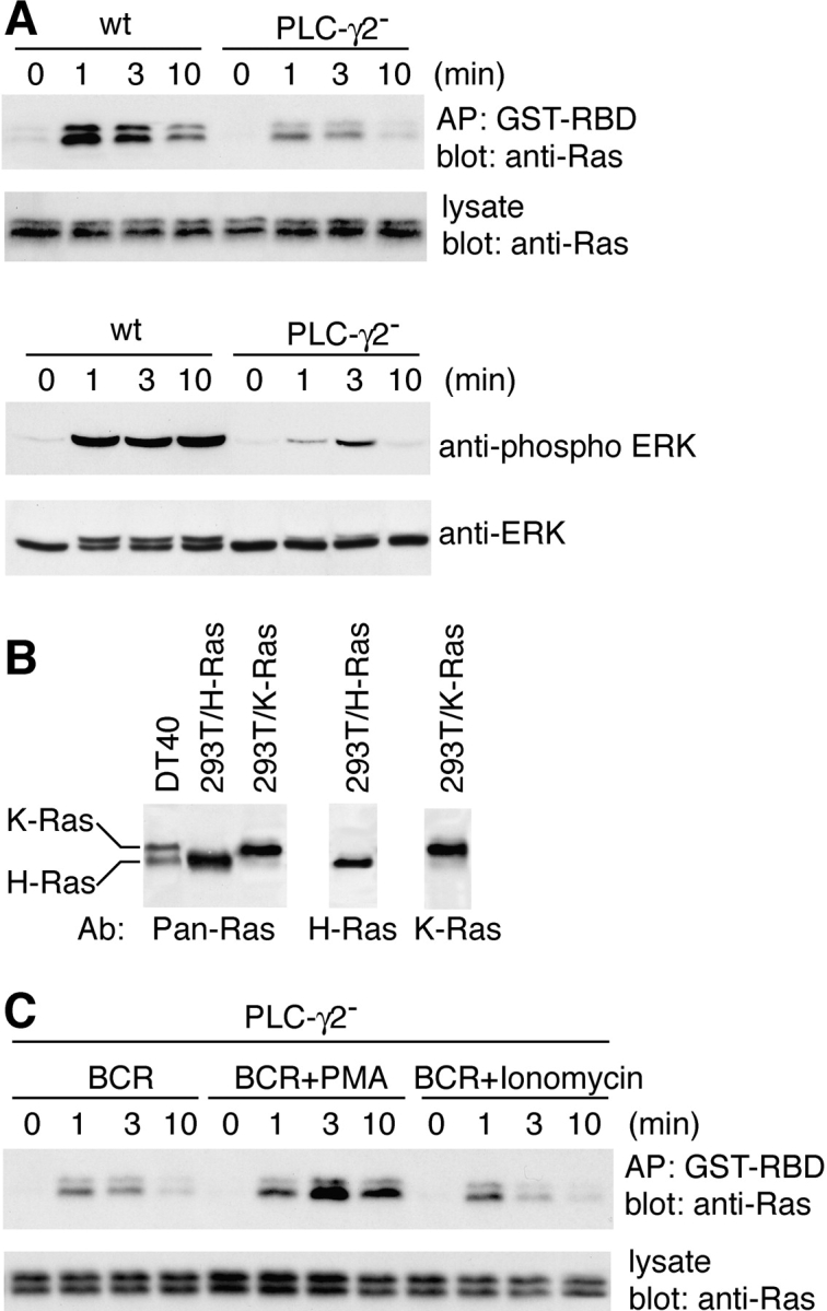
Figure 1. Requirement for PLC-γ2 in BCR-mediated Ras activation. (A) BCR-mediated Ras and ERK activation in wild-type, PLC-γ2–deficient DT40 B cells. For Ras activation, 107 cells/lane were stimulated with 5 μg/ml M4, and cell lysates were mixed with beads containing the Ras binding domain of Raf. Elutes of the beads (top) or cell lysates (bottom) were subjected to Western blot analysis with anti–pan-Ras mAb. AP, affinity precipitation. For ERK activation, 107 cells were stimulated with 5 μg/ml M4, and cell lysates (2 × 10⁶ cells/lane) were analyzed by Western blotting with anti-phospho–p44/p42 MAPK polyclonal Ab (top) and anti-p44/p42 MAPK polyclonal Ab (bottom). (B) Protein expression analysis of chicken H-Ras and K-Ras. Total cell lysates of DT40 cells (107 cells/lane) and 293T cells transfected with chicken H-Ras or K-Ras cDNA were prepared and analyzed by Western blotting using anti–pan-Ras, anti–H-Ras, and anti–K-Ras Abs. Although the bottom band was the major product of transfected chicken H-Ras in 293T cells, the faint top band, which has the same electrophoretic mobility as K-Ras, was detected when overexposed. Thus, the minor portion of the top band observed in DT40 B cells is likely to be composed of H-Ras. (C) Complementation of Ras activation after costimulation of BCR with PMA or ionomycin. PLC-γ2–deficient DT40 B cells were stimulated and analyzed as in A with 50 ng/ml PMA or 100 nM ionomycin. This concentration of ionomycin can induce calcium mobilization to the same extent as induced by M4 stimulation on wild-type DT40 B cells.
Figure 1. Requirement for PLC-γ2 in BCR-mediated Ras activation. (A) BCR-mediated Ras and ERK activation in wild-type, PLC-γ2–deficient DT40 B cells. For Ras activation, 107 cells/lane were stimulated with 5 μg/ml M4, and cell lysates were mixed with beads containing the Ras binding domain of Raf. Elutes of the beads (top) or cell lysates (bottom) were subjected to Western blot analysis with anti–pan-Ras mAb. AP, affinity precipitation. For ERK activation, 107 cells were stimulated with 5 μg/ml M4, and cell lysates (2 × 10⁶ cells/lane) were analyzed by Western blotting with anti-phospho–p44/p42 MAPK polyclonal Ab (top) and anti-p44/p42 MAPK polyclonal Ab (bottom). (B) Protein expression analysis of chicken H-Ras and K-Ras. Total cell lysates of DT40 cells (107 cells/lane) and 293T cells transfected with chicken H-Ras or K-Ras cDNA were prepared and analyzed by Western blotting using anti–pan-Ras, anti–H-Ras, and anti–K-Ras Abs. Although the bottom band was the major product of transfected chicken H-Ras in 293T cells, the faint top band, which has the same electrophoretic mobility as K-Ras, was detected when overexposed. Thus, the minor portion of the top band observed in DT40 B cells is likely to be composed of H-Ras. (C) Complementation of Ras activation after costimulation of BCR with PMA or ionomycin. PLC-γ2–deficient DT40 B cells were stimulated and analyzed as in A with 50 ng/ml PMA or 100 nM ionomycin. This concentration of ionomycin can induce calcium mobilization to the same extent as induced by M4 stimulation on wild-type DT40 B cells.CC BY-NC-SA · PubMed Central
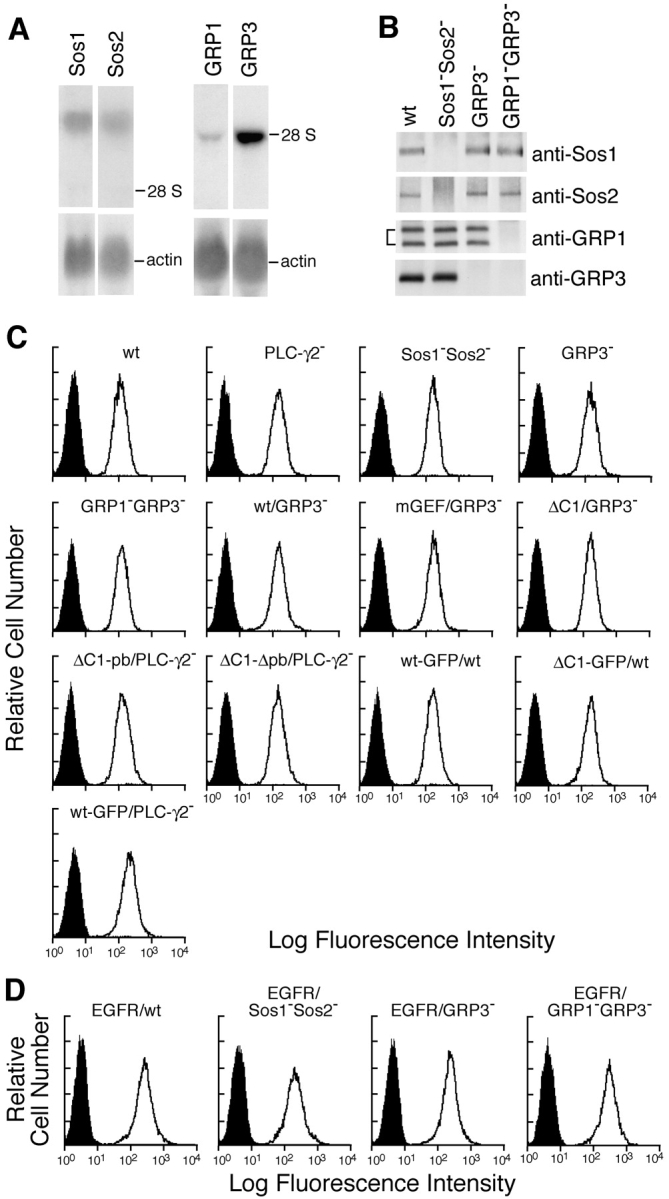
Figure 2. Generation of Sos1/Sos2 double-, RasGRP1/RasGRP3 double-, and RasGRP3-deficient DT40 B cells. (A) RNA expression of Sos1, Sos2, RasGRP1, and RasGRP3 was analyzed by Northern blot analysis using each chicken cDNA probe (top) or β-actin (bottom). The specific radio activity of each probe was roughly the same. Positions of 28S rRNA are shown. (B) Sos1, Sos2, RasGRP1, and RasGRP3 protein expression in wild-type and various mutant DT40 cells. Each protein was immunoprecipitated and detected by Western blotting with each specific Abs. Sos1−Sos2−, Sos1/Sos2 double-deficient DT40 cells. GRP3−, RasGRP3-deficient DT40 cells. GRP1−GRP3−, RasGRP1/RasGRP3 double-deficient DT40 cells. (C) Cell surface expression of BCR on wild-type and various DT40 mutants (see Fig. 6 A for designation). (D) Cell surface expression of transfected EGFR on wild-type and mutant DT40 cells.
Figure 2. Generation of Sos1/Sos2 double-, RasGRP1/RasGRP3 double-, and RasGRP3-deficient DT40 B cells. (A) RNA expression of Sos1, Sos2, RasGRP1, and RasGRP3 was analyzed by Northern blot analysis using each chicken cDNA probe (top) or β-actin (bottom). The specific radio activity of each probe was roughly the same. Positions of 28S rRNA are shown. (B) Sos1, Sos2, RasGRP1, and RasGRP3 protein expression in wild-type and various mutant DT40 cells. Each protein was immunoprecipitated and detected by Western blotting with each specific Abs. Sos1−Sos2−, Sos1/Sos2 double-deficient DT40 cells. GRP3−, RasGRP3-deficient DT40 cells. GRP1−GRP3−, RasGRP1/RasGRP3 double-deficient DT40 cells. (C) Cell surface expression of BCR on wild-type and various DT40 mutants (see Fig. 6 A for designation). (D) Cell surface expression of transfected EGFR on wild-type and mutant DT40 cells.CC BY-NC-SA · PubMed Central
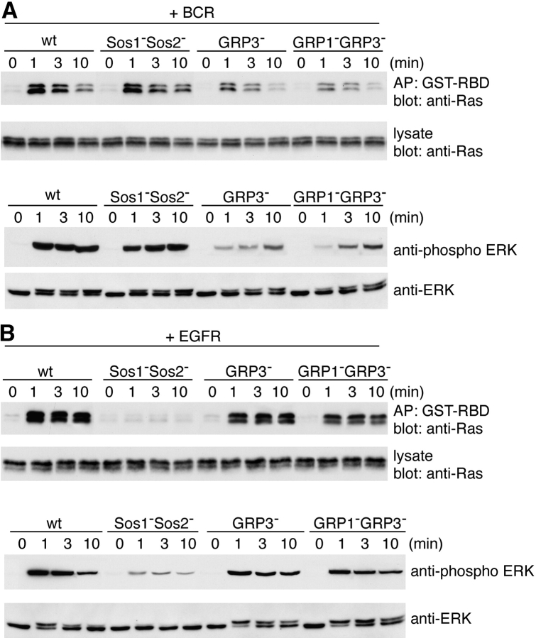
Figure 3. Differential requirement for RasGRP and Sos family in BCR- or EGFR-mediated Ras activation. (A) BCR-mediated Ras and ERK activation in wild-type, Sos1/Sos2 double-, RasGRP3-, and RasGRP1/RasGRP3 double-deficient DT40 cells. Ras and ERK activation was measured as described in Fig. 1. (B) EGFR-mediated Ras and ERK activation in each deficient DT40 cell. Cells were stimulated by 50 ng/ml EGF and analyzed as in Fig. 1. These experiments were performed at least three times and the representative results are shown.
Figure 3. Differential requirement for RasGRP and Sos family in BCR- or EGFR-mediated Ras activation. (A) BCR-mediated Ras and ERK activation in wild-type, Sos1/Sos2 double-, RasGRP3-, and RasGRP1/RasGRP3 double-deficient DT40 cells. Ras and ERK activation was measured as described in Fig. 1. (B) EGFR-mediated Ras and ERK activation in each deficient DT40 cell. Cells were stimulated by 50 ng/ml EGF and analyzed as in Fig. 1. These experiments were performed at least three times and the representative results are shown.CC BY-NC-SA · PubMed Central
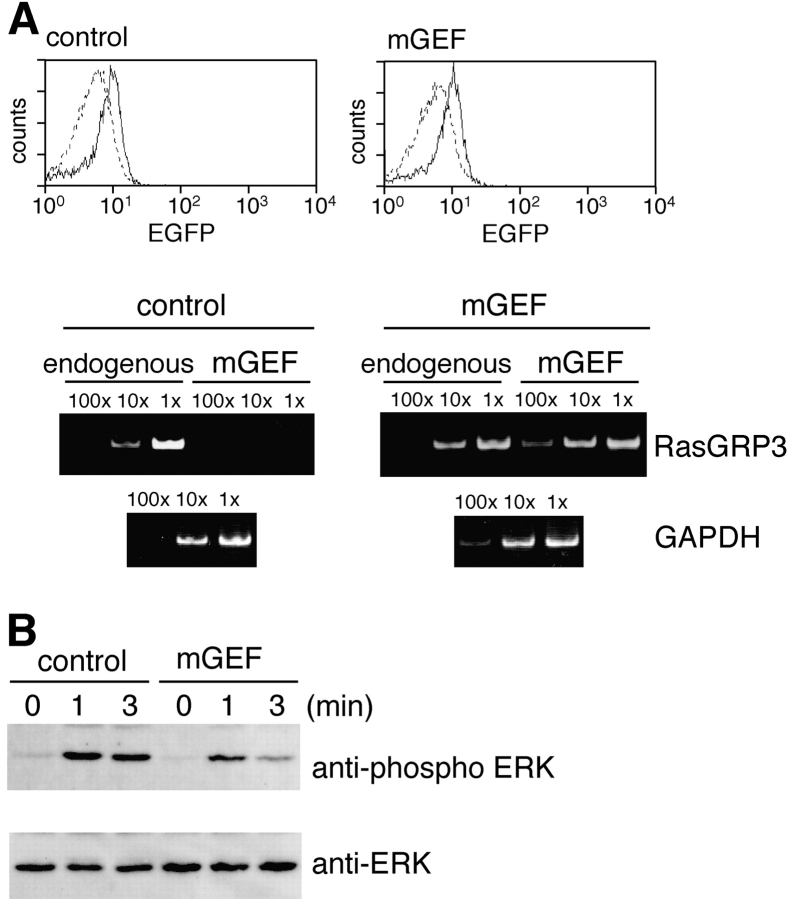
Figure 4. Requirement for RasGRP3 in BCR-mediated ERK activation in primary B cells. (A) Expression of mGEFRasGRP3 in infected B cells. Splenic B cells from C57BL/6J mice were transduced with a GEF mutant of mouse RasGRP3 (mGEFRasGRP3) by lentivirus. Transduction efficiencies were monitored by EGFP expression. Fluorescence intensities of untransduced cells are shown in dashed lines (top). Expression levels of endogenous RasGRP3 and infected mGEFRasGRP3 were analyzed by RT-PCR as described in the Materials and Methods (bottom). (B) Attenuation of Erk activation by the expression of mGEFRasGRP3. Infected cells were stimulated with 12 μg/ml anti–mouse IgM and ERK activation was measured using anti-phospho–p44/p42 MAPK mAb (E10) (top) and anti-p44/p42 MAPK Ab (bottom).
Figure 4. Requirement for RasGRP3 in BCR-mediated ERK activation in primary B cells. (A) Expression of mGEFRasGRP3 in infected B cells. Splenic B cells from C57BL/6J mice were transduced with a GEF mutant of mouse RasGRP3 (mGEFRasGRP3) by lentivirus. Transduction efficiencies were monitored by EGFP expression. Fluorescence intensities of untransduced cells are shown in dashed lines (top). Expression levels of endogenous RasGRP3 and infected mGEFRasGRP3 were analyzed by RT-PCR as described in the Materials and Methods (bottom). (B) Attenuation of Erk activation by the expression of mGEFRasGRP3. Infected cells were stimulated with 12 μg/ml anti–mouse IgM and ERK activation was measured using anti-phospho–p44/p42 MAPK mAb (E10) (top) and anti-p44/p42 MAPK Ab (bottom).CC BY-NC-SA · PubMed Central
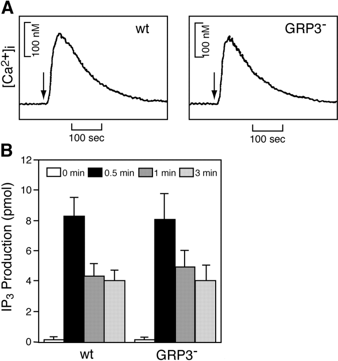
Figure 5. BCR-mediated PLC-γ2 activation is not affected by loss of RasGRP3. Cells were stimulated by 1.5 μg/ml M4 for indicated time periods and subjected to the following analyses. (A) Calcium mobilization. Intracellular-free calcium levels in Fura-2–loaded cells were monitored by a fluorescence spectrophotometer. (B) IP3 generation. Soluble IP3 was extracted from 2 × 10⁶ cells, and IP3 generation was measured by a Biotrak competitive binding assay system. The results were shown by mean ± standard error of three independent experiments.
Figure 5. BCR-mediated PLC-γ2 activation is not affected by loss of RasGRP3. Cells were stimulated by 1.5 μg/ml M4 for indicated time periods and subjected to the following analyses. (A) Calcium mobilization. Intracellular-free calcium levels in Fura-2–loaded cells were monitored by a fluorescence spectrophotometer. (B) IP3 generation. Soluble IP3 was extracted from 2 × 10⁶ cells, and IP3 generation was measured by a Biotrak competitive binding assay system. The results were shown by mean ± standard error of three independent experiments.CC BY-NC-SA · PubMed Central
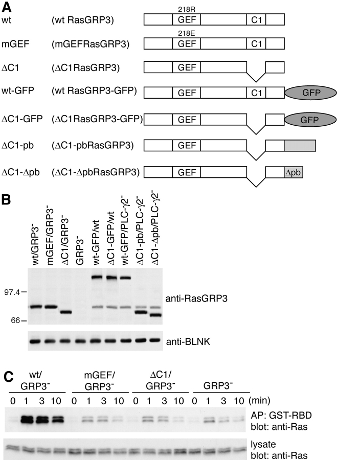
Figure 6. Both catalytic activity and C1 domain of RasGRP3 are required in BCR-mediated Ras activation. (A) Schematic diagram of various mutant RasGRP3 constructs. (B) Protein expression analyses of various mutant DT40 cell lines. Wild-type, RasGRP3-, or PLC-γ2–deficient DT40 B cells expressing various RasGRP3 mutants are shown. Endogenous RasGRP3 is also shown in wild-type or PLC-γ2–deficient DT40 cells. Whole cell lysates prepared from 2 × 10⁶ cells were analyzed by Western blotting using anti-RasGRP3 Ab or anti-BLNK Ab. (C) Analysis of BCR-mediated Ras activation in RasGRP3-deficient cells expressing wild-type RasGRP3 and its mutants was performed as described in Fig. 1. Each experiment was repeated at least three times.
Figure 6. Both catalytic activity and C1 domain of RasGRP3 are required in BCR-mediated Ras activation. (A) Schematic diagram of various mutant RasGRP3 constructs. (B) Protein expression analyses of various mutant DT40 cell lines. Wild-type, RasGRP3-, or PLC-γ2–deficient DT40 B cells expressing various RasGRP3 mutants are shown. Endogenous RasGRP3 is also shown in wild-type or PLC-γ2–deficient DT40 cells. Whole cell lysates prepared from 2 × 10⁶ cells were analyzed by Western blotting using anti-RasGRP3 Ab or anti-BLNK Ab. (C) Analysis of BCR-mediated Ras activation in RasGRP3-deficient cells expressing wild-type RasGRP3 and its mutants was performed as described in Fig. 1. Each experiment was repeated at least three times.CC BY-NC-SA · PubMed Central
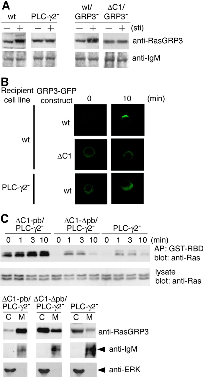
Figure 7. Membrane localization of RasGRP3 is necessary for Ras activation after BCR ligation. (A) Subcellular analysis of RasGRP3. Wild-type, PLC-γ2–deficient, wt/RasGRP3−, and ΔC1/RasGRP3− DT40 cells were stimulated by M4 for 10 min (+) and left unstimulated (−). Membrane fractions were resolved by SDS-PAGE and immunoblotted with anti-RasGRP3 Ab or anti-IgM Ab. (B) Impaired membrane translocation of RasGRP3 in PLC-γ2–deficient DT40 cells. Wild-type DT40 cells expressing RasGRP3-GFP (top) or its C1 deletion mutant (middle, ΔC1RasGRP3-GFP) were stimulated with anti–chicken IgM (M4) for indicated periods. PLC-γ2–deficient DT40 cells expressing RasGRP3-GFP were stimulated with M4 under the same condition (bottom). After the stimulation, cells were fixed and subcellular localization of RasGRP3-GFP fusion protein was visualized by confocal microscope as described in Materials and Methods. Among 100 cells counted for membrane localization, the numbers that showed clear membrane localization were 68 ± 16 for wild-type DT40 expressing RasGRP3-GFP, 8 ± 1 for wild-type DT40 cells expressing ΔC1RasGRP3-GFP, and 7 ± 1 for PLC-γ2–deficient DT40 cells expressing RasGRP3-GFP from three independent experiments. The representative results are shown. (C) Restoration of Ras activation by expression of a membrane-attached form of RasGRP3 in PLC-γ2–deficient DT40 cells. PLC-γ2–deficient DT40 cells expressing chimeras are shown as in Fig. 6 A. ΔC1RasGRP3 harboring the polybasic carboxyl terminus of K-Ras (ΔC1-pbRasGRP3/PLC-γ2−) or its polybasic amino acid–defective mutant (ΔC1-ΔpbRasGRP3/PLC-γ2−) were stimulated and analyzed with Ras activation as in Fig. 1 (top, GTP-bound Ras; middle, total Ras). Subcellular analyses of transfected RasGRP3 and endogenous RasGRP3 were also carried out before BCR stimulation. Samples were immunoblotted with anti-RasGRP3. Anti-IgM that recognized a μ heavy chain was used as a control for membrane fraction, and anti-ERK was used as a control for cytosolic fraction (bottom). C, cytosolic fraction. M, membrane fraction.
Figure 7. Membrane localization of RasGRP3 is necessary for Ras activation after BCR ligation. (A) Subcellular analysis of RasGRP3. Wild-type, PLC-γ2–deficient, wt/RasGRP3−, and ΔC1/RasGRP3− DT40 cells were stimulated by M4 for 10 min (+) and left unstimulated (−). Membrane fractions were resolved by SDS-PAGE and immunoblotted with anti-RasGRP3 Ab or anti-IgM Ab. (B) Impaired membrane translocation of RasGRP3 in PLC-γ2–deficient DT40 cells. Wild-type DT40 cells expressing RasGRP3-GFP (top) or its C1 deletion mutant (middle, ΔC1RasGRP3-GFP) were stimulated with anti–chicken IgM (M4) for indicated periods. PLC-γ2–deficient DT40 cells expressing RasGRP3-GFP were stimulated with M4 under the same condition (bottom). After the stimulation, cells were fixed and subcellular localization of RasGRP3-GFP fusion protein was visualized by confocal microscope as described in Materials and Methods. Among 100 cells counted for membrane localization, the numbers that showed clear membrane localization were 68 ± 16 for wild-type DT40 expressing RasGRP3-GFP, 8 ± 1 for wild-type DT40 cells expressing ΔC1RasGRP3-GFP, and 7 ± 1 for PLC-γ2–deficient DT40 cells expressing RasGRP3-GFP from three independent experiments. The representative results are shown. (C) Restoration of Ras activation by expression of a membrane-attached form of RasGRP3 in PLC-γ2–deficient DT40 cells. PLC-γ2–deficient DT40 cells expressing chimeras are shown as in Fig. 6 A. ΔC1RasGRP3 harboring the polybasic carboxyl terminus of K-Ras (ΔC1-pbRasGRP3/PLC-γ2−) or its polybasic amino acid–defective mutant (ΔC1-ΔpbRasGRP3/PLC-γ2−) were stimulated and analyzed with Ras activation as in Fig. 1 (top, GTP-bound Ras; middle, total Ras). Subcellular analyses of transfected RasGRP3 and endogenous RasGRP3 were also carried out before BCR stimulation. Samples were immunoblotted with anti-RasGRP3. Anti-IgM that recognized a μ heavy chain was used as a control for membrane fraction, and anti-ERK was used as a control for cytosolic fraction (bottom). C, cytosolic fraction. M, membrane fraction.CC BY-NC-SA · PubMed Central

Verification

Claims the paper makes

The claims and statistics of this paper haven’t been checked yet.

Run the check — free, no account needed. It reads the open-access text and checks the sources it cites.

A few free checks a day · papers already verified open instantly · results are public

Claims other papers make about this one

Each is checked against this paper’s own text: does it say what the citing paper says it does?

Self-check

Percentages, sums and reported differences re-computed from the paper’s own numbers.

The self-check needs the paper’s full text.

Citations

Cites

Show all 69 references

Research works printed but not linked: 2

  1. 1992, Mol. Cell. Biol., 12, 3305
  2. 2002, Nat. Rev. Mol. Cell Biol., 3, 339

Cited by

Sorted by citations

See all 113

Metadata

Authors
Oh-hora, M., Johmura, S., Hashimoto, A., Hikida, M., & Kurosaki, T.
Affiliations
  • 1Department of Molecular Genetics, Institute for Liver Research, Kansai Medical University, Moriguchi 570-8506, Japan
  • Kansai Medical University (JP)
Venue
The Journal of Experimental Medicine
Published
2003-12-15
Volume
198
Issue
12
Pages
1841-1851
ISSN
  • 0022-1007 (ISSN-L)
  • 1540-9538
OpenAlex
W2131566341
Cited by
113
References
69
Citations by year
2026: 2 · 2025: 2 · 2024: 2 · 2023: 2 · 2022: 2 · 2021: 2 · 2020: 4 · 2019: 4 · 2018: 1 · 2017: 2 · 2016: 4 · 2015: 4 · 2014: 3 · 2013: 6 · 2012: 4
FWCI
3.57
Type
journal article
Language
en
Open access
Licence
cc-by-nc-sa
Locations
  • The Journal of Experimental Medicine (publishedVersion)
  • PubMed Central (publishedVersion, cc-by-nc-sa)
  • PubMed (publishedVersion)
Funders
Uehara Memorial Foundation
Topics
  • Life Sciences › Biochemistry, Genetics and Molecular Biology › Molecular Biology › Protein Kinase Regulation and GTPase Signaling
  • Life Sciences › Biochemistry, Genetics and Molecular Biology › Cell Biology › Cellular transport and secretion
  • Life Sciences › Biochemistry, Genetics and Molecular Biology › Molecular Biology › Protein Tyrosine Phosphatases
Keywords
RasGRP3, SOS1, SOS2, Ras guanine nucleotide exchange factor, Ras activation, B cell receptor signaling, PLCγ2, epidermal growth factor receptor, membrane recruitment, B-cell lines
MeSH terms
Amino Acid Sequence, Animals, Mice, Inbred C57BL, Molecular Sequence Data, Type C Phospholipases, Receptors, Antigen, B-Cell, Signal Transduction, ras Proteins, Guanine Nucleotide Exchange Factors, ras Guanine Nucleotide Exchange Factors, Son of Sevenless Proteins, SOS1 Protein, Mice, Phospholipase C gamma

About these checks

Registry, retraction, Cites and statistics results are computed, not judged, with one exception: a printed reference that no index lists is read once by a model before it counts as a citation error. Claims are extracted by a model and kept only when their quote appears verbatim in the text; each cited source is then checked, with a model’s help, for whether it says what the claim relies on. Those verdicts can be wrong — read the claim’s quote and the cited source before relying on one.

Statistics and arithmetic are recomputed, not judged.

Confidence bands are shown only for fields calibrated on our annotated test set. None is calibrated yet, so no band is shown.

What counts as a claim ›How we measure confidence ›How we check this ›

Are you an author, or do you see a problem with this page? Request a review