

The DOI matches its Crossref record; 33 of the 34 references it cites were found in a registry or an index.
Blockade of CTLA-4 on both effector and regulatory T cell compartments contributes to the antitumor activity of anti-CTLA-4 antibodies
Open accessAbstract
Cytotoxic T lymphocyte–associated antigen 4 (CTLA-4) is a critical negative regulator of immune responses. Uniquely among known inhibitory receptors, its genetic ablation results in a fulminating and fatal lymphoproliferative disorder. This central regulatory role led to the development of antibodies designed to block CTLA-4 activity in vivo, aiming to enhance immune responses against cancer. Despite their preclinical efficacy and promising clinical activity against late stage metastatic melanoma, the critical cellular targets for their activity remains unclear. In particular, debate has focused on whether the effector T cell (Teff) or regulatory T cell (T reg cell) compartment is the primary target of antibody-mediated blockade. We developed a mouse expressing human instead of mouse CTLA-4, allowing us to evaluate the independent contributions of CTLA-4 blockade of each T cell compartment during cancer immunotherapy in an in vivo model of mouse melanoma. The data show that although blockade on effector cells significantly improves tumor protection, unicompartmental blockade on regulatory cells completely fails to enhance antitumor responses. However, concomitant blockade of both compartments leads to a synergistic effect and maximal antitumor activity. We conclude that the combination of direct enhancement of Teff cell function and concomitant inhibition of T reg cell activity through blockade of CTLA-4 on both cell types is essential for mediating the full therapeutic effects of anti–CTLA-4 antibodies during cancer immunotherapy.
Overview
The claims and statistics of this paper haven’t been checked yet.
Run the check — free, no account needed. It reads the open-access text and checks the sources it cites.
Integrity signals
Registry
Cites
Statistics
Figures
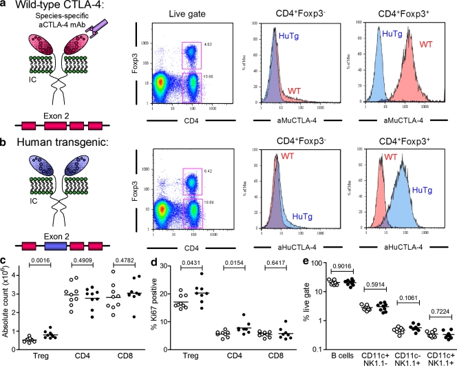
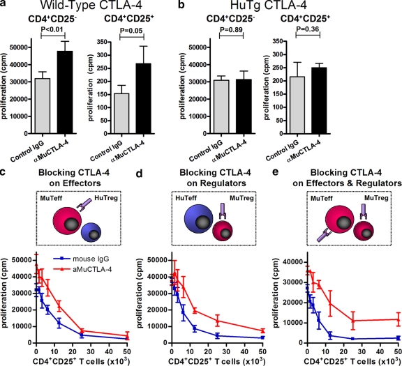
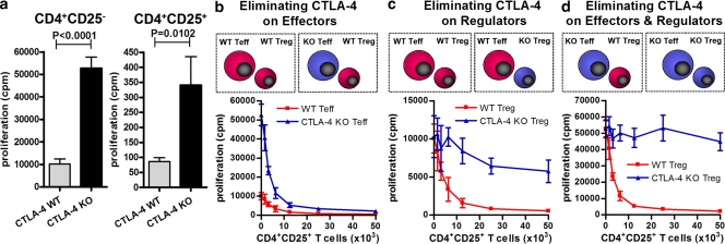
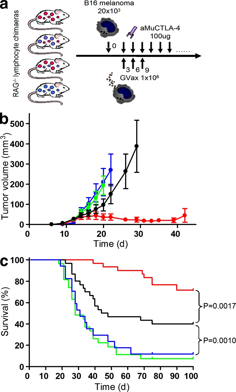
Verification
Claims the paper makes
The claims and statistics of this paper haven’t been checked yet.
Run the check — free, no account needed. It reads the open-access text and checks the sources it cites.
Claims other papers make about this one
Each is checked against this paper’s own text: does it say what the citing paper says it does?
In the citing paper
“However, depletion of Tregs alone does not enhance anti-tumor responses; rather, a combination of enhanced effector T cell function and the inhibition of Treg activity is necessary for maximal anti-tumor activity [119].”
From Current and Future Trends of Colorectal Cancer Treatment: Exploring Advances in Immunotherapy (2024) · cites this paper as [119]
In the citing paper
“Blocking CTLA-4 with antibodies increases CD8+ lymphocytes and depletes Tregs.”
From Current and Future Trends of Colorectal Cancer Treatment: Exploring Advances in Immunotherapy (2024) · cites this paper as [119]
Self-check
Percentages, sums and reported differences re-computed from the paper’s own numbers.
The self-check needs the paper’s full text.
Citations
Cites
Enhancement of Antitumor Immunity by CTLA-4 Blockade
Leach, D. R., Krummel, M. F., & Allison, J. P. (1996) · Science · doi:10.1126/science.271.5256.1734 · Cited by 3,955
Self-citationCTLA-4 Control over Foxp3 + Regulatory T Cell Function
Wing, K., Onishi, Y., Prieto-Martin, P., Yamaguchi, T., Miyara, M., Fehervari, Z., Nomura, T., & Sakaguchi, S. (2008) · Science · doi:10.1126/science.1160062 · Cited by 2,956
Lymphoproliferative Disorders with Early Lethality in Mice Deficient in Ctla-4
Waterhouse, P., Penninger, J. M., Timms, E., Wakeham, A., Shahinian, A., Lee, K. P., Thompson, C. B., Griesser, H., & Mak, T. W. (1995) · Science · doi:10.1126/science.270.5238.985 · Cited by 2,766
Takahashi, T., Tagami, T., Yamazaki, S., Uede, T., Shimizu, J., Sakaguchi, N., Mak, T. W., & Sakaguchi, S. (2000) · The Journal of Experimental Medicine · doi:10.1084/jem.192.2.303 · Cited by 2,233
Read, S., Malmström, V., & Powrie, F. M. (2000) · The Journal of Experimental Medicine · doi:10.1084/jem.192.2.295 · Cited by 2,206
Butte, M. J., Keir, M. E., Phamduy, T. B., Sharpe, A. H., & Freeman, G. J. (2007) · Immunity · doi:10.1016/j.immuni.2007.05.016 · Cited by 1,785
Phan, G. Q., Yang, J. C., Sherry, R. M., Hwu, P., Topalian, S. L., Schwartzentruber, D. J., Restifo, N. P., Haworth, L. R., Seipp, C. A., Freezer, L. J., et al. (2003) · Proceedings of the National Academy of Sciences · doi:10.1073/pnas.1533209100 · Cited by 1,575
Self-citationModulation of tryptophan catabolism by regulatory T cells
Fallarino, F., Grohmann, U., Hwang, K. W., Orabona, C., Vacca, C., Bianchi, R., Belladonna, M. L., Fioretti, M. C., Alegre, M., & Puccetti, P. (2003) · Nature Immunology · doi:10.1038/ni1003 · Cited by 1,375
Onizuka, S., Tawara, I., Shimizu, J., Sakaguchi, S., Fujita, T., & Nakayama, E. (1999) · PubMed · Cited by 1,062
van Elsas, A., Hurwitz, A. A., & Allison, J. P. (1999) · The Journal of Experimental Medicine · doi:10.1084/jem.190.3.355 · Cited by 1,020
Self-citation
Cited by
Sorted by citations
Items 101–200 of 967
Pathways and therapeutic targets in melanoma
Shtivelman, E., Davies, M. A., Hwu, P., Yang, J. C.-H., Lotem, M., Oren, M., Flaherty, K. T., & Fisher, D. E. (2014) · Oncotarget · doi:10.18632/oncotarget.1892 · Cited by 226
Kumar, P., Bhattacharya, P., & Prabhakar, B. S. (2018) · Journal of Autoimmunity · doi:10.1016/j.jaut.2018.08.007 · Cited by 222
Curran, M. A., Kim, M., Montalvo-Ortiz, W., Al‐Shamkhani, A., & Allison, J. P. (2011) · PLoS ONE · doi:10.1371/journal.pone.0019499 · Cited by 213
Self-citationImmune Evasion in Pancreatic Cancer: From Mechanisms to Therapy
Martínez‐Bosch, N., Vinaixa, J., & Navarro, P. (2018) · Cancers · doi:10.3390/cancers10010006 · Cited by 216
Willsmore, Z. N., Coumbe, B. G. T., Crescioli, S., Reci, S., Gupta, A., Harris, R. J. C., Chenoweth, A. M., Chauhan, J., Bax, H. J., McCraw, A. J., et al. (2021) · European Journal of Immunology · doi:10.1002/eji.202048747 · Cited by 216
Lussier, D. M., Johnson, J. L., Hingorani, P., & Blattman, J. N. (2015) · Journal for ImmunoTherapy of Cancer · doi:10.1186/s40425-015-0067-z · Cited by 206
ENTPD1/CD39 is a promising therapeutic target in oncology
Bastid, J., Regairaz, A., Alberici, G. F., Bonnefoy, N., Eliaou, J.-F., & Bensussan, A. (2012) · Oncogene · doi:10.1038/onc.2012.269 · Cited by 213
Park, H. J., Park, J. S., Jeong, Y. H., Son, J., Ban, Y. H., Lee, B.-H., Chen, L., Chang, J., Chung, D. H., Choi, I., et al. (2015) · The Journal of Immunology · doi:10.4049/jimmunol.1401936 · Cited by 206
Fransen, M. F., van der Sluis, T. C., OSSENDORP, F. A., Arens, R., & Melief, C. J. M. (2013) · Clinical Cancer Research · doi:10.1158/1078-0432.ccr-12-0781 · Cited by 198
T cell exhaustion: from pathophysiological basics to tumor immunotherapy
Catakovic, K., Klieser, E., Neureiter, D., & Geisberger, R. (2017) · Cell Communication and Signaling · doi:10.1186/s12964-016-0160-z · Cited by 207
Immunoregulatory functions of VISTA
Nowak, E. C., Lines, J. L., Varn, F. S., Deng, J., Sarde, A., Mabaera, R., Kuta, A., Mercier, I. L., Cheng, C., & Noelle, R. J. (2017) · Immunological Reviews · doi:10.1111/imr.12525 · Cited by 200
Tregs: Where We Are and What Comes Next?
Hai, Z., Liao, X., & Kang, Y. (2017) · Frontiers in Immunology · doi:10.3389/fimmu.2017.01578 · Cited by 200
Tumor Necrosis Factor α and Regulatory T Cells in Oncoimmunology
Salomon, B. L., Leclerc, M., Tosello, J., Ronin, É., Piaggio, E., & Cohen, J. L. (2018) · Frontiers in Immunology · doi:10.3389/fimmu.2018.00444 · Cited by 198
Reprogramming the immunological microenvironment through radiation and targeting Axl
Aguilera, T. A., Rafat, M., Castellini, L., Shehade, H., Kariolis, M. S., Hui, A. B., Stehr, H., von Eyben, R., Jiang, D., Ellies, L. G., et al. (2016) · Nature Communications · doi:10.1038/ncomms13898 · Cited by 197
Current state of immunotherapy for non-small cell lung cancer
Malhotra, J. D., Jabbour, S. K., & Aisner, J. (2007) · Translational Lung Cancer Research · doi:10.21037/tlcr.2017.03.01 · Cited by 193
Recent Developments in Cancer Vaccines
Palucka, K., Ueno, H., & Banchereau, J. (2011) · The Journal of Immunology · doi:10.4049/jimmunol.0902539 · Cited by 193
Exploiting CTLA-4, PD-1 and PD-L1 to reactivate the host immune response against cancer
Quezada, S. A., & Peggs, K. S. (2013) · British Journal of Cancer · doi:10.1038/bjc.2013.117 · Cited by 192
Self-citationThe cGAS/STING Pathway: A Novel Target for Cancer Therapy
Gan, Y., Li, X., Han, S., Liang, Q., Ma, X., Rong, P., Wang, W., & Li, W. (2022) · Frontiers in Immunology · doi:10.3389/fimmu.2021.795401 · Cited by 194
Dovedi, S. J., Elder, M. J., Yang, C., Sitnikova, S. I., Irving, L., Hansen, A., Hair, J., Jones, D. C., Hasani, S., Wang, B., et al. (2021) · Cancer Discovery · doi:10.1158/2159-8290.cd-20-1445 · Cited by 186
Pike, L., Bang, A., Mahal, B. A. V., Taylor, A., Krishnan, M. S., Spektor, A., Cagney, D. N., Aizer, A. A., Alexander, B. M., Rahma, O. E., et al. (2018) · International Journal of Radiation Oncology*Biology*Physics · doi:10.1016/j.ijrobp.2018.09.010 · Cited by 194
Cell-autonomous and -non-autonomous roles of CTLA-4 in immune regulation
Wing, K., Yamaguchi, T., & Sakaguchi, S. (2011) · Trends in Immunology · doi:10.1016/j.it.2011.06.002 · Cited by 188
Kabacaoğlu, D., Ciecielski, K. J., Ruess, D. A., & Algül, H. (2018) · Frontiers in Immunology · doi:10.3389/fimmu.2018.01878 · Cited by 181
Nanomedicine-based tumor photothermal therapy synergized immunotherapy
Shang, T., Yu, X., Han, S., & Yang, B. (2020) · Biomaterials Science · doi:10.1039/d0bm01158d · Cited by 180
Immunotherapy advances for glioblastoma
Reardon, D. A., Freeman, G. J., Wu, C. J., Chiocca, E. A., Wucherpfennig, K. W., Wen, P. Y., Fritsch, E. F., Curry, W. T., Sampson, J. H., & Dranoff, G. (2014) · Neuro-Oncology · doi:10.1093/neuonc/nou212 · Cited by 181
Critical role of the gut microbiota in immune responses and cancer immunotherapy
Li, Z., Xiong, W., Zhu, L. W., Wang, J., Zeng, Z., Kołat, D., Li, X., Zhou, D., Xu, X., & Zhao, L.-Y. (2024) · Journal of Hematology & Oncology · doi:10.1186/s13045-024-01541-w · Cited by 197
Cytotoxic T lymphocyte antigen-4 and immune checkpoint blockade
Buchbinder, E. L., & Hodi, F. S. (2015) · Journal of Clinical Investigation · doi:10.1172/jci80012 · Cited by 181
Interaction between natural killer cells and regulatory T cells: perspectives for immunotherapy
Pedroza‐Pacheco, I., Madrigal, J. A., & Saudemont, A. (2013) · Cellular and Molecular Immunology · doi:10.1038/cmi.2013.2 · Cited by 180
Mizukoshi, E., Nakamoto, Y., Arai, K., Yamashita, T., Sakai, A., Sakai, Y., Kagaya, T., Yamashita, T., Honda, M., & Kaneko, S. (2011) · Hepatology · doi:10.1002/hep.24149 · Cited by 175
Development of immune checkpoint therapy for cancer
Fritz, J. M., & Lenardo, M. J. (2019) · The Journal of Experimental Medicine · doi:10.1084/jem.20182395 · Cited by 179
Emerging strategies for treating metastasis
Esposito, M., Ganesan, S., & Kang, Y. (2021) · Nature Cancer · doi:10.1038/s43018-021-00181-0 · Cited by 175
Single dose of anti–CTLA-4 enhances CD8 + T-cell memory formation, function, and maintenance
Pedicord, V. A., Montalvo-Ortiz, W., Leiner, I. M., & Allison, J. P. (2010) · Proceedings of the National Academy of Sciences · doi:10.1073/pnas.1016791108 · Cited by 174
Self-citationMechanisms of action of therapeutic antibodies for cancer
Redman, J. M., Hill, E., Aldeghaither, D. S., & Weiner, L. M. (2015) · Molecular Immunology · doi:10.1016/j.molimm.2015.04.002 · Cited by 179
Ise, W., Kohyama, M., Nutsch, K., Lee, H. M., Suri, A., Unanue, E. R., Murphy, T. L., & MURPHY, K. M. M. (2009) · Nature Immunology · doi:10.1038/ni.1835 · Cited by 177
Redmond, W. L., Linch, S. N., & Kasiewicz, M. J. (2013) · Cancer Immunology Research · doi:10.1158/2326-6066.cir-13-0031-t · Cited by 174
Péré, H., Montier, Y., Bayry, J., Quintin‐Colonna, F., Merillon, N., Dransart, E., Badoual, C., Gey, A., Ravel, P., Marcheteau, E., et al. (2011) · Blood · doi:10.1182/blood-2011-01-329656 · Cited by 185
Ipilimumab: A novel immunostimulatory monoclonal antibody for the treatment of cancer
Graziani, G., Tentori, L., & Navarra, P. (2011) · Pharmacological Research · doi:10.1016/j.phrs.2011.09.002 · Cited by 166
Immune Checkpoint Inhibitors in Non-Small Cell Lung Cancer
Herzberg, B., Campo, M. J., & Gainor, J. F. (2016) · The Oncologist · doi:10.1634/theoncologist.2016-0189 · Cited by 165
Laurent, S., Queirolo, P., Boero, S., Salvi, S., Piccioli, P., Boccardo, S., Minghelli, S., Morabito, A., Fontana, V., Pietra, G., et al. (2013) · Journal of Translational Medicine · doi:10.1186/1479-5876-11-108 · Cited by 163
Structural basis for cancer immunotherapy by the first-in-class checkpoint inhibitor ipilimumab
Ramagopal, U. A., Liu, W., Garrett-Thomson, S. C., Bonanno, J. B., Yan, Q., Srinivasan, M., Wong, S. C., Bell, A., Mankikar, S., Rangan, V. S., et al. (2017) · Proceedings of the National Academy of Sciences · doi:10.1073/pnas.1617941114 · Cited by 170
Self-citationCombining epigenetic and immune therapy to overcome cancer resistance
Gomez, S., Tabernacki, T., Kobyra, J. A., Roberts, P. A., & Chiappinelli, K. B. (2019) · Seminars in Cancer Biology · doi:10.1016/j.semcancer.2019.12.019 · Cited by 170
Dendritic cells and immunity against cancer
Palucka, K., Ueno, H., Fay, J. W., & Banchereau, J. (2010) · Journal of Internal Medicine · doi:10.1111/j.1365-2796.2010.02317.x · Cited by 164
Hannani, D., Vétizou, M., Enot, D. P., Rusakiewicz, S., Chaput, N., Klatzmann, D., Desbois, M., Jacquelot, N., Vimond, N., Chouaı̈b, S., et al. (2015) · Cell Research · doi:10.1038/cr.2015.3 · Cited by 166
Self-citationAdvances in immunotherapy for melanoma
Redman, J. M., Gibney, G. T., & Atkins, M. B. (2016) · BMC Medicine · doi:10.1186/s12916-016-0571-0 · Cited by 165
PD-1 pathway inhibitors: The next generation of immunotherapy for advanced melanoma
Luke, J. J., & Ott, P. A. (2015) · Oncotarget · doi:10.18632/oncotarget.2980 · Cited by 159
Directing Dendritic Cell Immunotherapy Towards Successful Cancer Treatment
Sabado, R. L., & Bhardwaj, N. (2009) · Immunotherapy · doi:10.2217/imt.09.43 · Cited by 155
Immune checkpoint inhibition in ovarian cancer
Hamanishi, J., Mandai, M., & Konishi, I. (2016) · International Immunology · doi:10.1093/intimm/dxw020 · Cited by 158
Treg(s) in Cancer: Friends or Foe?
Wolf, D. G. F., Sopper, S., Pircher, A., Gastl, G. A., & Wolf, A. M. (2015) · Journal of Cellular Physiology · doi:10.1002/jcp.25016 · Cited by 157
CTLA4 aptamer delivers STAT3 siRNA to tumor-associated and malignant T cells
Herrmann, A., Priceman, S. J., Kujawski, M., Xin, H., Cherryholmes, G. A., Zhang, W., Zhang, C., Lahtz, C., Kowolik, C. M., Forman, S. J., et al. (2014) · Journal of Clinical Investigation · doi:10.1172/jci73174 · Cited by 156
Arruga, F., Gyau, B. B., Iannello, A., Vitale, N., Vaisitti, T., & Deaglio, S. (2020) · International Journal of Molecular Sciences · doi:10.3390/ijms21051825 · Cited by 160
Rationally Combining Anti-VEGF Therapy with Checkpoint Inhibitors in Hepatocellular Carcinoma
Hato, T., Zhu, A. X., & Duda, D. G. (2016) · Immunotherapy · doi:10.2217/imt.15.126 · Cited by 155
The Influence of Tumor Microenvironment on Immune Escape of Melanoma
Simiczyjew, A., Dratkiewicz, E., Mazurkiewicz, J., Ziętek, M., Matkowski, R., & Nowak, D. (2020) · International Journal of Molecular Sciences · doi:10.3390/ijms21218359 · Cited by 157
The interplay of effector and regulatory T cells in cancer
Roychoudhuri, R., Eil, R. L., & Restifo, N. P. (2015) · Current Opinion in Immunology · doi:10.1016/j.coi.2015.02.003 · Cited by 154
Rios‐Doria, J., Harper, J. W., Rothstein, R. P., Wetzel, L. K., Chesebrough, J. W., Marrero, A. M., Chen, C., Strout, P., Mulgrew, K. A., McGlinchey, K., et al. (2017) · Cancer Research · doi:10.1158/0008-5472.can-16-2854 · Cited by 150
Sufficiency of CD40 activation and immune checkpoint blockade for T cell priming and tumor immunity
Morrison, A. H., Diamond, M. S., Hay, C., Byrne, K. T., & Vonderheide, R. H. (2020) · Proceedings of the National Academy of Sciences · doi:10.1073/pnas.1918971117 · Cited by 154
EMT and Treatment Resistance in Pancreatic Cancer
Gaianigo, N., Melisi, D., & Carbone, C. (2017) · Cancers · doi:10.3390/cancers9090122 · Cited by 149
Plitas, G., & Rudensky, A. Y. (2020) · Annual Review of Cancer Biology · doi:10.1146/annurev-cancerbio-030419-033428 · Cited by 148
Ali, O. A., Lewin, S. A., Dranoff, G., & Mooney, D. J. (2016) · Cancer Immunology Research · doi:10.1158/2326-6066.cir-14-0126 · Cited by 150
Cano‐Mejia, J., Burga, R. A., Sweeney, E. E., Fisher, J. P., Bollard, C. M., Sandler, A. D., Cruz, C. C., & Fernandes, R. (2016) · Nanomedicine Nanotechnology Biology and Medicine · doi:10.1016/j.nano.2016.10.015 · Cited by 146
Magnuson, A. M., Kiner, E., Ergün, A., Park, J. S., Asinovski, N., Ortiz-Lopez, A., Kilcoyne, A., Paoluzzi-Tomada, E., Weissleder, R., Mathis, D. J., et al. (2018) · Proceedings of the National Academy of Sciences · doi:10.1073/pnas.1810580115 · Cited by 151
Immunotherapy in Bladder Cancer: Current Methods and Future Perspectives
Wołącewicz, M., Hrynkiewicz, R., Grywalska, E., Suchojad, T., Leksowski, T., Roliński, J., & Niedźwiedzka‐Rystwej, P. (2020) · Cancers · doi:10.3390/cancers12051181 · Cited by 152
The opposing roles of CD 4+ T cells in anti‐tumour immunity
Ahrends, T., & Borst, J. G. (2018) · Immunology · doi:10.1111/imm.12941 · Cited by 144
Combination Approaches with Immune-Checkpoint Blockade in Cancer Therapy
Swart, M., Verbrugge, I., & Beltman, J. B. (2016) · Frontiers in Oncology · doi:10.3389/fonc.2016.00233 · Cited by 141
Zhuo, Q., Yu, B., Zhou, J., Zhang, J., Zhang, R., Xie, J., Wang, Q., & Zhao, S. (2019) · Scientific Reports · doi:10.1038/s41598-019-56661-y · Cited by 138
Kondo, Y., Ohno, T., Nishii, N., Harada, K., Yagita∥, H., & Azuma, M. (2016) · Oral Oncology · doi:10.1016/j.oraloncology.2016.04.005 · Cited by 141
Protein kinase C-η controls CTLA-4–mediated regulatory T cell function
Kong, K., Fu, G., Zhang, Y., Yokosuka, T., Casas, J., Canonigo-Balancio, A. J., Bécart, S., Kim, G., Yates, J. R., Kronenberg, M., et al. (2014) · Nature Immunology · doi:10.1038/ni.2866 · Cited by 139
Immunosuppressive Tumor Microenvironment in Cervical Cancer Patients
Piersma, S. J. (2011) · Cancer Microenvironment · doi:10.1007/s12307-011-0066-7 · Cited by 140
Mitsui, J., Nishikawa, H., Muraoka, D., Wang, L., Noguchi, T., Sato, E., Kondo, S., Allison, J. P., Sakaguchi, S., Old, L. J., et al. (2010) · Clinical Cancer Research · doi:10.1158/1078-0432.ccr-09-3243 · Cited by 135
Self-citationImmunotherapy earns its spot in the ranks of cancer therapy
Pardoll, D. M., & Drake, C. G. (2012) · The Journal of Experimental Medicine · doi:10.1084/jem.20112275 · Cited by 134
Tumor-conditional anti-CTLA4 uncouples antitumor efficacy from immunotherapy-related toxicity
Pai, C.-C. S., Simons, D. M., Lu, X., Evans, M. J., Wei, J., Wang, Y.-H., Chen, M., Huang, J., Park, C., Chang, A., et al. (2018) · Journal of Clinical Investigation · doi:10.1172/jci123391 · Cited by 134
Immune checkpoint inhibitors in hepatocellular carcinoma: emerging challenges in clinical practice
Pinter, M., Scheiner, B., & Pinato, D. J. (2023) · The Lancet. Gastroenterology & hepatology · doi:10.1016/s2468-1253(23)00147-4 · Cited by 139
Role of immune cells in pancreatic cancer from bench to clinical application
Chang, J. H., Jiang, Y., & Pillarisetty, V. G. (2016) · Medicine · doi:10.1097/md.0000000000005541 · Cited by 137
Hannani, D., Vétizou, M., Enot, D. P., Rusakiewicz, S., Chaput, N., Klatzmann, D., Desbois, M., Jacquelot, N., Vimond, N., Chouaı̈b, S., et al. (2015) · Cell Research · doi:10.1038/cr.2015.28 · Cited by 135
Self-citationThe soluble isoform of CTLA‐4 as a regulator of T‐cell responses
Ward, F. J., Dahal, L. N., Wijesekera, S. K., Abdul-Jawad, S., Kaewarpai, T., Xu, H., Vickers, M. A., & Barker, R. N. (2013) · European Journal of Immunology · doi:10.1002/eji.201242529 · Cited by 133
Baksh, K. A., & Weber, J. S. (2015) · Seminars in Oncology · doi:10.1053/j.seminoncol.2015.02.015 · Cited by 132
Orchestrating immune check-point blockade for cancer immunotherapy in combinations
Perez‐Gracia, J. L., Labiano, S., Rodriguez-Ruiz, M. E., Sanmamed, M. F., & Melero, I. (2014) · Current Opinion in Immunology · doi:10.1016/j.coi.2014.01.002 · Cited by 131
Patel, S. S., Weirather, J. L., Lipschitz, M., Lako, A., Chen, P.-H., Griffin, G. K., Armand, P. A., Shipp, M. A., & Rodig, S. J. (2019) · Blood · doi:10.1182/blood.2019002206 · Cited by 130
Montler, R. D., Bell, R. B., Thalhofer, C., Leidner, R. S., Feng, Z., Fox, B. A., Cheng, A. C., Bui, T. G., Tucker, C., Hoen, H. M., et al. (2016) · Clinical & Translational Immunology · doi:10.1038/cti.2016.16 · Cited by 128
Prospects for TIM3-Targeted Antitumor Immunotherapy
Ngiow, S. F., Teng, M. W. L., & Smyth, M. J. (2011) · Cancer Research · doi:10.1158/0008-5472.can-11-1487 · Cited by 127
Tumor microenvironment (TME)-driven immune suppression in B cell malignancy
Nicholas, N. S., Apollonio, B., & Ramsay, A. G. (2015) · Biochimica et Biophysica Acta (BBA) - Molecular Cell Research · doi:10.1016/j.bbamcr.2015.11.003 · Cited by 131
Yoshimoto, Y., Suzuki, Y., Mimura, K., Ando, K., Oike, T., Sato, H., Okonogi, N., Maruyama, T., Izawa, S., Noda, S., et al. (2014) · PLoS ONE · doi:10.1371/journal.pone.0092572 · Cited by 124
An updated overview of HPV-associated head and neck carcinomas
Zaravinos, A. (2014) · Oncotarget · doi:10.18632/oncotarget.1934 · Cited by 125
CTLA-4 is expressed by human monocyte-derived dendritic cells and regulates their functions
Laurent, S., Carrega, P., Saverino, D., Piccioli, P., Camoriano, M., Morabito, A., Dozin, B., Fontana, V., Simone, R., Mortara, L., et al. (2010) · Human Immunology · doi:10.1016/j.humimm.2010.07.007 · Cited by 121
The expanding role of immunotherapy
Martín-Liberal, J., de Olza, M. O., Hierro, C., Gros, A., Rodon, J. A., & Tabernero, J. M. (2017) · Cancer Treatment Reviews · doi:10.1016/j.ctrv.2017.01.008 · Cited by 125
Galectin-1 as a potent target for cancer therapy: role in the tumor microenvironment
Ito, K., Stannard, K. A., Gabutero, E., Clark, A. M., Neo, S., Onturk, S., Blanchard, H., & Ralph, S. J. (2012) · Cancer and Metastasis Reviews · doi:10.1007/s10555-012-9388-2 · Cited by 131
Intratumoral regulatory T cells: markers, subsets and their impact on anti-tumor immunity
Yano, H., Andrews, L. P., Workman, C. J., & Vignali, D. A. A. (2019) · Immunology · doi:10.1111/imm.13067 · Cited by 116
Agarwalla, P. K., Barnard, Z. R., Fecci, P. E., Dranoff, G., & Curry, W. T. (2012) · Journal of Immunotherapy · doi:10.1097/cji.0b013e3182562d59 · Cited by 119
Jing, W., Gershan, J. A., Weber, J. J., Tlomak, D., McOlash, L., Sabatos-Peyton, C. A., & Johnson, B. D. (2015) · Journal for ImmunoTherapy of Cancer · doi:10.1186/s40425-014-0043-z · Cited by 120
Paradoxical Activation of T Cells via Augmented ERK Signaling Mediated by a RAF Inhibitor
Callahan, M. K., Masters, G. R., Pratilas, C. A., Ariyan, C. E., Katz, J. B., Kitano, S., Russell, V., Gordon, R. A., Vyas, S., Yuan, J. D., et al. (2013) · Cancer Immunology Research · doi:10.1158/2326-6066.cir-13-0160 · Cited by 116
Impact of tumour microenvironment and Fc receptors on the activity of immunomodulatory antibodies
Furness, A. J. S., Vargas, F. A., Peggs, K. S., & Quezada, S. A. (2014) · Trends in Immunology · doi:10.1016/j.it.2014.05.002 · Cited by 119
Self-citationImmunologic biomarkers as correlates of clinical response to cancer immunotherapy
Disis, M. L. (2011) · Cancer Immunology Immunotherapy · doi:10.1007/s00262-010-0960-8 · Cited by 119
Immune evasion in acute myeloid leukemia: current concepts and future directions
Teague, R. M., & Kline, J. P. (2013) · Journal for ImmunoTherapy of Cancer · doi:10.1186/2051-1426-1-13 · Cited by 119
Immune checkpoint blockade immunotherapy to activate anti‐tumour T ‐cell immunity
Ramsay, A. G. (2013) · British Journal of Haematology · doi:10.1111/bjh.12380 · Cited by 123
Immune modulation by hypofractionated stereotactic radiation therapy: Therapeutic implications
Popp, I., Grosu, A., Niedermann, G., & Duda, D. G. (2016) · Radiotherapy and Oncology · doi:10.1016/j.radonc.2016.07.013 · Cited by 118
Immunotherapy in Urothelial Cancer: Recent Results and Future Perspectives
Farina, M. S., Lundgren, K. T., & Joaquim Bellmunt, B. (2017) · Drugs · doi:10.1007/s40265-017-0748-7 · Cited by 118
CD103 is a hallmark of tumor‐infiltrating regulatory T cells
Anz, D., Mueller, W., Golić, M., Kunz, W. G., Rapp, M., Koelzer, V. H., Ellermeier, J., Ellwart, J. W., Schnurr, M. M., Bourquin, C., et al. (2011) · International Journal of Cancer · doi:10.1002/ijc.25902 · Cited by 121
Promising systemic immunotherapies in head and neck squamous cell carcinoma
Gildener‐Leapman, N., Ferris, R. L., & Bauman, J. E. (2013) · Oral Oncology · doi:10.1016/j.oraloncology.2013.09.009 · Cited by 116
Cancer vaccines: Enhanced immunogenic modulation through therapeutic combinations
Gatti-Mays, M. E., Redman, J. M., Collins, J. M., & Bilušić, M. (2017) · Human Vaccines & Immunotherapeutics · doi:10.1080/21645515.2017.1364322 · Cited by 118
Wistuba-Hamprecht, K., Martens, A., Heubach, F., Romano, E., Geukes Foppen, M., Yuan, J., Postow, M., Wong, P., Mallardo, D., Schilling, B., et al. (2017) · European Journal of Cancer · doi:10.1016/j.ejca.2016.12.011 · Cited by 108
Therapeutic use of anti-CTLA-4 antibodies
Blank, C. U., & Enk, A. (2014) · International Immunology · doi:10.1093/intimm/dxu076 · Cited by 115
Wang, K., Coutifaris, P., Brocks, D., Wang, G., Azar, T., Solis, S., Nandi, A., Anderson, S., Han, N. A., Manne, S., et al. (2024) · Cancer Cell · doi:10.1016/j.ccell.2024.08.007 · Cited by 127
Metadata
- Authors
- Peggs, K. S., Quezada, S. A., Chambers, C. A., Korman, A. J., & Allison, J. P.
- Affiliations
- Memorial Sloan Kettering Cancer Center (US)
- University of Massachusetts Chan Medical School (US)
- Medarex, Inc., Milpitas, CA 95035 3
- Corresponding author
- Allison, James P.
- Venue
- Journal of Experimental Medicine
- Publisher
- Rockefeller University Press
- Published
- 2009-07-06
- Volume
- 206
- Issue
- 8
- Pages
- 1717-1725
- ISSN
- 0022-1007 (ISSN-L)
- 1540-9538
- PMC
- PMC2722174
- OpenAlex
- W2013043293
- Cited by
- 967
- References
- 34
- Citations by year
- 2026: 17 · 2025: 46 · 2024: 57 · 2023: 56 · 2022: 51 · 2021: 54 · 2020: 59 · 2019: 64 · 2018: 78 · 2017: 82 · 2016: 78 · 2015: 81 · 2014: 56 · 2013: 60 · 2012: 39
- FWCI
- 20.80
- Type
- journal article
- Language
- en
- Open access
- bronze
- doi.org
- Locations
- The Journal of Experimental Medicine (publishedVersion)
- PubMed Central (publishedVersion, cc-by-nc-sa)
- PubMed (publishedVersion)
- UCL Discovery (University College London) (submittedVersion)
- Funders
- Howard Hughes Medical Institute
- Cancer Research Institute
- Bristol-Myers Squibb
- Memorial Sloan-Kettering Cancer Center
- University College London
- Topics
- Health Sciences › Medicine › Oncology › Cancer Immunotherapy and Biomarkers
- Life Sciences › Immunology and Microbiology › Immunology › T-cell and B-cell Immunology
- Life Sciences › Immunology and Microbiology › Immunology › Immunotherapy and Immune Responses
- Keywords
- CTLA-4, anti-CTLA-4 antibody, regulatory T cells, effector T cells, cancer immunotherapy, melanoma, immune checkpoint blockade, humanized mice, antitumor immunity, T cell tolerance
- MeSH terms
- Animals, Antibodies, Neoplasm, Humans, Immunosuppression Therapy, Immunotherapy, Melanoma, Experimental, Mice, Inbred C57BL, Mice, Transgenic, Recombinant Proteins, Antigens, CD, T-Lymphocyte Subsets, Mice, Knockout, Cell Line, Tumor, T-Lymphocytes, Regulatory, Mice, CTLA-4 Antigen, In Vitro Techniques
About these checks
Registry, retraction, Cites and statistics results are computed, not judged, with one exception: a printed reference that no index lists is read once by a model before it counts as a citation error. Claims are extracted by a model and kept only when their quote appears verbatim in the text; each cited source is then checked, with a model’s help, for whether it says what the claim relies on. Those verdicts can be wrong — read the claim’s quote and the cited source before relying on one.
Statistics and arithmetic are recomputed, not judged.
Confidence bands are shown only for fields calibrated on our annotated test set. None is calibrated yet, so no band is shown.
What counts as a claim ›How we measure confidence ›How we check this ›
Are you an author, or do you see a problem with this page? Request a review
