

The DOI matches its Crossref record; 29 of the 29 references it cites were found in a registry or an index.
Mutually exclusive redox forms of HMGB1 promote cell recruitment or proinflammatory cytokine release
Open accessAbstract
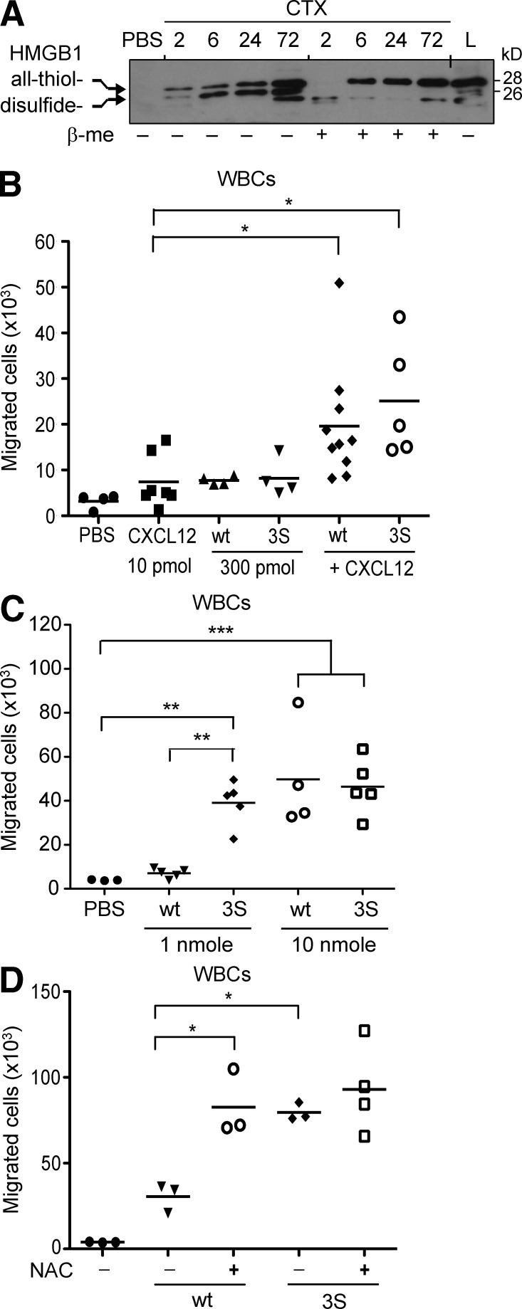
Tissue damage causes inflammation, by recruiting leukocytes and activating them to release proinflammatory mediators. We show that high-mobility group box 1 protein (HMGB1) orchestrates both processes by switching among mutually exclusive redox states. Reduced cysteines make HMGB1 a chemoattractant, whereas a disulfide bond makes it a proinflammatory cytokine and further cysteine oxidation to sulfonates by reactive oxygen species abrogates both activities. We show that leukocyte recruitment and activation can be separated. A nonoxidizable HMGB1 mutant in which serines replace all cysteines (3S-HMGB1) does not promote cytokine production, but is more effective than wild-type HMGB1 in recruiting leukocytes in vivo. BoxA, a HMGB1 inhibitor, interferes with leukocyte recruitment but not with activation. We detected the different redox forms of HMGB1 ex vivo within injured muscle. HMGB1 is completely reduced at first and disulfide-bonded later. Thus, HMGB1 orchestrates both key events in sterile inflammation, leukocyte recruitment and their induction to secrete inflammatory cytokines, by adopting mutually exclusive redox states.
Overview
The claims and statistics of this paper haven’t been checked yet.
Run the check — free, no account needed. It reads the open-access text and checks the sources it cites.
Integrity signals
Registry
Cites
Statistics
Figures
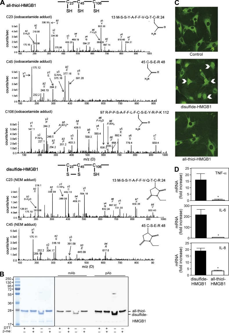
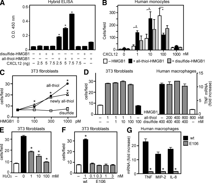
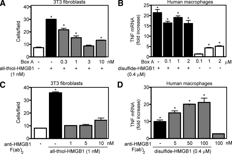
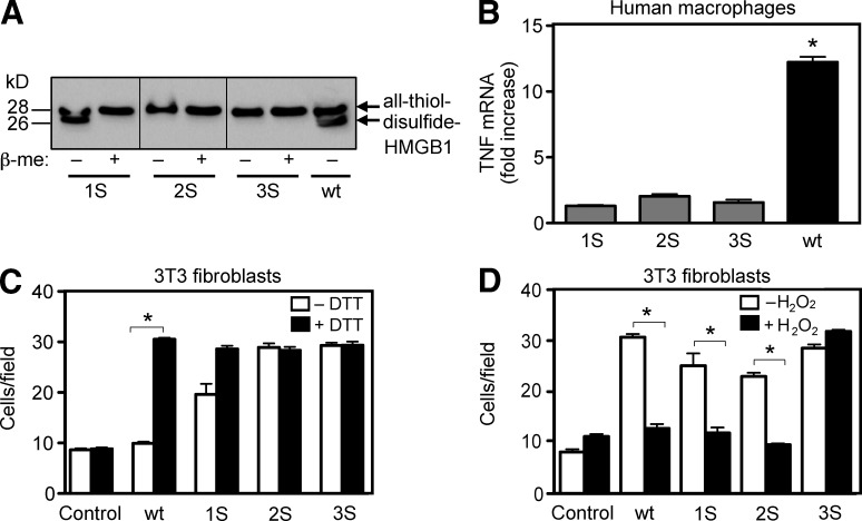
Verification
Claims the paper makes
The claims and statistics of this paper haven’t been checked yet.
Run the check — free, no account needed. It reads the open-access text and checks the sources it cites.
Claims other papers make about this one
Each is checked against this paper’s own text: does it say what the citing paper says it does?
In the citing paper
“That said, it should be noted that biological activities of HMGB1 may flip from pro-inflammatory to anti-inflammatory depending on multiple variables including its oxidation state [917–923].”
From Molecular mechanisms of cell death: recommendations of the nomenclature committee on cell death 2018. (2018) · cites this paper as [918]
In the citing paper
“The disulfide form mediates the proinflammatory effects of HMGB1 by signaling through TLR4, whereas the reduced form mediates the chemotactic properties of HMGB1 by forming a complex with the chemokine CXCL12 and signaling through CXCR4.”
From Danger Signals and Inflammasomes: Stress-Evoked Sterile Inflammation in Mood Disorders (2017) · cites this paper as Venereau et al, 2012
In the citing paper
“HMGB1 can function either as a chemotactic or proinflammatory mediator depending on the redox state of three critical cysteines (Venereau et al, 2012).”
From Danger Signals and Inflammasomes: Stress-Evoked Sterile Inflammation in Mood Disorders (2017) · cites this paper as Venereau et al, 2012
In the citing paper
“The disulfide form mediates the proinflammatory effects of HMGB1 by signaling through TLR4, whereas the reduced form mediates the chemotactic properties of HMGB1 by forming a complex with the chemokine CXCL12 and signaling through CXCR4.”
From Danger Signals and Inflammasomes: Stress-Evoked Sterile Inflammation in Mood Disorders (2017) · cites this paper as Venereau et al, 2012
Self-check
Percentages, sums and reported differences re-computed from the paper’s own numbers.
The self-check needs the paper’s full text.
Citations
Cites
1 retracted
Yang, H., Lundbäck, P., Ottosson, L., Erlandsson-Harris, H., Venereau, E., Bianchi, M. E., Al-Abed, Y., Andersson, U., Tracey, K. J., & Antoine, D. J. (2012) · Molecular Medicine · doi:10.2119/molmed.2011.00389 · Cited by 407
Self-citationRetractedDAMPs, PAMPs and alarmins: all we need to know about danger
Bianchi, M. E. (2007) · Journal of Leukocyte Biology · doi:10.1189/jlb.0306164 · Cited by 2,794 · cited 1×
“High-mobility group box 1 (HMGB1) is a nuclear protein that signals tissue damage when released into the extracellular medium, and thus works as a damage-associated molecular pattern (DAMP; Bianchi, 2007).”
BackgroundSelf-citationHMGB1 is a therapeutic target for sterile inflammation and infection
Andersson, U., & Tracey, K. J. (2011) · Annual Review of Immunology · doi:10.1146/annurev-immunol-030409-101323 · Cited by 1,437 · cited 1×
“Notably, immune cells secrete HMGB1 when activated by infection or tissue damage (Andersson and Tracey, 2011); mesothelioma and other cancer cells secrete HMGB1 constitutively (Jube et al., 2012).”
BackgroundSelf-citationAndersson, U., Wang, H., Palmblad, K., Aveberger, A.-C., Bloom, O., Erlandsson-Harris, H., Janson, A., Kokkola, R., Zhang, M., Yang, H., et al. (2000) · The Journal of Experimental Medicine · doi:10.1084/jem.192.4.565 · Cited by 1,401
Self-citationGardella, S., Andrei, C., Ferrera, D., Lotti, L. V., Torrisi, M. R., Bianchi, M. E., & Rubartelli, A. (2002) · EMBO Reports · doi:10.1093/embo-reports/kvf198 · Cited by 926 · cited 1×
“HMGB1 is released passively into the extracellular medium during necrosis and is secreted actively by monocytes and macrophages after stimulation with LPS and ATP (Gardella et al., 2002).”
BackgroundSelf-citationYang, H., Hreggvidsdottir, H. S., Palmblad, K., Wang, H., Ochani, M., Li, J., Lü, B., Chavan, S. S., Rosas‐Ballina, M., Al‐Abed, Y., et al. (2010) · Proceedings of the National Academy of Sciences · doi:10.1073/pnas.1003893107 · Cited by 812
Self-citationSchiraldi, M., Raucci, A., Martínez‐Muñoz, L., Livoti, E., Celona, B., Vénéreau, E., Apuzzo, T., Marchis, F. D., Pedotti, M., Bachi, A., et al. (2012) · The Journal of Experimental Medicine · doi:10.1084/jem.20111739 · Cited by 676
Self-citationHigh-Mobility Group Box-1 in Ischemia-Reperfusion Injury of the Heart
Andrassy, M., Volz, H. C., Igwe, J. C., Funke, B., Eichberger, S. N., Kaya, Z., Buss, S. J., Autschbach, F., Pleger, S. T., Lukić, I. K., et al. (2008) · Circulation · doi:10.1161/circulationaha.108.769331 · Cited by 635 · cited 1×
“…effects of BoxA in models of hepatitis (Sitia et al., 2007), peritonitis (Orlova et al., 2007), ischemia/reperfusion of the heart and the brain (Andrassy et al., 2008; Muhammad et al., 2008), and other ailments, may be entirely attributable to the reduced recruitment of inflammatory cells in…”
BackgroundSelf-citationKazama, H., Ricci, J.-E., Herndon, J. M., Hoppe, G., Green, D. R., & Ferguson, T. A. (2008) · Immunity · doi:10.1016/j.immuni.2008.05.013 · Cited by 627 · cited 1×
“High ROS levels produced during apoptosis cause terminal oxidation of HMGB1, redirecting its ability to activate acquired immune responses toward tolerance (Kazama et al., 2008).”
BackgroundBianchi, M. E. (2009) · Journal of Leukocyte Biology · doi:10.1189/jlb.1008585 · Cited by 419 · cited 2×
“, 2000), several studies could not reproduce these results, leading us to conclude that HMGB1 can only act in synergy with other inflammatory mediators (Bianchi, 2009).”
ResultSelf-citation
Cited by
Sorted by citations
Items 501–600 of 723
An ongoing search for potential targets and therapies for lethal sepsis
Bao, G., He, L., Lee, D., D’Angelo, J. K., & Wang, H. (2015) · Military Medical Research · doi:10.1186/s40779-015-0047-0 · Cited by 11
Xie, Y., Zhang, K., Zhang, K., Zhang, J., Wang, L., Wang, X., Hu, X., Liang, Z., & Li, J. (2019) · Journal of Cellular Physiology · doi:10.1002/jcp.29234 · Cited by 11
Reed, K. R., Song, F., Young, M. A., Hassan, N., Antoine, D. J., Gemici, N.-P. B., Clarke, A. R., & Jenkins, J. R. (2016) · Oncotarget · doi:10.18632/oncotarget.10076 · Cited by 11
Self-citationLi, D., Bao, X.-Q., Wang, X., Sun, H., & Zhang, D. (2017) · Naunyn-Schmiedeberg s Archives of Pharmacology · doi:10.1007/s00210-017-1363-6 · Cited by 11
Das, S. K., Yuan, Y. F., & Li, M. Q. (2017) · Cellular Reprogramming · doi:10.1089/cell.2016.0050 · Cited by 11
Leo, F. D., Quilici, G., Marchis, F. D., Mantonico, M. V., Bianchi, M. E., & Musco, G. (2020) · Frontiers in Chemistry · doi:10.3389/fchem.2020.598710 · Cited by 11
Self-citationMulti-omic stratification of the missense variant cysteinomePreprint
Desai, H. S., Ofori, S., Boatner, L. M., Yu, F., Villanueva, M., Ung, N., Nesvizhskii, A. I., & Backus, K. M. (2023) · bioRxiv (Cold Spring Harbor Laboratory) · doi:10.1101/2023.08.12.553095 · Cited by 11
Date, S., & Bhatt, L. K. (2024) · Inflammopharmacology · doi:10.1007/s10787-024-01586-w · Cited by 11
Intrinsic disorder and fuzzy interactions drive multiple functions of HMGB1
Ghitti, M., Colley, L. S., Mantonico, M. V., Musco, G., & Bianchi, M. E. (2025) · Trends in Biochemical Sciences · doi:10.1016/j.tibs.2025.08.001 · Cited by 12
Self-citationMardente, S., Aventaggiato, M., Splendiani, E., Mari, E., Zicari, A., Catanzaro, G., Pò, A., Coppola, L., & Tafani, M. (2023) · International Journal of Molecular Sciences · doi:10.3390/ijms24032754 · Cited by 10
The Condition of Neuropathic Pain
Toth, C. C., & Moulin, D. E. (2013) · Cambridge University Press eBooks · doi:10.1017/cbo9781139152211.005 · Cited by 10
Yates, K. P., Deppe, R. B., Comerford, M., Masuoka, H. C., Cummings, O. W., Tonascia, J., Chalasani, N., Vuppalanchi, R., & CRN, F. T. N. (2017) · PLoS ONE · doi:10.1371/journal.pone.0185813 · Cited by 10
Reduced-HMGB1 suppresses poly(I:C)-induced inflammation in keratinocytes
Mori, H., Murakami, M., Tsuda, T., Kameda, K., Utsunomiya, R., Masuda, K., Shiraishi, K., Dai, X., Tohyama, M., Nakaoka, H., et al. (2018) · Journal of Dermatological Science · doi:10.1016/j.jdermsci.2018.01.007 · Cited by 10
Chikhirzhina, E. V., Starkova, T. Y., & Polyanichko, A. M. (2021) · BIOPHYSICS · doi:10.1134/s0006350921030039 · Cited by 10
Sharafkhah, M., Mosayebi, G., Massoudifar, A., Seddigh, S. H., Abdolrazaghnejad, A., Alamdara, M. A., Mokarian, P., Mohammadbeigi, A., & Ebrahimi-Monfared, M. (2022) · Neurology India · doi:10.4103/0028-3886.338707 · Cited by 10
Nonoxid-HMGB1 Attenuates Cognitive Impairment After Traumatic Brain Injury in Rats
Chen, J.-Q., Gao, S., Luo, L., Jiang, Z., Liang, C., He, H., & Guo, Y. (2022) · Frontiers in Medicine · doi:10.3389/fmed.2022.827585 · Cited by 10
Edye, M. E., Walker, L. E., Sills, G. J., Allan, S. M., & Brough, D. (2014) · Inflammasome · doi:10.2478/infl-2014-0004 · Cited by 11
Kršek, A., Schleicher, L. M. S., Jagodic, A., & Batičić, L. (2025) · International Journal of Molecular Sciences · doi:10.3390/ijms26189178 · Cited by 13
Battista, C., Howell, B. A., Siler, S. Q., & Watkins, P. B. (2018) · Methods in pharmacology and toxicology · doi:10.1007/978-1-4939-7677-5_6 · Cited by 9
High Mobility Group Box 1: An Immune-regulatory Protein
Jingjing, Z., Sun, T., Wu, S., & Liu, Y. (2019) · Current Gene Therapy · doi:10.2174/1566523219666190621111604 · Cited by 9
HMGB1 in Septic Muscle Atrophy: Roles and Therapeutic Potential for Muscle Atrophy and Regeneration
Qi, S., Wu, Q., Xiang, P., Hou, C., Kang, Z., Chen, M., Yi, C., Bai, X., Li, T., Li, Z., et al. (2025) · Journal of Cachexia Sarcopenia and Muscle · doi:10.1002/jcsm.13711 · Cited by 11
Oncolytic Virotherapy and Immunogenic Cell Death: Mechanisms, Platforms, and Clinical Translation
Inoue, H. (2026) · Viruses · doi:10.3390/v18040461 · Cited by 10
A Murine Model of Vesicant-Induced Acute Lung Injury
Zafar, I., Manzoor, S., Mariappan, N., et al. (2023) · Journal of Pharmacology and Experimental Therapeutics · doi:10.1124/jpet.123.001780 · Cited by 8
Foelsch, K., Pelczar, P., Zierz, E., Kondratowicz, S., Qi, M., Mueller, C., Alawi, M., Hübener, S., Clauditz, T. S., Gagliani, N., et al. (2024) · Journal of Crohn s and Colitis · doi:10.1093/ecco-jcc/jjae017 · Cited by 8
Rouhiainen, A., Nykänen, N.-P., Kuja‐Panula, J., Vanttola, P., Huttunen, H. J., & Rauvala, H. (2018) · Medicines · doi:10.3390/medicines5030079 · Cited by 8
Kishi, S., Fujiwara‐Tani, R., Honoki, K., et al. (2022) · Cancer Science · doi:10.1111/cas.15400 · Cited by 8
Cribb, P., Perdomo, V., Alonso, V. L., et al. (2017) · PLoS neglected tropical diseases · doi:10.1371/journal.pntd.0005350 · Cited by 8
Liming, S., Li, G., Wenxin, S., & Guirong, T. (2018) · Experimental Lung Research · doi:10.1080/01902148.2018.1505976 · Cited by 7
Contribution of Connexin Hemichannels to the Pathogenesis of Acute Lung Injury
Wang, S., Sun, Y., Bai, Y., Zhou, N., Chen, N., Miller, E. J., Zhang, Y., & Li, W. (2020) · Mediators of Inflammation · doi:10.1155/2020/8094347 · Cited by 8
Zhang, J., Shimozaki, K., Hattori, S., Pastukh, V., Maloney, D., Hogan, M. V., & Wang, J. H. (2024) · PLoS ONE · doi:10.1371/journal.pone.0311147 · Cited by 9
Hao, W., Yan, A., Guo, X., Chen, Z., Chen, N., Li, H., Wu, F., Sun, P., Zhao, Y., Zhao, G., et al. (2025) · Journal of Controlled Release · doi:10.1016/j.jconrel.2025.114199 · Cited by 7
Apodaca, E., Demichelis‐Gómez, R., Rosas‐López, A., Mejía‐Domínguez, N. R., Galvan-López, I., Addorosio, M., Tracey, K. J., & Valdés‐Ferrer, S. I. (2022) · Therapeutic Advances in Hematology · doi:10.1177/20406207221125990 · Cited by 7
Self-citationYe, J., Gao, S., Liu, Z.-C., Chen, X., He, J., & Hu, Z. (2024) · Brain and Behavior · doi:10.1002/brb3.3457 · Cited by 7
Zeng, X., Luo, D., Zhang, S., et al. (2024) · PLoS ONE · doi:10.1371/journal.pone.0307024 · Cited by 7
Zhang, S., Feng, F., Dai, J., Li, J., Bu, X., & Xie, X. (2021) · Oxidative Medicine and Cellular Longevity · doi:10.1155/2021/9993240 · Cited by 8
Cheng, K. J., Mohamed, E. H. M., Syafruddin, S. E., & Ibrahim, Z. A. (2022) · Journal of Cell Communication and Signaling · doi:10.1007/s12079-022-00681-3 · Cited by 6
Ex vivo activation of dendritic cells via coacervate-mediated exogenous tumor cell lysate delivery
Seong, J., Jeong, S., Kim, S., Yun, S., Baek, Y., & Kim, K. (2023) · Biomaterials Science · doi:10.1039/d3bm00234a · Cited by 7
HMGB1 cleavage by complement C1s and its potent anti-inflammatory product
Lorvellec, M., Chouquet, A., Koch, J., Bally, I., Signor, L., Vigne, J., Dalonneau, F., Thielens, N. M., Rabilloud, T., Dalzon, B., et al. (2023) · Frontiers in Immunology · doi:10.3389/fimmu.2023.1151731 · Cited by 7
Barozzi, I., Slaven, N., Canale, E., Lopes, R., Barbosa, I. A. M., Bleu, M., Ivanoiu, D., Pacini, C., Mensa’, E., Chambers, A., et al. (2024) · Cancer Discovery · doi:10.1158/2159-8290.cd-23-1157 · Cited by 7
Tripisciano, C., Eichhorn, T., Harm, S., & Weber, V. (2014) · BioMed Research International · doi:10.1155/2014/238160 · Cited by 7
Wang, S., Zhao, J., Wang, C., Yao, Y., Song, Z., Li, L., & Jiang, J. (2024) · Neurochemical Research · doi:10.1007/s11064-024-04174-0 · Cited by 6
Bianchi, M. E., Rubartelli, A., & Sitia, R. (2024) · Antioxidants and Redox Signaling · doi:10.1089/ars.2024.0554 · Cited by 6
Self-citationHigh Mobility Group Box 1 and Cardiovascular Diseases: Study of Act and Connect
Wasim, R., Singh, A., Islam, A., Mohammed, S., Anwar, A., & Mahmood, T. (2024) · Cardiovascular Toxicology · doi:10.1007/s12012-024-09919-5 · Cited by 7
RIPK3-MLKL dependent necroptosis mediates depressive-like behavior by facilitating neuroinflammation
Zheng, H., Xu, Z.-Y., Hu, T., Wu, Y., Huang, C.-W., Li, J., Cao, Z., Wang, W., Jiang, C., & Su, W. (2025) · Journal of Neuroimmunology · doi:10.1016/j.jneuroim.2025.578643 · Cited by 6
Shi, Q., Wu, Q., Fang, F., et al. (2025) · Biochemical and Biophysical Research Communications · doi:10.1016/j.bbrc.2025.152392 · Cited by 6
HMGB1 as an emerging key modulator of bone remodeling: a narrative review
Tang, J., Li, M., & Huang, L. C. (2025) · Stem Cell Research & Therapy · doi:10.1186/s13287-025-04543-8 · Cited by 7
Mechanism of action of HMGB1 in urologic malignancies
Li, D., Lei, X., Zhao, L., Fu, Q., Chen, Y., & Yang, X. (2025) · Frontiers in Oncology · doi:10.3389/fonc.2025.1593157 · Cited by 7
Neuro-immune regulation of sepsis-associated delirium via the PBN-CeA-spleen axis
Dong, M., Zou, Y. F., Ye, Z., Zou, W., Zhang, L., Ai, Y., & Peng, Q. (2025) · Journal of Neuroinflammation · doi:10.1186/s12974-025-03527-y · Cited by 9
Lu, F., Wang, X., Xue, X., Liu, L. Z., Li, D., Liu, A. F., Qin, S., & Liu, L. (2026) · Cells · doi:10.3390/cells15060545 · Cited by 7
Oxidative stress and metabolic dysfunction in oral lichen planus pathogenesis
Song, H.-J., Kang, K.-H., Byun, J., & Kim, D. H. (2026) · Animal Cells and Systems · doi:10.1080/19768354.2026.2648907 · Cited by 6
Xie, Y., Zhang, K., Zhang, J., Zhang, K., Hu, X., Wang, L., Wang, X., & Li, J. (2022) · Animal Reproduction Science · doi:10.1016/j.anireprosci.2022.107152 · Cited by 6
Wang, B., Feng, S., Jiang, Y., et al. (2024) · Molecular Immunology · doi:10.1016/j.molimm.2024.10.001 · Cited by 6
Morioka, N., Nakamura, Y., Hisaoka‐Nakashima, K., & Nakata, Y. (2024) · Biochemical Pharmacology · doi:10.1016/j.bcp.2024.116058 · Cited by 6
Turan, A., Tarique, M., Zhang, L., Kazmi, S., Ulker, V., Tedla, M. G., Badal, D., Yolcu, E. S., & Shirwan, H. (2024) · The Journal of Immunology · doi:10.4049/jimmunol.2300743 · Cited by 6
Kang, R., & Tang, D. (2016) · Springer eBooks · doi:10.1007/978-3-319-30079-5_5 · Cited by 6
Increased Lacrimal Fluid Level of HMGB1 in Vernal Keratoconjunctivitis
Caputo, R., Pasti, M., de Libero, C., et al. (2018) · Ocular Immunology and Inflammation · doi:10.1080/09273948.2018.1467465 · Cited by 6
Appavoo, E., Hajam, I. A., Muneeswaran, N. S., Kondabattula, G., Bhanuprakash, V., & Kishore, S. (2015) · Microbiology and Immunology · doi:10.1111/1348-0421.12350 · Cited by 6
Alruwaili, F. N., Nour, O. A., & Ibrahim, T. M. (2025) · Pharmaceuticals · doi:10.3390/ph18081164 · Cited by 5
Salminen, A. (2026) · Neurochemistry International · doi:10.1016/j.neuint.2026.106115 · Cited by 5
Zhou, L., Liu, Q., He, Y., Tang, Y., Liu, S., Wang, X., Shen, S., Ji, J., Liu, Z., Zeng, X., et al. (2026) · Frontiers in Immunology · doi:10.3389/fimmu.2026.1737661 · Cited by 7
Toprak, S. F., Koçana, C. Ç., Demirtaş, E. N., et al. (2025) · Thrombosis Research · doi:10.1016/j.thromres.2025.109407 · Cited by 5
Sirait, R. H., Hatta, M., Ramli, M., Simanjuntak, T. P., Utomo, B. S. R., Islam, A. A., & Arief, S. K. (2017) · Open Journal of Anesthesiology · doi:10.4236/ojanes.2017.72004 · Cited by 5
HMGB1: a potential new target for tendinopathy treatment
Lu, P., Li, Y., Dai, G.-C., Zhang, Y., Shi, L., Zhang, M., Wang, H., & Rui, Y. (2023) · Connective Tissue Research · doi:10.1080/03008207.2023.2199089 · Cited by 5
Croca, S. C., Bassett, P., Pericleous, C., Alber, K. F., Latchman, D. S., Isenberg, D. A., Giles, I., Rahman, A., & Ioannou, Y. (2014) · Arthritis Research & Therapy · doi:10.1186/ar4477 · Cited by 5
Panneerselvam, S., Durai, P., Yesudhas, D., Achek, A., Kwon, H., & Choi, S. (2016) · RSC Advances · doi:10.1039/c6ra16343b · Cited by 5
Upregulation of High Mobility Group Box 1 May Contribute to the Pathogenesis of Biliary Atresia
Ye, C. J., Wang, J., Yang, Y., Shen, Z. Z., Chen, G., Huang, Y. L., Zheng, Y., Dong, R., & Zheng, S. (2018) · European Journal of Pediatric Surgery · doi:10.1055/s-0038-1660447 · Cited by 5
The challenges of long-term sepsis survivors: when surviving is just the beginning.
Valdés‐Ferrer, S. I. (2014) · PubMed · Cited by 5
Co-Targeting IL-6 and EGFR signaling for the treatment of schwannomatosis and associated painPreprint
Yin, Z., Wu, L., Zhang, Y., Sun, Y., Chen, J. W., Subudhi, S., Ho, W. W., Lee, G. Y., Wang, A. C., Gao, X., et al. (2023) · bioRxiv (Cold Spring Harbor Laboratory) · doi:10.1101/2023.02.06.527377 · Cited by 5
Correlation of HMGB1, TLR2 and TLR4 with left ventricular diastolic dysfunction in sepsis patients
Lv, X., Shi, X., Maihemuti, M., et al. (2023) · Scandinavian Journal of Immunology · doi:10.1111/sji.13260 · Cited by 4
Post-translational modifications drive the effects of HMGB1 in alcohol-associated liver disease
Ge, X., Nithyananthan, S., Song, Z., Désert, R., Han, H., Das, S., Komakula, S. S. B., Wang, C., Lantvit, D. D., Ge, Z., et al. (2024) · Hepatology Communications · doi:10.1097/hc9.0000000000000549 · Cited by 4
Autoimmune skin disease pathogenesis: a chronological immune cascade and multi-hit model
Chen, J., Yin, H., Li, S., & Li, C. (2026) · Immunity & Inflammation · doi:10.1007/s44466-025-00017-x · Cited by 5
Fassi, E. M. A., Pirani, E., Cecchinato, V., et al. (2025) · Computational and Structural Biotechnology Journal · doi:10.1016/j.csbj.2025.04.023 · Cited by 4
Self-citationDualistic Roles of High Mobility Group Box 1 in Cancer and Inflammation
Zeng, W., Zhang, X., Jiang, Y., Luo, Y., Wang, Z., Du, X., & Chen, L. (2025) · Cancer Medicine · doi:10.1002/cam4.71455 · Cited by 4
Perish in the Attempt: Regulated Cell Death in Regenerative and Nonregenerative Tissue
Li, S., & Tao, G. (2023) · Antioxidants and Redox Signaling · doi:10.1089/ars.2022.0166 · Cited by 5
Liver Transplantation: A Test of Cellular Physiology, Preservation, and Injury
Martins, B., Mossemann, J., Aguilar, F., Zhao, S., Bilan, P. J., & Sayed, B. A. (2024) · Physiology · doi:10.1152/physiol.00020.2024 · Cited by 4
HMGB1-BoxA gene therapy in reversing cisplatin resistance in non-small cell lung cancer
Puthdee, N., Mai, V.-H., Settayanon, S., et al. (2025) · PLoS ONE · doi:10.1371/journal.pone.0327144 · Cited by 4
Sirait, R. H., Hatta, M., Arief, S. K., Simanjuntak, T. P., & Utomo, B. S. R. (2018) · Pharmacognosy Journal · doi:10.5530/pj.2018.3.96 · Cited by 4
Liu, L., Zhao, Z., Lu, L., Liu, J., Wu, X. F., Sun, J., Wei, Y., & Dong, J. (2018) · Traditional Medicine and Modern Medicine · doi:10.1142/s2575900018300035 · Cited by 4
Self-citationTargeting HMGB1 in the Treatment of Non-Small Cell Lung Adenocarcinoma
Anderson, B. J., Vue, M., Gayluak, N., Brown, S. J., Bemis, L., & Simmons, G. E., Jr. (2021) · Onco · doi:10.3390/onco1010004 · Cited by 4
Lucas, K., Ackermann, M., Leifke, A. L., Li, W. W., Pöschl, U., & Fröhlich‐Nowoisky, J. (2021) · bioRxiv (Cold Spring Harbor Laboratory) · doi:10.1101/2021.06.16.448642 · Cited by 4
Stem/progenitor cell-based therapy for Duchenne muscular dystrophy
Tominari, T., Sathyaprakash, C., & Aoki, Y. (2025) · Frontiers in Cell and Developmental Biology · doi:10.3389/fcell.2025.1640275 · Cited by 5
Öner, F., Özgenç, G., Aslan, H., Guzeltan, O., & Karasu, Y. Ö. (2025) · BMC Oral Health · doi:10.1186/s12903-025-06954-8 · Cited by 3
HMGB1 and mitochondrial dysfunction: a double-edged sword in ageing
Wahid, M., & Cunningham, M. (2025) · Open Biology · doi:10.1098/rsob.240376 · Cited by 3
Oxycodone induces HMGB1-mediated neuroimmune crosstalk between oligodendrocytes and microglia
Maulik, M., Rose, C., Mallick, S., Jones, A., Dodge, A., Chakraborty, S., Carter, A., Jeffers, A., Ford, M., Zhang, E., et al. (2026) · Neuroscience · doi:10.1016/j.neuroscience.2026.02.037 · Cited by 3
Schmitz, E. G., Ridout, A., Smith, A. L., Eiken, A. P., Skupa, S. A., Drengler, E. M., Singh, S., Rana, S., Natarajan, A., & El-Gamal, D. (2024) · Biomedicines · doi:10.3390/biomedicines12122857 · Cited by 3
Festoff, B. W., Sajja, R. K., & Cucullo, L. (2017) · BioMed Research International · doi:10.1155/2017/1549194 · Cited by 3
5-Aminosalicylic Acid Modulates the Immune Response in Chronic Beryllium Disease Subjects
Day, B. J., Huang, J., Barkes, B. Q., et al. (2017) · Lung · doi:10.1007/s00408-017-0062-x · Cited by 3
Gacaferi, H., Mimpen, J. Y., Baldwin, M. J., Snelling, S. J. B., Nelissen, R. G. H. H., Carr, A. J., & Dakin, S. G. (2020) · Translational Sports Medicine · doi:10.1002/tsm2.175 · Cited by 3
Oncolytic Viruses as Immunotherapeutic Agents
Trehub, Y., & Havrilov, A. (2020) · Springer eBooks · doi:10.1007/978-3-030-50287-4_27 · Cited by 3
Kümmel, A., Groß, S., Feldtmann, R., Chamling, B., Strohbach, A., Lehnert, K., Bahls, M., Loerzer, L., Moormann, K., Witte, J., et al. (2022) · The American Journal of Cardiology · doi:10.1016/j.amjcard.2022.05.014 · Cited by 3
The C-Terminal Acidic Tail Modulates the Anticancer Properties of HMGB1
Borde, C., Dillard, C., L’honoré, A., Quignon, F., Hamon, M., Marchand, C. H., Faccion, R. S., Costa, M. G. S., Pramil, É., Larsen, A. K., et al. (2022) · International Journal of Molecular Sciences · doi:10.3390/ijms23147865 · Cited by 3
Thanksgiving to Yeast, the HMGB Proteins History from Yeast to Cancer
Maceiras, M. L., Vizoso‐Vázquez, Á., Barreiro-Alonso, A., Cámara-Quílez, M., & Cerdán, M. E. (2023) · Microorganisms · doi:10.3390/microorganisms11040993 · Cited by 4
Arteaga, A., Biguetti, C. C., Chandrashekar, B. L., et al. (2023) · ACS Biomaterials Science & Engineering · doi:10.1021/acsbiomaterials.3c00367 · Cited by 3
Arteaga, A., Biguetti, C. C., Chandrashekar, B. L., et al. (2023) · Bone · doi:10.1016/j.bone.2023.116917 · Cited by 3
From Genome to Geroscience: How DNA Damage Shapes Systemic Decline
Siametis, A., & Garinis, G. A. (2025) · BioEssays · doi:10.1002/bies.70051 · Cited by 5
Tang, D., & Kang, R. (2015) · InTech eBooks · doi:10.5772/61208 · Cited by 3
Lotsberg, M. L., Chen, S., Dhakal, S., Lorens, J. B., Baguley, B. C., Chouaı̈b, S., & Engelsen, A. S. T. (2020) · Elsevier eBooks · doi:10.1016/b978-0-12-819609-0.00008-0 · Cited by 3
Antoine, D. J. (2018) · Methods in pharmacology and toxicology · doi:10.1007/978-1-4939-7677-5_26 · Cited by 3
Self-citationTian, Y., Jiang, L., Zhang, S., Cao, Y., Liu, F., Xia, L., & Su, Z. (2022) · doi:10.22541/au.166126875.56478352/v1 · Cited by 3
Natural compounds and programmed necrosis: pioneering a new frontier in cancer treatments
Amin, M. A., Bouhenni, H., Zehravi, M., Sweilam, S. H., Durgawale, T. P., Qureshi, M. S., Durgapal, S., Haque, M. A., Vodeti, R., Urs, D., et al. (2025) · Naunyn-Schmiedeberg s Archives of Pharmacology · doi:10.1007/s00210-025-04050-w · Cited by 3
Metadata
- Authors
- Venereau, E., Casalgrandi, M., Schiraldi, M., Antoine, D. J., Cattaneo, A., De Marchis, F., Liu, J., Antonelli, A., Preti, A., Raeli, L., Shams, S. S., Yang, H., Varani, L., Andersson, U., Tracey, K. J., Bachi, A., Uguccioni, M., & Bianchi, M. E.
- Affiliations
- Division of Genetics and Cell Biology, San Raffaele Scientific Institute, 20132 Milan, Italy 1
- HMGBiotech Srl, 20133 Milan, Italy 2
- Institute for Research in Biomedicine (CH)
- University of Liverpool (GB)
- IRCCS Ospedale San Raffaele (IT)
- Vita-Salute San Raffaele University (IT)
- Feinstein Institute for Medical Research (US)
- Karolinska University Hospital (SE)
- Corresponding author
- Bianchi, Marco E.
- ORCID
- Venue
- Journal of Experimental Medicine
- Publisher
- Rockefeller University Press
- Published
- 2012-08-06
- Volume
- 209
- Issue
- 9
- Pages
- 1519-1528
- ISSN
- 0022-1007 (ISSN-L)
- 1540-9538
- PMC
- PMC3428943
- OpenAlex
- W2141438911
- Semantic Scholar
- 17d0978fd742005f41f9a10b50083eec9ce6ff3d
- Cited by
- 720
- References
- 29
- Citations by year
- 2026: 48 · 2025: 38 · 2024: 54 · 2023: 46 · 2022: 38 · 2021: 44 · 2020: 56 · 2019: 40 · 2018: 60 · 2017: 58 · 2016: 52 · 2015: 64 · 2014: 66 · 2013: 46 · 2012: 10
- FWCI
- 38.20
- Type
- journal article
- Language
- en
- Open access
- bronze
- doi.org
- Locations
- The Journal of Experimental Medicine (publishedVersion)
- Europe PMC (PubMed Central) (publishedVersion, cc-by-nc-sa)
- PubMed Central (publishedVersion, cc-by-nc-sa)
- PubMed (publishedVersion)
- reroDoc Digital Library (submittedVersion)
- Funders
- European Commission (281608, 280873)
- National Institute of General Medical Sciences (R01GM062508)
- Topics
- Life Sciences › Biochemistry, Genetics and Molecular Biology › Clinical Biochemistry › Advanced Glycation End Products research
- Life Sciences › Immunology and Microbiology › Immunology › Immune Response and Inflammation
- Life Sciences › Immunology and Microbiology › Immunology › Neutrophil, Myeloperoxidase and Oxidative Mechanisms
- Keywords
- HMGB1, high mobility group box 1, redox regulation, cysteine oxidation, disulfide bonds, leukocyte recruitment, sterile inflammation, tissue damage, muscle damage, reactive oxygen species, chemoattractants, leukocyte activation
- MeSH terms
- Animals, Antibodies, Monoclonal, Cell Movement, Chemotactic Factors, Cysteine, Disulfides, Inflammation, Leukocytes, Male, Mice, Inbred C57BL, Mutation, Recombinant Proteins, Cytokines, Reactive Oxygen Species, Muscle, Skeletal, HMGB1 Protein, Mice, Rats
About these checks
Registry, retraction, Cites and statistics results are computed, not judged, with one exception: a printed reference that no index lists is read once by a model before it counts as a citation error. Claims are extracted by a model and kept only when their quote appears verbatim in the text; each cited source is then checked, with a model’s help, for whether it says what the claim relies on. Those verdicts can be wrong — read the claim’s quote and the cited source before relying on one.
Statistics and arithmetic are recomputed, not judged.
Confidence bands are shown only for fields calibrated on our annotated test set. None is calibrated yet, so no band is shown.
What counts as a claim ›How we measure confidence ›How we check this ›
Are you an author, or do you see a problem with this page? Request a review
