

The DOI matches its Crossref record; 54 of the 54 references it cites were found in a registry or an index.
Impact of naturally forming human α/β-tryptase heterotetramers in the pathogenesis of hereditary α-tryptasemia
Open accessAbstract
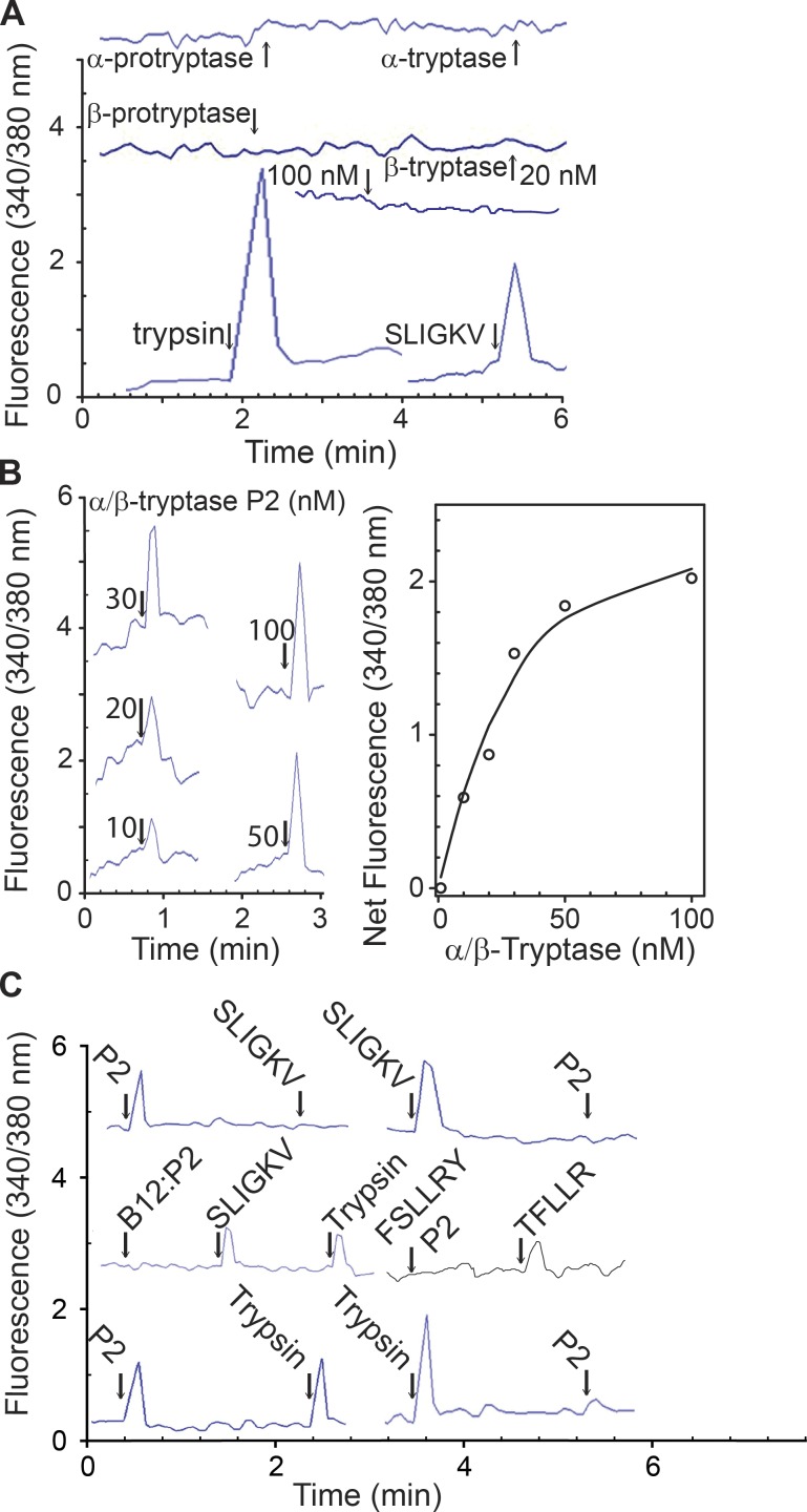
Both α-tryptase and β-tryptase are preferentially expressed by human mast cells, but the purpose of α-tryptase is enigmatic, because its tetramers lack protease activity, whereas β-tryptase tetramers are active proteases. The monogenic disorder called hereditary α-tryptasemia, due to increased α-tryptase gene copies and protein expression, presents with clinical features such as vibratory urticaria and dysautonomia. We show that heterotetramers composed of 2α- and 2β-tryptase protomers (α/β-tryptase) form naturally in individuals who express α-tryptase. α/β-Tryptase, but not homotetramer, activates protease-activated receptor-2 (PAR2), which is expressed on cell types such as smooth muscle, neurons, and endothelium. Also, only α/β-tryptase makes mast cells susceptible to vibration-triggered degranulation by cleaving the α subunit of the EGF-like module-containing mucin-like hormone receptor-like 2 (EMR2) mechanosensory receptor. Allosteric effects of α-tryptase protomers on neighboring β-tryptase protomers likely result in the novel substrate repertoire of α/β-tryptase tetramers that in turn cause some of the clinical features of hereditary α-tryptasemia and of other disorders involving mast cells.
Overview
The claims and statistics of this paper haven’t been checked yet.
Run the check — free, no account needed. It reads the open-access text and checks the sources it cites.
Integrity signals
Registry
Cites
Statistics
Figures
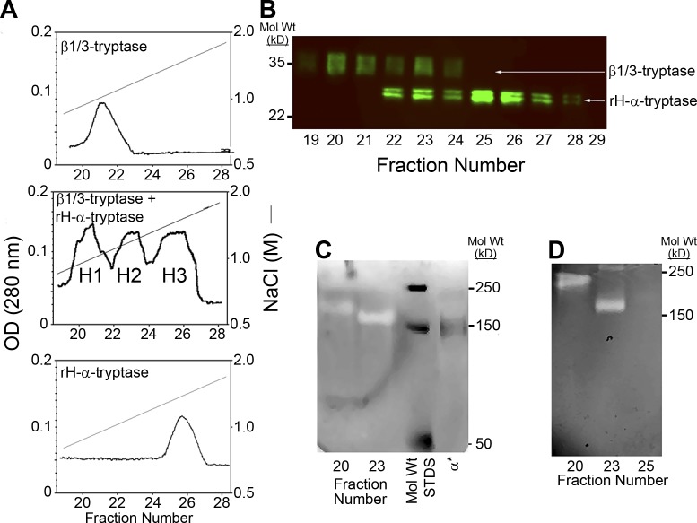
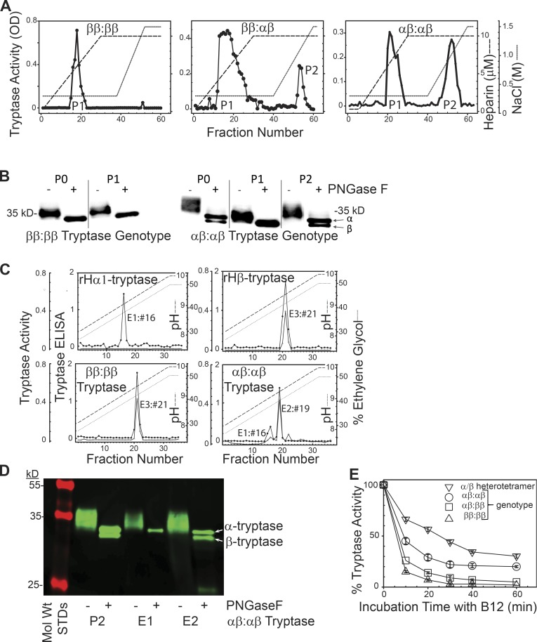
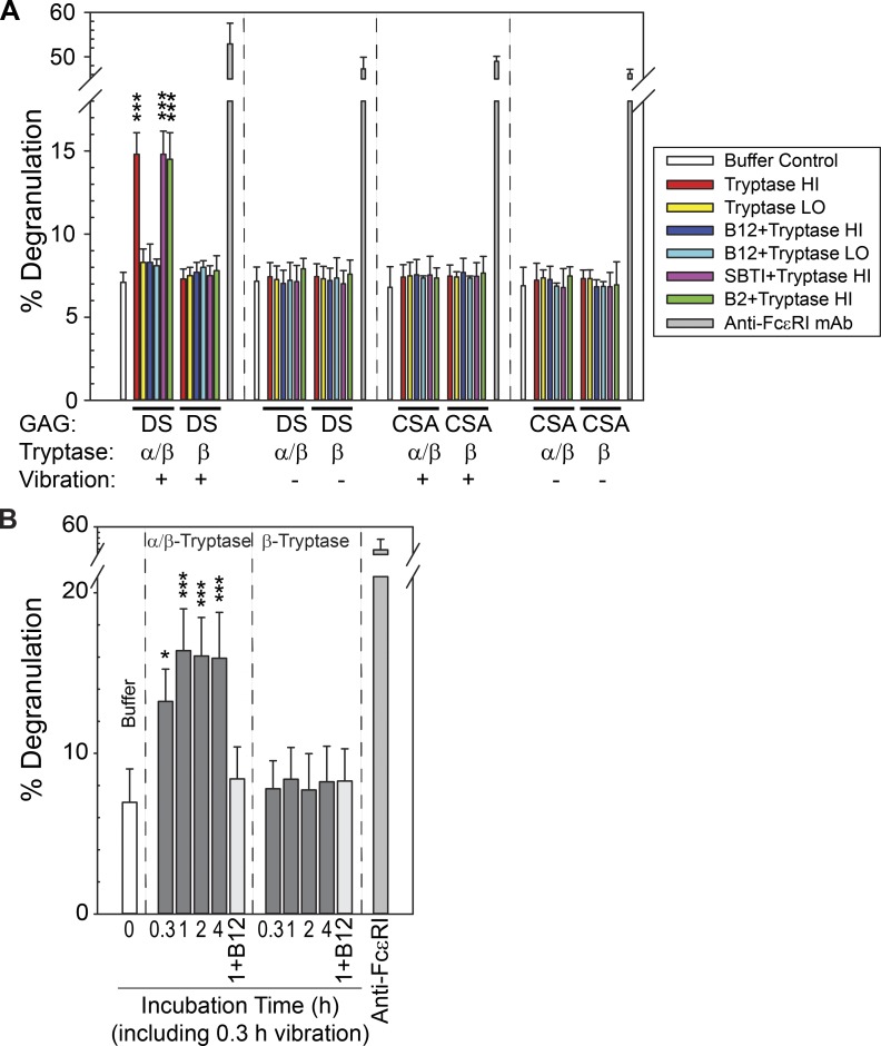
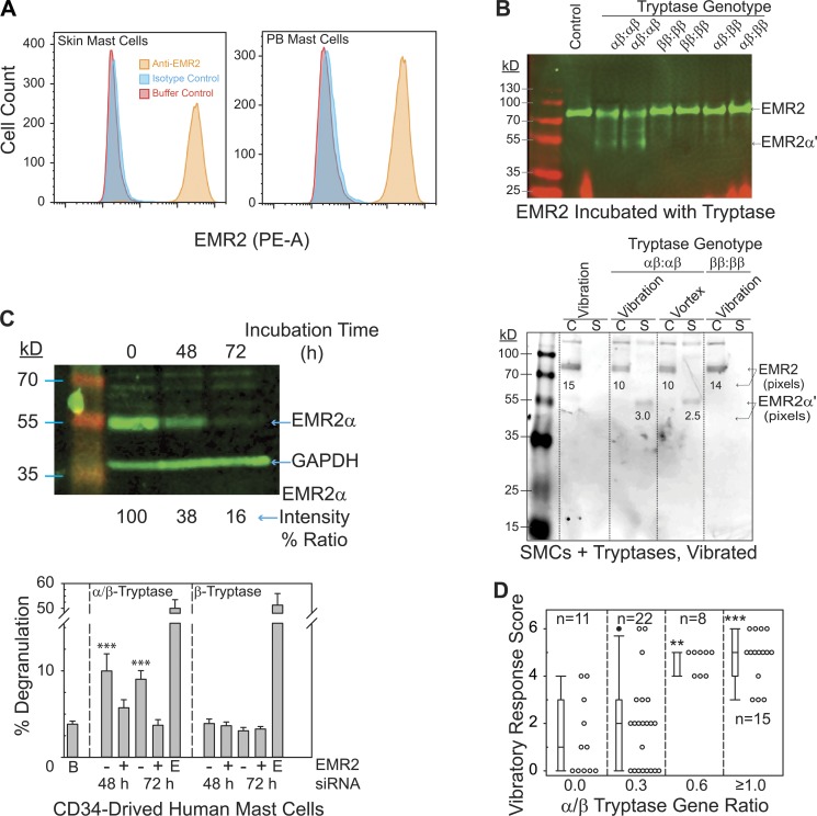
Verification
Claims the paper makes
The claims and statistics of this paper haven’t been checked yet.
Run the check — free, no account needed. It reads the open-access text and checks the sources it cites.
Claims other papers make about this one
Each is checked against this paper’s own text: does it say what the citing paper says it does?
The source backs part of the claim, not all of it — or backs it for a different population, condition or comparison.
In the citing paper
“Recent studies have shown that in individuals with HαT, these heterotetramers increase vascular permeability by cleaving and activating protease-activated receptor-2 (PAR2) on endothelial cells, resulting in increased vascular leakage (14, 15).”
From Defining “Normal” basal serum tryptase levels: a context-dependent approach to improve diagnostics in systemic mastocytosis (2025) · cites this paper as [14]
Experimental — accuracy not yet measuredThe cited source says what the claim attributes to it.
In the citing paper
“A recent description of a / b -tryptase heterotetrameric tryptases, which have expanded biologic functions relative to mature b -tryptases, may offer a possible mechanism explaining MCA in these individuals. 128,129”
From Selecting the Right Criteria and Proper Classification to Diagnose Mast Cell Activation Syndromes: A Critical Review (2021) · cites this paper as [128]
Self-check
Percentages, sums and reported differences re-computed from the paper’s own numbers.
The self-check needs the paper’s full text.
Citations
Cites
Mast-Cell Infiltration of Airway Smooth Muscle in Asthma
Brightling, C. E., Bradding, P. H., Symon, F. A., Holgate, S. T., Wardlaw, A. J., & Pavord, I. D. (2002) · New England Journal of Medicine · doi:10.1056/nejmoa012705 · Cited by 1,240
Identification of a mast cell specific receptor crucial for pseudo-allergic drug reactions
McNeil, B. D., Pundir, P., Meeker, S., Han, L., Undem, B., Kulka, M., & Dong, X.-Z. (2014) · Nature · doi:10.1038/nature14022 · Cited by 1,164
Agonists of proteinase-activated receptor 2 induce inflammation by a neurogenic mechanism
Steinhoff, M. S., Vergnolle, N., Young, S. H., Tognetto, M., Amadesi, S., Ennes, H. S., Trevisani, M., Hollenberg, M. D., Wallace, J. L., Caughey, G. H., et al. (2000) · Nature Medicine · doi:10.1038/72247 · Cited by 936
Tryptase levels as an indicator of mast-cell activation in systemic anaphylaxis and mastocytosis
Schwartz, L. B., Metcalfe, D. D., Miller, J. S., Earl, H., & Sullivan, T. (1987) · New England Journal of Medicine · doi:10.1056/nejm198706253162603 · Cited by 784
Self-citationProteinase-Activated Receptor-2 Mediates Itch: A Novel Pathway for Pruritus in Human Skin
Steinhoff, M. S., Neisius, U., Ikoma, A., Fartasch, M., Heyer, G., Skov, P. S., Luger, T. A., & Schmelz, M. (2003) · Journal of Neuroscience · doi:10.1523/jneurosci.23-15-06176.2003 · Cited by 652
Interactions of Mast Cell Tryptase with Thrombin Receptors and PAR-2
Molino, M., Barnathan, E. S., Numerof, R. P., Clark, J., Dreyer, M. L., Cumashi, A., Hoxie, J. A., Schechter, N. M., Woolkalís, M. J., & Brass, L. F. (1997) · Journal of Biological Chemistry · doi:10.1074/jbc.272.7.4043 · Cited by 592
Quantitation of histamine, tryptase, and chymase in dispersed human T and TC mast cells.
Schwartz, L. B., Irani, A. M., Roller, K., Castells, M., & Schechter, N. M. (1987) · The Journal of Immunology · doi:10.4049/jimmunol.138.8.2611 · Cited by 500
Proteinase-activated receptor-2 and hyperalgesia: A novel pain pathway
Vergnolle, N., Bunnett, N. W., Sharkey, K. A., Brussee, V., Compton, S. J., Grady, E. F., Cirino, G., Gerard, N. P., Basbaum, A. I., Andrade‐Gordon, P., et al. (2001) · Nature Medicine · doi:10.1038/89945 · Cited by 465
Schwartz, L. B., Yunginger, J. W., Miller, J., Bokhari, R., & Dull, D. (1989) · Journal of Clinical Investigation · doi:10.1172/jci114051 · Cited by 456
Fujisawa, D., Kashiwakura, J., Kita, H., Kikukawa, Y., Fujitani, Y., Sasaki-Sakamoto, T., Kuroda, K., Nunomura, S., Hayama, K., Terui, T., et al. (2014) · Journal of Allergy and Clinical Immunology · doi:10.1016/j.jaci.2014.05.004 · Cited by 410
Cited by
Sorted by citations
Lyons, J. J., Chovanec, J., O’Connell, M. P., Liu, Y., Šelb, J., Zanotti, R., Bai, Y., Kim, J., Le, Q. T., DiMaggio, T., et al. (2021) · Journal of Allergy and Clinical Immunology · doi:10.1016/j.jaci.2020.06.035 · Cited by 230
Self-citationPlum, T., Wang, X., Rettel, M., Krijgsveld, J., Feyerabend, T. B., & Rodewald, H.-R. (2020) · Immunity · doi:10.1016/j.immuni.2020.01.012 · Cited by 200
Greiner, G., Sprinzl, B., Górska, A., Ratzinger, F., Gurbisz, M., Witzeneder, N., Schmetterer, K. G., Gisslinger, B., Uyanik, G., Hadzijusufovic, E., et al. (2021) · Blood · doi:10.1182/blood.2020006157 · Cited by 189
The ingenious mast cell: Contemporary insights into mast cell behavior and function
Dahlin, J. S., Maurer, M., Metcalfe, D. D., Pejler, G., Sagi‐Eisenberg, R., & Nilsson, G. P. (2021) · Allergy · doi:10.1111/all.14881 · Cited by 159
Weiler, C. R., Austen, K. F., Akin, C., Barkoff, M. S., Bernstein, J. A., Bonadonna, P., Butterfield, J. H., Carter, M., Fox, C. C., Maitland, A., et al. (2019) · Journal of Allergy and Clinical Immunology · doi:10.1016/j.jaci.2019.08.023 · Cited by 123
Metadata
- Authors
- Le, Q. T., Lyons, J. J., Naranjo, A. N., Olivera, A., Lazarus, R. A., Metcalfe, D. D., Milner, J. D., & Schwartz, L. B.
- Affiliations
- Virginia Commonwealth University (US)
- National Institutes of Health (US)
- Genentech (US)
- Corresponding author
- Schwartz, Lawrence B.
- ORCID
- Venue
- Journal of Experimental Medicine
- Publisher
- Rockefeller University Press
- Published
- 2019-07-23
- Volume
- 216
- Issue
- 10
- Pages
- 2348-2361
- ISSN
- 0022-1007 (ISSN-L)
- 1540-9538
- PMC
- PMC6780998
- OpenAlex
- W2964163570
- Cited by
- 126
- References
- 54
- Citations by year
- 2026: 12 · 2025: 17 · 2024: 13 · 2023: 18 · 2022: 17 · 2021: 24 · 2020: 16 · 2019: 8
- FWCI
- 6.48
- Type
- journal article
- Language
- en
- Open access
- hybrid
- doi.org
- Licence
- Locations
- The Journal of Experimental Medicine (publishedVersion, cc-by-nc-sa)
- Europe PMC (PubMed Central) (publishedVersion, cc-by-nc-sa)
- PubMed Central (publishedVersion, cc-by-nc-sa)
- PubMed (publishedVersion)
- Funders
- Charles Thomas Research Fund
- Medical College of Virginia Foundation
- National Institutes of Health (U18AI77435, R0127517)
- Division of Intramural Research of the National Institute of Allergy and Infectious Diseases
- Topics
- Life Sciences › Immunology and Microbiology › Immunology › Mast cells and histamine
- Life Sciences › Biochemistry, Genetics and Molecular Biology › Cancer Research › Protease and Inhibitor Mechanisms
- Health Sciences › Medicine › Hematology › Coagulation, Bradykinin, Polyphosphates, and Angioedema
- Keywords
- hereditary alpha-tryptasemia, beta-tryptase, heterotetramer, mast cells, protease-activated receptor 2, mast cell degranulation, autonomic dysfunction, allosteric effects, protease specificity, mast cell disease
- MeSH terms
- Adult, Allosteric Regulation, Female, Humans, Male, Mast Cells, Vibration, Cell Degranulation, Genetic Diseases, Inborn, Receptors, G-Protein-Coupled, Receptor, PAR-2, Tryptases, Protein Multimerization
About these checks
Registry, retraction, Cites and statistics results are computed, not judged, with one exception: a printed reference that no index lists is read once by a model before it counts as a citation error. Claims are extracted by a model and kept only when their quote appears verbatim in the text; each cited source is then checked, with a model’s help, for whether it says what the claim relies on. Those verdicts can be wrong — read the claim’s quote and the cited source before relying on one.
Statistics and arithmetic are recomputed, not judged.
Confidence bands are shown only for fields calibrated on our annotated test set. None is calibrated yet, so no band is shown.
What counts as a claim ›How we measure confidence ›How we check this ›
Are you an author, or do you see a problem with this page? Request a review
