

The DOI matches its Crossref record.
Colloid osmotic parameterization and measurement of subcellular crowding
Abstract
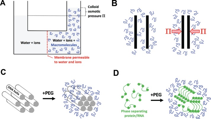
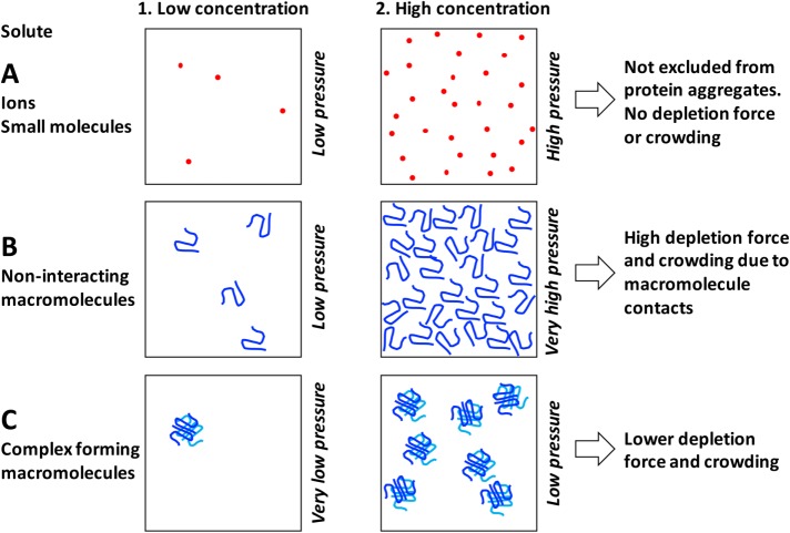
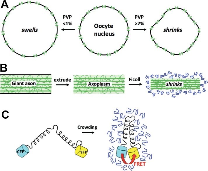
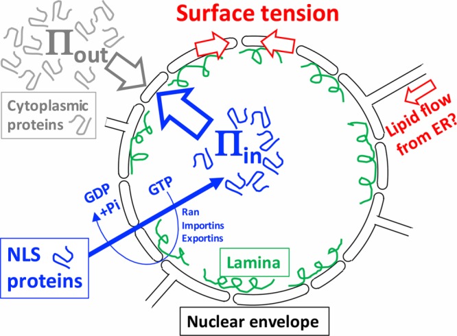
Crowding of the subcellular environment by macromolecules is thought to promote protein aggregation and phase separation. A challenge is how to parameterize the degree of crowding of the cell interior or artificial solutions that is relevant to these reactions. Here I review colloid osmotic pressure as a crowding metric. This pressure is generated by solutions of macromolecules in contact with pores that are permeable to water and ions but not macromolecules. It generates depletion forces that push macromolecules together in crowded solutions and thus promotes aggregation and phase separation. I discuss measurements of colloid osmotic pressure inside cells using the nucleus, the cytoplasmic gel, and fluorescence resonant energy transfer (FRET) biosensors as osmometers, which return a range of values from 1 to 20 kPa. I argue for a low value, 1–2 kPa, in frog eggs and perhaps more generally. This value is close to the linear range on concentration–pressure curves and is thus not crowded from an osmotic perspective. I discuss the implications of a low crowding pressure inside cells for phase separation biology, buffer design, and proteome evolution. I also discuss a pressure–tension model for nuclear shape, where colloid osmotic pressure generated by nuclear protein import inflates the nucleus.
Overview
The claims and statistics of this paper haven’t been checked yet.
Run the check — free, no account needed. It reads the open-access text and checks the sources it cites.
Integrity signals
Registry
Cites
Statistics
Figures
Verification
Claims the paper makes
The claims and statistics of this paper haven’t been checked yet.
Run the check — free, no account needed. It reads the open-access text and checks the sources it cites.
Claims other papers make about this one
Each is checked against this paper’s own text: does it say what the citing paper says it does?
In the citing paper
“Notably, a colloidal pressure between 1–2 kPa and 20 kPa has been suggested for the cytoplasm (reviewed in Mitchison, 2019), which would correspond to the osmotic pressure exerted by ≤ 2.5% PEG8000 (Stanley & Strey, 2003).”
From Molecular crowding and RNA synergize to promote phase separation, microtubule interaction, and seeding of Tau condensates (2022) · cites this paper as Mitchison, 2019
Self-check
Percentages, sums and reported differences re-computed from the paper’s own numbers.
The self-check needs the paper’s full text.
Citations
Cites
No references could be resolved yet.
Research works printed but not linked: 41
- Adair GS, 1928, Proc R Soc Lond A, 120, 573, 10.1098/rspa.1928.0167 · DOI
- Alberti S, 2018, J Mol Biol, 430, 4806, 10.1016/j.jmb.2018.06.038 · DOI
- Asakura S, 1954, J Chem Phys, 22, 1255, 10.1063/1.1740347 · DOI
- Boersma AJ, 2015, Nat Methods, 12, 227, 10.1038/nmeth.3257 · DOI
- Brangwynne CP, 2009, Science, 324, 1729, 10.1126/science.1172046 · DOI
- Delarue M, 2018, Cell, 174, 338, 10.1016/j.cell.2018.05.042 · DOI
- Dick DA, 1959, Int Rev Cytol, 8, 387, 10.1016/S0074-7696(08)62736-9 · DOI
- Dill KA, 2011, Proc Natl Acad Sci USA, 108, 17876, 10.1073/pnas.1114477108 · DOI · Paper page
- Eisenberg H, 1976, Biological Macromolecules and Polyelectrolytes in Solution
- Enyedi B, 2016, Cell, 165, 1160, 10.1016/j.cell.2016.04.016 · DOI
- Finan JD, 2010, J Cell Biochem, 109, 460, 10.1002/jcb.22437 · DOI
- Hall D, 2002, Biophys Chem, 98, 93, 10.1016/S0301-4622(02)00087-X · DOI
- Harding CV, 1959, J Gen Physiol, 42, 1155, 10.1085/jgp.42.6.1155 · DOI
- Hyman AA, 2014, Annu Rev Cell Dev Biol, 30, 39, 10.1146/annurev-cellbio-100913-013325 · DOI · Paper page
- Jin L, 2006, J Colloid Interface Sci, 304, 77, 10.1016/j.jcis.2006.08.046 · DOI
- Kim D-H, 2016, J Cell Sci, 129, 457, 10.1242/jcs.185173 · DOI
- Lee H, 2008, Biophys J, 95, 1590, 10.1529/biophysj.108.133025 · DOI
- Levy DL, 2010, Cell, 143, 288, 10.1016/j.cell.2010.09.012 · DOI
- Liu B, 2017, Biophys J, 112, 1929, 10.1016/j.bpj.2017.04.004 · DOI
- Luby-Phelps K, 1986, J Cell Biol, 102, 2015, 10.1083/jcb.102.6.2015 · DOI
- Magid AD, 1992, Exp Eye Res, 55, 615, 10.1016/S0014-4835(05)80174-5 · DOI
- Marenduzzo D, 2006, J Cell Biol, 175, 681, 10.1083/jcb.200609066 · DOI
- Minton AP, 2013, Biopolymers, 99, 239, 10.1002/bip.22163 · DOI
- Money NP, 1989, Plant Physiol, 91, 766, 10.1104/pp.91.2.766 · DOI
- Newport JW, 1990, J Cell Biol, 111, 2247, 10.1083/jcb.111.6.2247 · DOI
- Ogston AG, 1970, Biochem J, 119, 67, 10.1042/bj1190067 · DOI
- Parsegian VA, 2000, Proc Natl Acad Sci USA, 97, 3987, 10.1073/pnas.97.8.3987 · DOI
- Podgornik R, 1995, Biophys Chem, 57, 111, 10.1016/0301-4622(95)00058-6 · DOI
- Rand RP, 2000, Cell Mol Life Sci, 57, 1018, 10.1007/PL00000742 · DOI
- Rivas G, 2018, Biophys Rev, 10, 241, 10.1007/s12551-017-0347-6 · DOI
- Sama RRK, 2013, J Cell Physiol, 228, 2222, 10.1002/jcp.24395 · DOI
- Scatchard G, 1946, J Am Chem Soc, 68, 2315, 10.1021/ja01215a054 · DOI
- Schmidt HB, 2016, Trends Biochem Sci, 41, 46, 10.1016/j.tibs.2015.11.001 · DOI
- Schummel PH, 2017, Chemphyschem Eur J Chem Phys Phys Chem, 18, 189, 10.1002/cphc.201601032 · DOI
- Soranno A, 2014, Proc Natl Acad Sci USA, 111, 4874, 10.1073/pnas.1322611111 · DOI
- Spyropoulos CS, 1979, J Membr Biol, 47, 195, 10.1007/BF01869079 · DOI
- Starling EH, 1896, J Physiol, 19, 312, 10.1113/jphysiol.1896.sp000596 · DOI
- Vink H, 1971, Eur Polym J, 7, 1411, 10.1016/0014-3057(71)90035-8 · DOI
- Wang J, 2018, Cell, 174, 688, 10.1016/j.cell.2018.06.006 · DOI
- Wühr M, 2014, Curr Biol, 24, 1467, 10.1016/j.cub.2014.05.044 · DOI
- Wühr M, 2015, Curr Biol, 25, 2663, 10.1016/j.cub.2015.08.047 · DOI
Cited by
Mechanics and functional consequences of nuclear deformations
Kalukula, Y., Stephens, A. D., Lammerding, J., & Gabriele, S. (2022) · Nature Reviews Molecular Cell Biology · doi:10.1038/s41580-022-00480-z · Cited by 475
Hochmair, J., Exner, C., Franck, M., Dominguez‐Baquero, A., Diez, L., Brognaro, H., Kraushar, M. L., Mielke, T., Radbruch, H., Kaniyappan, S., et al. (2022) · The EMBO Journal · doi:10.15252/embj.2021108882 · Cited by 106
Tau liquid–liquid phase separation: At the crossroads of tau physiology and tauopathy
Islam, M., Shen, F., Regmi, D., Petersen, K., Karim, M. R. U., & Du, D. (2024) · Journal of Cellular Physiology · doi:10.1002/jcp.30853 · Cited by 28
Metadata
- Authors
- Mitchison, T. J.
- Venue
- Molecular Biology of the Cell
- Published
- 2019-01-15
- Volume
- 30
- Issue
- 2
- Pages
- 173-180
- ISSN
- 1059-1524
- 1939-4586
- PMC
- PMC6589563
- Cited by
- 96
- References
- 0
- Language
- en
About these checks
Registry, retraction, Cites and statistics results are computed, not judged, with one exception: a printed reference that no index lists is read once by a model before it counts as a citation error. Claims are extracted by a model and kept only when their quote appears verbatim in the text; each cited source is then checked, with a model’s help, for whether it says what the claim relies on. Those verdicts can be wrong — read the claim’s quote and the cited source before relying on one.
Statistics and arithmetic are recomputed, not judged.
Confidence bands are shown only for fields calibrated on our annotated test set. None is calibrated yet, so no band is shown.
What counts as a claim ›How we measure confidence ›How we check this ›
Are you an author, or do you see a problem with this page? Request a review
Data sources
Bibliographic data: Semantic Scholar, OpenAlex, Crossref.
Contains information from the Semantic Scholar Academic Graph Datasets, made available under the ODC Attribution License (ODC-BY 1.0).
Semantic Scholar’s data is described in: Kinney, R., et al. (2023). The Semantic Scholar Open Data Platform. arXiv:2301.10140.
