

The DOI matches its Crossref record; 27 of the 27 references it cites were found in a registry or an index.
Clinical efficacy of haematopoietic stem cell transplantation for adult adrenoleukodystrophy
Abstract
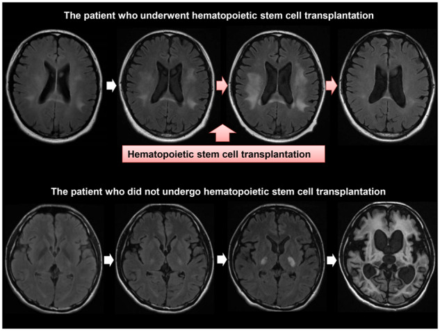
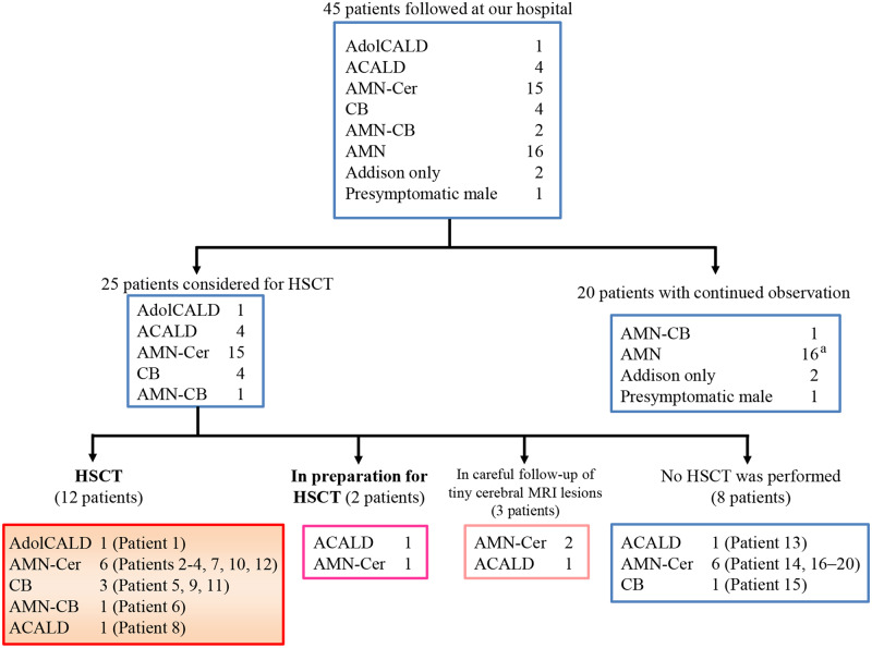
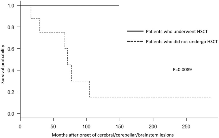
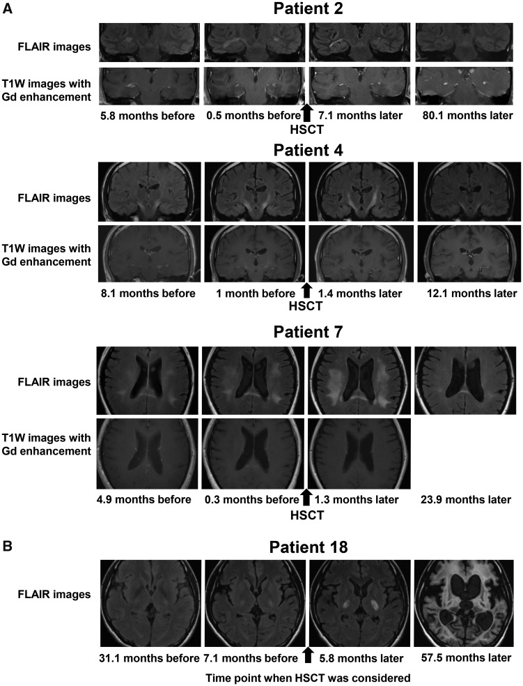
Accumulated experience supports the efficacy of allogenic haematopoietic stem cell transplantation in arresting the progression of childhood-onset cerebral form of adrenoleukodystrophy in early stages. For adulthood-onset cerebral form of adrenoleukodystrophy, however, there have been only a few reports on haematopoietic stem cell transplantation and the clinical efficacy and safety of that for adulthood-onset cerebral form of adrenoleukodystrophy remain to be established. To evaluate the clinical efficacy and safety of haematopoietic stem cell transplantation, we conducted haematopoietic stem cell transplantation on 12 patients with adolescent-/adult-onset cerebral form/cerebello-brainstem form of adrenoleukodystrophy in a single-institution-based prospective study. Through careful prospective follow-up of 45 male adrenoleukodystrophy patients, we aimed to enrol patients with adolescent-/adult-onset cerebral form/cerebello-brainstem form of adrenoleukodystrophy at early stages. Indications for haematopoietic stem cell transplantation included cerebral form of adrenoleukodystrophy or cerebello-brainstem form of adrenoleukodystrophy with Loes scores up to 13, the presence of progressively enlarging white matter lesions and/or lesions with gadolinium enhancement on brain MRI. Clinical outcomes of haematopoietic stem cell transplantation were evaluated by the survival rate as well as by serial evaluation of clinical rating scale scores and neurological and MRI findings. Clinical courses of eight patients who did not undergo haematopoietic stem cell transplantation were also evaluated for comparison of the survival rate. All the patients who underwent haematopoietic stem cell transplantation survived to date with a median follow-up period of 28.6 months (4.2–125.3 months) without fatality. Neurological findings attributable to cerebral/cerebellar/brainstem lesions became stable or partially improved in all the patients. Gadolinium-enhanced brain lesions disappeared or became obscure within 3.5 months and the white matter lesions of MRI became stable or small. The median Loes scores before haematopoietic stem cell transplantation and at the last follow-up visit were 6.0 and 5.25, respectively. Of the eight patients who did not undergo haematopoietic stem cell transplantation, six patients died 69.1 months (median period; range 16.0–104.1 months) after the onset of the cerebral/cerebellar/brainstem lesions, confirming that the survival probability was significantly higher in patients with haematopoietic stem cell transplantation compared with that in patients without haematopoietic stem cell transplantation (P = 0.0089). The present study showed that haematopoietic stem cell transplantation was conducted safely and arrested the inflammatory demyelination in all the patients with adolescent-/adult-onset cerebral form/cerebello-brainstem form of adrenoleukodystrophy when haematopoietic stem cell transplantation was conducted in the early stages. Further studies are warranted to optimize the procedures of haematopoietic stem cell transplantation for adolescent-/adult-onset cerebral form/cerebello-brainstem form of adrenoleukodystrophy.
Overview
The claims and statistics of this paper haven’t been checked yet.
Run the check — free, no account needed. It reads the open-access text and checks the sources it cites.
Integrity signals
Registry
Cites
Statistics
Figures
Verification
Claims the paper makes
The claims and statistics of this paper haven’t been checked yet.
Run the check — free, no account needed. It reads the open-access text and checks the sources it cites.
Claims other papers make about this one
Each is checked against this paper’s own text: does it say what the citing paper says it does?
In the citing paper
“In early disease stages, allogeneic hematopoietic stem-cell transplantation (allo-HSCT) can halt disease progression12–14.”
From Hematopoietic stem-cell gene therapy is associated with restored white matter microvascular function in cerebral adrenoleukodystrophy (2023) · cites this paper as [14]
Self-check
Percentages, sums and reported differences re-computed from the paper’s own numbers.
The self-check needs the paper’s full text.
Citations
Cites
Investigation of the freely available easy-to-use software ‘EZR’ for medical statistics
Kanda, Y. (2013) · Bone Marrow Transplantation · doi:10.1038/bmt.2012.244 · Cited by 18,767
Rating neurologic impairment in multiple sclerosis: An expanded disability status scale (EDSS)
Kurtzke, J. F. (1983) · Neurology · doi:10.1212/wnl.33.11.1444 · Cited by 15,011
(1965) · PubMed · Cited by 14,925
Functional evaluation : Barthel index
Mahonery, F. I. (1965) · Medical Entomology and Zoology · Cited by 12,762
1994 Consensus Conference on Acute GVHD Grading.
Przepiorka, D., Weisdorf, D., Martin, P., Hg, K., Beatty, P., Hows, J., & Thomas, E. D. (1995) · PubMed · Cited by 5,037
Putative X-linked adrenoleukodystrophy gene shares unexpected homology with ABC transporters
Mosser, J., Douar, A.-M., Sarde, C.-O., Kioschis, P., Feil, R., Moser, H., Poustka, A.-M., Mandel, J.-L., & Aubourg, P. (1993) · Nature · doi:10.1038/361726a0 · Cited by 1,202
Moser, H. W., Mahmood, A., & Raymond, G. V. (2007) · Nature Clinical Practice Neurology · doi:10.1038/ncpneuro0421 · Cited by 663
Hematopoietic stem-cell gene therapy for cerebral adrenoleukodystrophy
Eichler, F., Duncan, C., Musolino, P. L., Orchard, P. J., De Oliveira, S., Thrasher, A. J., Armant, M., Dansereau, C., Lund, T. C., Miller, W. P., et al. (2017) · New England Journal of Medicine · doi:10.1056/nejmoa1700554 · Cited by 604 · cited 1×
“The efficacy of the gene therapy for CCALD has recently been reported (Eichler et al. , 2017).”
Antilymphocyte Globulin for Prevention of Chronic Graft-versus-Host Disease
Kröger, N., Solano, C., Wolschke, C., Bandini, G., Patriarca, F., Pini, M., Nagler, A., Selleri, C., Risitano, A., Messina, G., et al. (2016) · New England Journal of Medicine · doi:10.1056/nejmoa1506002 · Cited by 573
Qualitative alteration in substrate specificity of mitochondrial monoamine oxidase in brain
Moskvitina, T. A., Kamyshanskaya, N. S., Kaverina, L. P., & Gorkin, V. Z. (1976) · Journal of Neurochemistry · doi:10.1111/j.1471-4159.1976.tb04462.x · Cited by 440
Cited by
Treatment of cerebral adrenoleukodystrophy: allogeneic transplantation and lentiviral gene therapy
Gupta, A. O., Raymond, G., Pierpont, E. I., Kemp, S., McIvor, R. S., Rayannavar, A., Miller, B., Lund, T. C., & Orchard, P. J. (2022) · Expert Opinion on Biological Therapy · doi:10.1080/14712598.2022.2124857 · Cited by 67
“older patients, recent reports suggest adults with cerebral disease may have outcomes comparable to a pediatric age group [90,91].”
BackgroundLauer, A., Speroni, S. L., Choi, M., Da, X., Duncan, C., McCarthy, S., Krishnan, V., Lusk, C. A., Rohde, D., Hansen, M. B., et al. (2023) · Nature Communications · doi:10.1038/s41467-023-37262-w · Cited by 26
Gene therapy for the leukodystrophies: From preclinical animal studies to clinical trials.
Metovic, J., Li, Y., Gong, Y., & Eichler, F. (2024) · Neurotherapeutics · doi:10.1016/j.neurot.2024.e00443 · Cited by 16
Metadata
- Authors
- Matsukawa, T., Yamamoto, T., Honda, A., Toya, T., Ishiura, H., Mitsui, J., Tanaka, M., Hao, A., Shinohara, A., Ogura, M., Kataoka, K., Seo, S., Kumano, K., Hosoi, M., Narukawa, K., Yasunaga, M., Maki, H., Ichikawa, M., Nannya, Y., Imai, Y., Takahashi, T., Takahashi, Y., Nagasako, Y., Yasaka, K., Mano, K. K., Matsukawa, M. K., Miyagawa, T., Hamada, M., Sakuishi, K., Hayashi, T., Iwata, A., Terao, Y., Shimizu, J., Goto, J., Mori, H., Kunimatsu, A., Aoki, S., Hayashi, S., Nakamura, F., Arai, S., Momma, K., Ogata, K., Yoshida, T., Abe, O., Inazawa, J., Toda, T., Kurokawa, M., & Tsuji, S.
- Affiliations
- Department of Neurology, Graduate School of Medicine, The University of Tokyo, Tokyo 113-8655, Japan
- Department of Hematology and Oncology, Graduate School of Medicine, The University of Tokyo, Tokyo 113-8655, Japan
- Department of Radiology, Graduate School of Medicine, The University of Tokyo, Tokyo 113-8655, Japan
- Department of Molecular Cytogenetics, Medical Research Institute and School of Biomedical Science, Tokyo Medical and Dental University, Tokyo 113-8519, Japan
- Department of Neurology, National Hospital Organization Higashisaitama National Hospital, Saitama 349-0196, Japan
- Department of Neurology, Fujimi Kogen Hospital, Nagano 399-0214, Japan
- Venue
- Brain Communications
- Publisher
- Oxford University Press (OUP)
- Published
- 2020-01-01
- Volume
- 2
- Issue
- 1
- ISSN
- 2632-1297
- PMC
- PMC7425345
- Cited by
- 26
- References
- 27
- Language
- en
- Funders
- Ministry of Education, Culture, Sports, Science and Technology of Japan
- Ministry of Health, Labour and Welfare, Japan
- Japan Agency for Medical Research and Development
About these checks
Registry, retraction, Cites and statistics results are computed, not judged, with one exception: a printed reference that no index lists is read once by a model before it counts as a citation error. Claims are extracted by a model and kept only when their quote appears verbatim in the text; each cited source is then checked, with a model’s help, for whether it says what the claim relies on. Those verdicts can be wrong — read the claim’s quote and the cited source before relying on one.
Statistics and arithmetic are recomputed, not judged.
Confidence bands are shown only for fields calibrated on our annotated test set. None is calibrated yet, so no band is shown.
What counts as a claim ›How we measure confidence ›How we check this ›
Are you an author, or do you see a problem with this page? Request a review
Data sources
Bibliographic data: Semantic Scholar, OpenAlex, Crossref.
Contains information from the Semantic Scholar Academic Graph Datasets, made available under the ODC Attribution License (ODC-BY 1.0).
Semantic Scholar’s data is described in: Kinney, R., et al. (2023). The Semantic Scholar Open Data Platform. arXiv:2301.10140.
