

The DOI matches its Crossref record; 56 of the 56 references it cites were found in a registry or an index.
Cortical Folding Patterns and Predicting Cytoarchitecture
Open accessAbstract
The human cerebral cortex is made up of a mosaic of structural areas, frequently referred to as Brodmann areas (BAs). Despite the widespread use of cortical folding patterns to perform ad hoc estimations of the locations of the BAs, little is understood regarding 1) how variable the position of a given BA is with respect to the folds, 2) whether the location of some BAs is more variable than others, and 3) whether the variability is related to the level of a BA in a putative cortical hierarchy. We use whole-brain histology of 10 postmortem human brains and surface-based analysis to test how well the folds predict the locations of the BAs. We show that higher order cortical areas exhibit more variability than primary and secondary areas and that the folds are much better predictors of the BAs than had been previously thought. These results further highlight the significance of cortical folding patterns and suggest a common mechanism for the development of the folds and the cytoarchitectonic fields.
Overview
The claims and statistics of this paper haven’t been checked yet.
Run the check — free, no account needed. It reads the open-access text and checks the sources it cites.
Integrity signals
Registry
Cites
Statistics
Figures
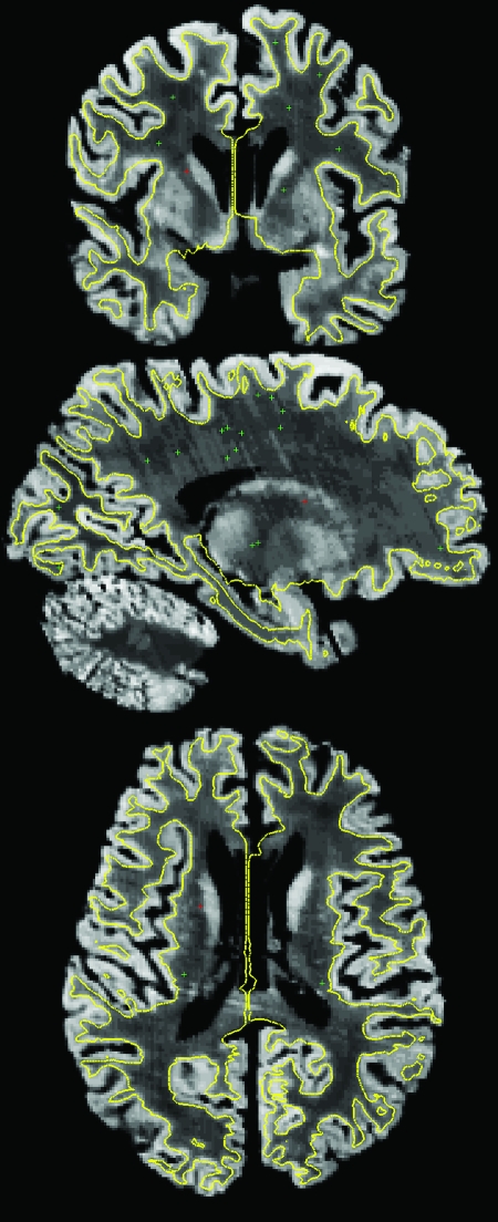
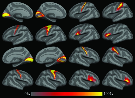
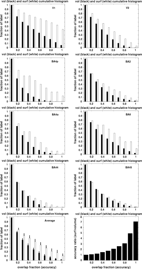
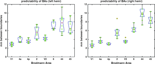
Verification
Claims the paper makes
The claims and statistics of this paper haven’t been checked yet.
Run the check — free, no account needed. It reads the open-access text and checks the sources it cites.
Claims other papers make about this one
Each is checked against this paper’s own text: does it say what the citing paper says it does?
In the citing paper
“functional areal boundaries (see Parcellation Choice) of heteromodal association areas are variable with respect to sulcal and gyral anatomy (Fischl et al., 2008).”
From Development of large-scale functional networks from birth to adulthood: A guide to the neuroimaging literature (2017) · cites this paper as Fischl et al., 2008
Self-check
Percentages, sums and reported differences re-computed from the paper’s own numbers.
The self-check needs the paper’s full text.
Citations
Cites
Co-Planar Stereotaxic Atlas of the Human Brain
Talairach, J., & Tournoux, P. (1988) · Medical Entomology and Zoology · Cited by 11,798
Cortical Surface-Based Analysis
Dale, A. M., Fischl, B., & Sereno, M. I. (1999) · NeuroImage · doi:10.1006/nimg.1998.0395 · Cited by 10,910
Self-citationDistributed hierarchical processing in the primate cerebral cortex
Felleman, D. J., & Van Essen, D. C. (1991) · Cerebral Cortex · doi:10.1093/cercor/1.1.1 · Cited by 7,783
Cortical Surface-Based Analysis
Fischl, B., Sereno, M. I., & Dale, A. M. (1999) · NeuroImage · doi:10.1006/nimg.1998.0396 · Cited by 6,147
Self-citationDISCONNEXION SYNDROMES IN ANIMALS AND MAN
Geschwind, N. (1965) · Brain · doi:10.1093/brain/88.2.237 · Cited by 3,419
Specification of Cerebral Cortical Areas
Rakic, P. (1988) · Science · doi:10.1126/science.3291116 · Cited by 3,127 · cited 2×
“…areas is that the 1978 Cortical Folding Patterns d Fischl et al. specification of the architectonic regions is present in a protomap in the proliferative ventricular zone in the form of radial columns that guide the formation and migration of cortical neurons during neurodevelopment (Rakic 1988).”
Background(1909) · Cited by 2,575
DISCONNEXION SYNDROMES IN ANIMALS AND MAN
Geschwind, N. (1965) · Brain · doi:10.1093/brain/88.3.585 · Cited by 1,840
Fischl, B. R., Liu, A., & Dale, A. M. (2001) · IEEE Transactions on Medical Imaging · doi:10.1109/42.906426 · Cited by 1,755
Self-citationAN ANATOMICAL STUDY OF CONVERGING SENSORY PATHWAYS WITHIN THE CEREBRAL CORTEX OF THE MONKEY
Jones, E. G., & Powell, T. P. S. (1970) · Brain · doi:10.1093/brain/93.4.793 · Cited by 1,717
Research works printed but not linked: 6
- 2007, PNAS, 108, 7652
- 1933, J PSYCHOL NEUROL, 45, 381
- 1988, COMP PRIMATE BIOL, 4, 39
- 1911, J PSYCHOL NEUROL, 18, 107
- 2004, Journal of Anatomy, 205, 217
- 1995, Journal of Anatomy, 187, 515
Cited by
Sorted by citations
Items 701–736 of 736
Evaluation of surface-based hippocampal registration using ground-truth subfield definitionsPreprint
DeKraker, J., Palomero‐Gallagher, N., Kedo, O., Bernasconi, N., Muenzing, S. E. A., Axer, M., Amunts, K., KHAN, A. R., Bernhardt, B. C., & Evans, A. C. (2023) · eLife · doi:10.7554/elife.88404.2
Self-citationBourke, J. D., Cooper, G. J., Forstmann, B. U., Schall, U., & Todd, J. (2024) · bioRxiv (Cold Spring Harbor Laboratory) · doi:10.1101/2024.04.03.587995
Mondragon‐Estrada, E., Newburger, J. W., DePalma, S. R., Brueckner, M. K., Cleveland, J., Chung, W. K., Gelb, B. D., Goldmuntz, E., Hagler, D. J., Huang, H., et al. (2024) · SSRN Electronic Journal · doi:10.2139/ssrn.4845174
Gorev, V., Gorbunov, A. V., Panikratova, Y. R., Tomyshev, A., Hatsenko, I., Kuleshov, N. Н., Salmasi, J., Hasanova, K., Balashova, L. M., Lobanova, E. I., et al. (2024) · Neuroscience and Behavioral Physiology · doi:10.1007/s11055-024-01703-w
Brain Cortical Functional Gradients Predict Cortical Folding Patterns via Attention Mesh Convolution
Li, Y., He, Z., Zhong, T., Li, C., Zhu, D., Han, J., Liu, T., & Zhang, T. (2024) · Lecture notes in computer science · doi:10.1007/978-3-031-72104-5_14
Pas, K. E., Saleem, K. S., Basser, P. J., & Avram, A. V. (2024) · Imaging Neuroscience · doi:10.1162/imag_a_00393
Zalesky, A., Sotiropoulos, S. N., Jbabdi, S., & Fornito, A. (2025) · Elsevier eBooks · doi:10.1016/b978-0-12-818894-1.00007-0
Türközer, H. B., Zeng, V., Hoang, D. T., Sritharan, J., Iska, N., Ivleva, E. I., Clementz, B. A., Pearlson, G. D., Keedy, S., Gershon, E. S., et al. (2026) · Biological Psychiatry · doi:10.1016/j.biopsych.2026.03.098
Morphometric Analysis of Human Paracentral Lobule: A Cadaveric Study with its Clinical Implications
Varalakshmi, K. L., Sangeeta, M., Muthiah, P., & Jain, B. S. H. (2025) · National Journal of Clinical Anatomy · doi:10.4103/njca.njca_149_24
Peelle, J. E. (2025) · Cambridge University Press eBooks · doi:10.1017/9781009245296.004
Gorbunov, A. V., Горев, В. В., Panikratova, Y. R., Tomyshev, A. S., Zavadenko, N. N., Khatsenko, I. E., Khasanova, K. A., Gorbunov, M. A., & Лебедева, И. С. (2025) · L O Badalyan Neurological Journal · doi:10.46563/2686-8997-2025-6-1-6-12
Richlan, F. (2001) · De Boeck Supérieur eBooks · doi:10.3917/dbu.kern.2020.01.0173
Neocortex Folding in Primates up to Human: Evolution and Mechanisms
Long, K. R., Huttner, W. Β., & Namba, T. (2025) · Developmental Neuroscience · doi:10.1159/000547228
Architectonic Organization of the Telencephalon Related to Glioma
Manilha, R., & Kadri, P. A. S. (2025) · Springer eBooks · doi:10.1007/978-3-031-95119-0_10
Functional cortical cartography
Frost, M. A. (2012) · Research Publications (Maastricht University) · doi:10.26481/dis.20120914mf
Mosaic evolution and increased integration underpin the evolvability of the dog brain
Balcarcel, A. M., Fabre, A., Orliac, M. J., Verger, K. L., Selba, M. C., Hipsley, C. A., & Evin, A. (2026) · Evolution · doi:10.1093/evolut/qpag002
Linking cortical morphology and neurophysiological dynamics in Parkinson’s disease
Mirpour, K., Alijanpourotaghsara, A., & Pouratian, N. (2026) · Scientific Reports · doi:10.1038/s41598-026-41274-z
Plasticité des représentations corticales motrices après accident vasculaire cérébral
Ahdab, R. (2010) · theses.fr (ABES) · doi:10.70675/e6191692z14b2z401dz9787z00908975d089
Carrière, N. (2015) · theses.fr (ABES) · doi:10.70675/08ca0910za340z4a0bzaf66z1be41e77e43e
Morphométrie de trois sillons d'intérêt dans la dyslexie développementale
di Covella, L. S. (2016) · theses.fr (ABES) · doi:10.70675/9c0dfe25z71a2z49faza745zb8fae1d55be2
Etude des bases neurales de la catégorisation chez les sujets sains et les patients cérébro-lésés
Garcin, B. (2017) · theses.fr (ABES) · doi:10.70675/592eb0eezc890z4234zaad6z38c8fb3724e8
Characterization of the Ferret Neocortical Development using Structural Magnetic Resonnance Imaging
Foubet, O. (2018) · theses.fr (ABES) · doi:10.70675/8ea07177z61f0z4d55zb217z6e36e24aa4df
Reading and math : neuronal constraints and how we adapt to them
Roëll, M. (2019) · theses.fr (ABES) · doi:10.70675/f41334ecza596z42e1zb181ze8b37f96afe5
Le cortex vocal chez le primate : une investigation anatomo-fonctionnelle
Bodin, C. (2019) · theses.fr (ABES) · doi:10.70675/94d67f43z2595z41abza479z96567259e48e
Poliva, O. (2026) · bioRxiv (Cold Spring Harbor Laboratory) · doi:10.64898/2026.05.05.722843
Long-term effects of preterm birth on cortical folding trajectories in early childhood
Jang, Y. H., Kim, J. M., Lee, B. G., Hoh, J., Lee, G. Y., Kim, H. H., Lyu, I., & Lee, H. J. (2026) · Brain Communications · doi:10.1093/braincomms/fcag097
Tyrovolas, S., Portella, M. J., Usall, J., Cambra-Martí, M. R., Callén, A., Haro, J. M., Núñez, C., & Stephan-Otto, C. (2026) · Brain Structure and Function · doi:10.1007/s00429-026-03206-5
Ersungur, E. B., Poyraz, B. Ç., Arslan, S., & Kargın, O. A. (2026) · Journal of Affective Disorders · doi:10.1016/j.jad.2026.122635
A novel and adaptable method to prepare samples for postmortem imaging with artifact reduction
Smith, E. N., Rosenblum, E. W., Stevens, A., Morgan, L., Mora, J., Vinke, L. N., Moreau, A. L., Tirrell, L. S., Zöllei, L., Edlow, B. L., et al. (2026) · Journal of Neuroscience Methods · doi:10.1016/j.jneumeth.2026.110941
Self-citationBrain wiring and neuronal dynamics : advances in MR imaging of focal epilepsy
Besseling, R. M. H. (2014) · Research Publications (Maastricht University) · doi:10.26481/dis.20140404rb
Automatic Labeling of the Human Cerebral Cortex
Yeo, B. T. T. (2015) · Elsevier eBooks · doi:10.1016/b978-0-12-397025-1.00306-7
Self-citation
Metadata
- Authors
- Fischl, B., Rajendran, N., Busa, E., Augustinack, J., Hinds, O., Yeo, B. T. T., Mohlberg, H., Amunts, K., & Zilles, K.
- Affiliations
- Harvard University (US)
- McGovern Institute for Brain Research (US)
- Massachusetts Institute of Technology (US)
- Forschungszentrum Jülich (DE)
- Corresponding author
- Fischl, B.
- Venue
- Cerebral Cortex
- Publisher
- Oxford University Press (OUP)
- Published
- 2007-12-12
- Volume
- 18
- Issue
- 8
- Pages
- 1973-1980
- ISSN
- 1047-3211 (ISSN-L)
- 1460-2199
- PMC
- PMC2474454
- OpenAlex
- W2154281919
- Cited by
- 737
- References
- 56
- Citations by year
- 2026: 18 · 2025: 22 · 2024: 34 · 2023: 38 · 2022: 45 · 2021: 58 · 2020: 52 · 2019: 44 · 2018: 55 · 2017: 56 · 2016: 41 · 2015: 40 · 2014: 32 · 2013: 48 · 2012: 42
- FWCI
- 19.50
- Type
- journal article
- Language
- en
- Open access
- hybrid
- doi.org
- Licence
- cc-by-nc
- Locations
- Cerebral Cortex (publishedVersion, cc-by-nc)
- Europe PMC (PubMed Central) (publishedVersion, cc-by-nc)
- PubMed Central (publishedVersion, cc-by-nc)
- PubMed (publishedVersion)
- Digital Access to Scholarship at Harvard (DASH) (Harvard University) (submittedVersion, cc-by)
- DSpace@MIT (Massachusetts Institute of Technology) (submittedVersion, cc-by-nc)
- JuSER (Forschungszentrum Jülich) (submittedVersion)
- RWTH Publications (RWTH Aachen) (submittedVersion)
- Funders
- Deutsche Forschungsgemeinschaft
- National Institutes of Health (BIRN002, RR14075, EB005149)
- National Institute of Mental Health
- National Institute of Neurological Disorders and Stroke (R01NS052585)
- National Institute of Biomedical Imaging and Bioengineering (R01EB001550, R01EB006758, U54EB005149)
- National Center for Research Resources (BIRN002, U24 RR021382, P41-RR-14075, RR 14075, U24RR021382, R01 RR1659401A1, R01-RR16594, P41RR014075, BIRN002, R01RR016594)
- Topics
- Health Sciences › Medicine › Radiology, Nuclear Medicine and Imaging › Advanced Neuroimaging Techniques and Applications
- Life Sciences › Neuroscience › Cognitive Neuroscience › Functional Brain Connectivity Studies
- Life Sciences › Neuroscience › Developmental Neuroscience › Neurogenesis and neuroplasticity mechanisms
- Keywords
- cortical folding, Brodmann areas, cytoarchitecture, cortical hierarchy, cortical development, postmortem brain, surface-based analysis
- MeSH terms
- Brain Mapping, Cerebral Cortex, Humans, Predictive Value of Tests
About these checks
Registry, retraction, Cites and statistics results are computed, not judged, with one exception: a printed reference that no index lists is read once by a model before it counts as a citation error. Claims are extracted by a model and kept only when their quote appears verbatim in the text; each cited source is then checked, with a model’s help, for whether it says what the claim relies on. Those verdicts can be wrong — read the claim’s quote and the cited source before relying on one.
Statistics and arithmetic are recomputed, not judged.
Confidence bands are shown only for fields calibrated on our annotated test set. None is calibrated yet, so no band is shown.
What counts as a claim ›How we measure confidence ›How we check this ›
Are you an author, or do you see a problem with this page? Request a review
