

The DOI matches its Crossref record; 2 of the 2 references it cites were found in a registry or an index.
Only a Single Taxonomically Restricted Gene Family in the Drosophila melanogaster Subgroup Can Be Identified with High Confidence
Abstract
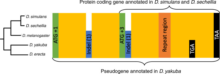
Taxonomically restricted genes (TRGs) are genes that are present only in one clade. Protein-coding TRGs may evolve de novo from previously noncoding sequences: functional ncRNA, introns, or alternative reading frames of older protein-coding genes, or intergenic sequences. A major challenge in studying de novo genes is the need to avoid both false-positives (nonfunctional open reading frames and/or functional genes that did not arise de novo) and false-negatives. Here, we search conservatively for high-confidence TRGs as the most promising candidates for experimental studies, ensuring functionality through conservation across at least two species, and ensuring de novo status through examination of homologous noncoding sequences. Our pipeline also avoids ascertainment biases associated with preconceptions of how de novo genes are born. We identify one TRG family that evolved de novo in the Drosophila melanogaster subgroup. This TRG family contains single-copy genes in Drosophila simulans and Drosophila sechellia. It originated in an intron of a well-established gene, sharing that intron with another well-established gene upstream. These TRGs contain an intron that predates their open reading frame. These genes have not been previously reported as de novo originated, and to our knowledge, they are the best Drosophila candidates identified so far for experimental studies aimed at elucidating the properties of de novo genes.
Overview
The claims and statistics of this paper haven’t been checked yet.
Run the check — free, no account needed. It reads the open-access text and checks the sources it cites.
Integrity signals
Registry
Cites
Statistics
Figures
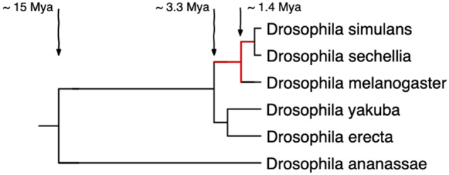
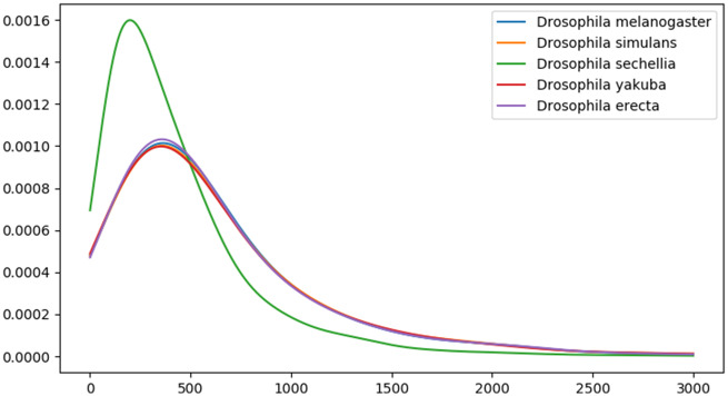
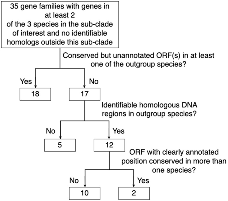
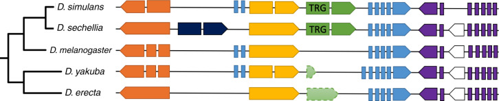

Verification
Claims the paper makes
The claims and statistics of this paper haven’t been checked yet.
Run the check — free, no account needed. It reads the open-access text and checks the sources it cites.
Claims other papers make about this one
Each is checked against this paper’s own text: does it say what the citing paper says it does?
The source backs part of the claim, not all of it — or backs it for a different population, condition or comparison.
In the citing paper
“It appears that most of the ab initio potential lineage-specific genes are artifacts, as the number of singletons is too high considering estimates of the rate of emergence of new genes (Casola 2018, Zhang et al. 2019, Zile et al. 2020, Vakirlis et al. 2024).”
From Annotation matters: the effect of structural gene annotation on orthology inference (2025) · cites this paper as Zile et al. 2020
Experimental — accuracy not yet measured
Self-check
Percentages, sums and reported differences re-computed from the paper’s own numbers.
The self-check needs the paper’s full text.
Citations
Cites
Hashimoto, T., Horikawa, D. D., Saito, Y., Kuwahara, H., Kozuka-Hata, H., Shin-I, T., Minakuchi, Y., Ohishi, K., Motoyama, A., Aizu, T., et al. (2016) · Nature Communications · doi:10.1038/ncomms12808 · Cited by 394
Origins of De Novo Genes in Human and Chimpanzee
Ruiz-Orera, J., Hernandez-Rodriguez, J., Chiva, C., Sabidó, E., Kondova, I., Bontrop, R., Marqués-Bonet, T., & Albà, M. M. (2015) · PLOS Genetics · doi:10.1371/journal.pgen.1005721 · Cited by 160
Research works printed but not linked: 69
- Abascal, 2018, Loose ends: almost one in five human genes still have unresolved coding status, Nucleic Acids Res, 46, 7070, 10.1093/nar/gky587 · DOI
- Albertin, 2015, The octopus genome and the evolution of cephalopod neural and morphological novelties, Nature, 524, 220, 10.1038/nature14668 · DOI
- Altenhoff, 2018, The OMA orthology database in 2018: retrieving evolutionary relationships among all domains of life through richer web and programmatic interfaces, Nucleic Acids Res, 46, D477, 10.1093/nar/gkx1019 · DOI · Paper page
- Ángyán, 2012, Estimating intrinsic structural preferences of de novo emerging random-sequence proteins: is aggregation the main bottleneck?, FEBS Lett, 586, 2468, 10.1016/j.febslet.2012.06.007 · DOI
- Arendsee, 2019, fagin: synteny-based phylostratigraphy and finer classification of young genes, BMC Bioinformatics, 20, 440, 10.1186/s12859-019-3023-y · DOI
- Baalsrud, 2018, De novo gene evolution of antifreeze glycoproteins in codfishes revealed by whole genome sequence data, Mol Biol Evol, 35, 593, 10.1093/molbev/msx311 · DOI · Paper page
- Balakirev, 2003, Pseudogenes: are they “junk” or functional DNA?, Annu Rev Genet, 37, 123, 10.1146/annurev.genet.37.040103.103949 · DOI
- Blevins, 2017, Comparative transcriptomics and ribo-seq: looking at de novo gene emergence in Saccharomycotina, PeerJ, 3030
- Brosius, 2005, Waste not, want not – transcript excess in multicellular eukaryotes, Trends Genet, 21, 287, 10.1016/j.tig.2005.02.014 · DOI
- Bungard, 2017, Foldability of a natural de novo evolved protein, Structure, 25, 1687, 10.1016/j.str.2017.09.006 · DOI
- Cai, 2008, De novo origination of a new protein-coding gene in Saccharomyces cerevisiae, Genetics, 179, 487, 10.1534/genetics.107.084491 · DOI
- Camacho, 2009, BLAST+: architecture and applications, BMC Bioinformatics, 10, 421, 10.1186/1471-2105-10-421 · DOI · Paper page
- Casola, 2018, From de novo to ‘de nono’: The majority of novel protein coding genes identified with phylostratigraphy are old genes or recent duplicates, Genome Biol Evol, 10, 2906
- Chen, 2010, New genes in Drosophila quickly become essential, Science, 330, 1682, 10.1126/science.1196380 · DOI
- Chen, 2015, Combinatorial DNA rearrangement facilitates the origin of new genes in ciliates, Genome Biol Evol, 7, 2859
- Cock, 2009, Biopython: freely available python tools for computational molecular biology and bioinformatics, Bioinformatics, 25, 1422, 10.1093/bioinformatics/btp163 · DOI · Paper page
- Domazet-Loso, 2003, An evolutionary analysis of orphan genes in Drosophila, Genome Res, 13, 2213, 10.1101/gr.1311003 · DOI
- Donoghue, 2011, Evolutionary origins of Brassicaceae specific genes in Arabidopsis thaliana, BMC Evol Biol, 11, 10.1186/1471-2148-11-47 · DOI
- 2007, Evolution of genes and genomes on the Drosophila phylogeny, Nature, 450, 203, 10.1038/nature06341 · DOI · Paper page
- Eddy, 2011, Accelerated profile HMM searches, PLoS Comput Biol, 7, e1002195, 10.1371/journal.pcbi.1002195 · DOI · Paper page
- Fernandez-Escamilla, 2004, Prediction of sequence-dependent and mutational effects on the aggregation of peptides and proteins, Nat Biotechnol, 22, 1302, 10.1038/nbt1012 · DOI
- Finn, 2016, The pfam protein families database: towards a more sustainable future, Nucleic Acids Res, 44, D279, 10.1093/nar/gkv1344 · DOI · Paper page
- Frigola, 2017, Reduced mutation rate in exons due to differential mismatch repair, Nat Genet, 49, 1684, 10.1038/ng.3991 · DOI
- Ghatak, 2019, The y-ome defines the 35% of Escherichia coli genes that lack experimental evidence of function, Nucleic Acids Res, 47, 2446, 10.1093/nar/gkz030 · DOI
- Graur, 2013, On the immortality of television sets: “function” in the human genome according to the evolution-free gospel of ENCODE, Genome Biol Evol, 5, 578, 10.1093/gbe/evt028 · DOI
- Guan, 2018, Gene refashioning through innovative shifting of reading frames in mosses, Nat Commun, 9, 10.1038/s41467-018-04025-x · DOI
- Guerzoni, 2016, De novo genes arise at a slow but steady rate along the primate lineage and have been subject to incomplete lineage sorting, Genome Biol Evol, 8, 1222, 10.1093/gbe/evw074 · DOI
- Haft, 2018, RefSeq: an update on prokaryotic genome annotation and curation, Nucleic Acids Res, 46, D851, 10.1093/nar/gkx1068 · DOI
- Heames, 2020, A Continuum of Evolving De Novo Genes Drives Protein-Coding Novelty in Drosophila, J Mol Evol, 88, 382, 10.1007/s00239-020-09939-z · DOI
- Hild, 2003, An integrated gene annotation and transcriptional profiling approach towards the full gene content of the Drosophila genome, Genome Biol, 5, R3, 10.1186/gb-2003-5-1-r3 · DOI
- Jain, 2019, The evolutionary traceability of a protein, Genome Biol Evol, 11, 531, 10.1093/gbe/evz008 · DOI
- Katoh, 2013, MAFFT multiple sequence alignment software version 7: improvements in performance and usability, Mol Biol Evol, 30, 772, 10.1093/molbev/mst010 · DOI · Paper page
- Knopp, 2019, De Novo Emergence of Peptides That Confer Antibiotic Resistance, mBio, 10
- Knopp, 2018, No beneficial fitness effects of random peptides, Nat Ecol Evol, 2, 1046, 10.1038/s41559-018-0585-4 · DOI
- Knowles, 2009, Recent de novo origin of human protein-coding genes, Genome Res, 19, 1752, 10.1101/gr.095026.109 · DOI · Paper page
- Levine, 2006, Novel genes derived from noncoding DNA in Drosophila melanogaster are frequently x-linked and exhibit testis-biased expression, Proc Natl Acad Sci U S A, 103, 9935, 10.1073/pnas.0509809103 · DOI
- Linquist, 2020, Getting clear about the F-word in genomics, PLoS Genet, 16, e1008702, 10.1371/journal.pgen.1008702 · DOI
- McLysaght, 2015, New genes from non-coding sequence: the role of de novo protein-coding genes in eukaryotic evolutionary innovation, Philos Trans R Soc B, 370, 20140332, 10.1098/rstb.2014.0332 · DOI
- McLysaght, 2016, Open questions in the study of de novo genes: what, how and why, Nat Rev Genet, 17, 567, 10.1038/nrg.2016.78 · DOI
- Neme, 2017, Random sequences are an abundant source of bioactive RNAs or peptides, Nat Ecol Evol, 1, 0127, 10.1038/s41559-017-0127 · DOI
- Neme, 2016, Fast turnover of genome transcription across evolutionary time exposes entire non-coding DNA to de novo gene emergence, eLife, 5, 09977, 10.7554/eLife.09977 · DOI
- Obbard, 2012, Estimating divergence dates and substitution rates in the Drosophila phylogeny, Mol Biol Evol, 29, 3459, 10.1093/molbev/mss150 · DOI
- O’Leary, 2016, Reference sequence (RefSeq) database at NCBI: current status, taxonomic expansion, and functional annotation, Nucleic Acids Res, 44, D733, 10.1093/nar/gkv1189 · DOI · Paper page
- Pearson, 2000, Flexible sequence similarity searching with the FASTA3 program package, Methods Mol Biol, 132, 185
- Reinhardt, 2013, De novo ORFs in Drosophila are important to organismal fitness and evolved rapidly from previously non-coding sequences, PLoS Genet, 9, e1003860, 10.1371/journal.pgen.1003860 · DOI
- Remmert, 2012, HHblits: lightning-fast iterative protein sequence searching by HMM-HMM alignment, Nat Methods, 9, 173, 10.1038/nmeth.1818 · DOI · Paper page
- Ruiz-Orera, 2018, Translation of neutrally evolving peptides provides a basis for de novo gene evolution, Nat Ecol Evol, 2, 890, 10.1038/s41559-018-0506-6 · DOI
- Savojardo, 2018, BUSCA: an integrative web server to predict subcellular localization of proteins, Nucleic Acids Res, 46, W459, 10.1093/nar/gky320 · DOI
- Schaffer, 2001, Improving the accuracy of PSI-BLAST protein database searches with composition-based statistics and other refinements, Nucleic Acids Res, 29, 2994, 10.1093/nar/29.14.2994 · DOI
- Schlötterer, 2015, Genes from scratch – the evolutionary fate of de novo genes, Trends Genet, 31, 215, 10.1016/j.tig.2015.02.007 · DOI
- Stewart, 2019, Chromosomal rearrangements as a source of new gene formation in Drosophila yakuba, PLoS Genet, 15, e1008314, 10.1371/journal.pgen.1008314 · DOI
- Sun, 2015, Identification and evolution of the orphan genes in the domestic silkworm, Bombyx mori, FEBS Lett, 589, 2731, 10.1016/j.febslet.2015.08.008 · DOI
- Toll-Riera, 2008, Origin of primate orphan genes: a comparative genomics approach, Mol Biol Evol, 26, 603, 10.1093/molbev/msn281 · DOI · Paper page
- Tretyachenko, 2017, Random protein sequences can form defined secondary structures and are well-tolerated in vivo, Sci Rep, 7, 15449, 10.1038/s41598-017-15635-8 · DOI
- Vakirlis, 2018, A molecular portrait of de novo genes in yeasts, Mol Biol Evol, 35, 631, 10.1093/molbev/msx315 · DOI
- Vakirlis, 2018, Methods in molecular biology, 63
- Veidenberg, 2016, Wasabi: an integrated platform for evolutionary sequence analysis and data visualization, Mol Biol Evol, 33, 1126, 10.1093/molbev/msv333 · DOI
- Waterhouse, 2018, BUSCO applications from quality assessments to gene prediction and phylogenomics, Mol Biol Evol, 35, 543, 10.1093/molbev/msx319 · DOI · Paper page
- Weisman, 2017, Gene evolution: getting something from nothing, Curr Biol, 27, R661, 10.1016/j.cub.2017.05.056 · DOI
- Werren, 2010, Functional and evolutionary insights from the genomes of three parasitoid Nasonia species, Science, 327, 343, 10.1126/science.1178028 · DOI
- Willis, 2018, Gene birth contributes to structural disorder encoded by overlapping genes, Genetics, 210, 303, 10.1534/genetics.118.301249 · DOI
- Wilson, 2017, Young genes are highly disordered as predicted by the preadaptation hypothesis of de novo gene birth, Nat Ecol Evol, 1, 0146, 10.1038/s41559-017-0146 · DOI
- Wilson, 2011, Putatively noncoding transcripts show extensive association with ribosomes, Genome Biol Evol, 3, 1245, 10.1093/gbe/evr099 · DOI
- Wissler, 2013, Mechanisms and dynamics of orphan gene emergence in insect genomes, Genome Biol Evol, 5, 439, 10.1093/gbe/evt009 · DOI
- Wu, 2018, Tracing the de novo origin of protein-coding genes in yeast, mBio, 9, 0102418, 10.1128/mBio.01024-18 · DOI
- Yang, 2011, De novo origin of new genes with introns in Plasmodium vivax, FEBS Lett, 585, 641, 10.1016/j.febslet.2011.01.017 · DOI
- Zhang, 2019, Rapid evolution of protein diversity by de novo origination in Oryza, Nat Ecol Evol, 3, 679, 10.1038/s41559-019-0822-5 · DOI · Paper page
- Zhao, 2014, Origin and spread of de novo genes in Drosophila melanogaster populations, Science, 343, 769, 10.1126/science.1248286 · DOI
- Zhou, 2008, On the origin of new genes in Drosophila, Genome Res, 18, 1446, 10.1101/gr.076588.108 · DOI
Cited by
Sorted by citations
Annotation matters: the effect of structural gene annotation on orthology inference
Prieto-Baños, S., Nevers, Y., Altenhoff, A., Warwick Vesztrocy, A., Dessimoz, C., & Glover, N. M. (2025) · Bioinformatics · doi:10.1093/bioinformatics/btaf365 · Cited by 19
Self-citation
Metadata
- Authors
- Zile, K., Dessimoz, C., Wurm, Y., & Masel, J.
- Affiliations
- Division of Biosciences, University College London, United Kingdom
- Swiss Institute of Bioinformatics, Lausanne, Switzerland
- School of Biological and Chemical Sciences, Queen Mary University of London, United Kingdom
- Department of Ecology and Evolutionary Biology, University of Arizona
- Venue
- Genome Biology and Evolution
- Publisher
- Oxford University Press (OUP)
- Published
- 2020-06-26
- Volume
- 12
- Issue
- 8
- Pages
- 1355-1366
- ISSN
- 1759-6653
- PMC
- PMC8059200
- Cited by
- 18
- References
- 2
- Language
- en
- Funders
- Biotechnology and Biological Sciences Research Council (BB/M009513/1)
- National Institutes of Health (GM104040)
- Swiss National Science Foundation (183723)
- Natural Environment Research Council (NE/L00626X/1)
- Department of Computer Science cluster at University College London
About these checks
Registry, retraction, Cites and statistics results are computed, not judged, with one exception: a printed reference that no index lists is read once by a model before it counts as a citation error. Claims are extracted by a model and kept only when their quote appears verbatim in the text; each cited source is then checked, with a model’s help, for whether it says what the claim relies on. Those verdicts can be wrong — read the claim’s quote and the cited source before relying on one.
Statistics and arithmetic are recomputed, not judged.
Confidence bands are shown only for fields calibrated on our annotated test set. None is calibrated yet, so no band is shown.
What counts as a claim ›How we measure confidence ›How we check this ›
Are you an author, or do you see a problem with this page? Request a review
