

The DOI matches its Crossref record; 7 of the 7 references it cites were found in a registry or an index.
TransHLA: a Hybrid Transformer model for HLA-presented epitope detection
Open accessPublished version of a preprint
Abstract
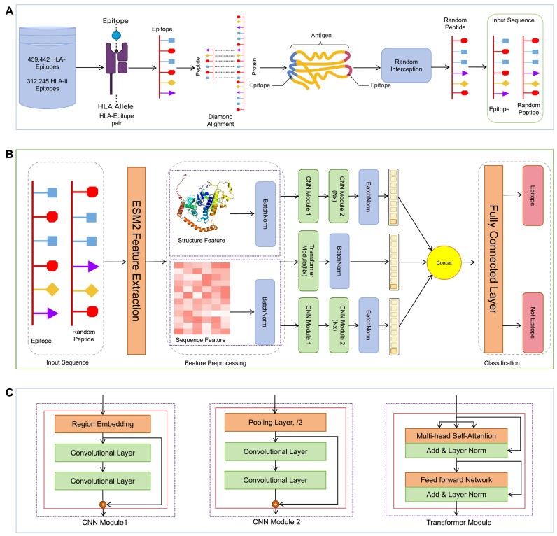
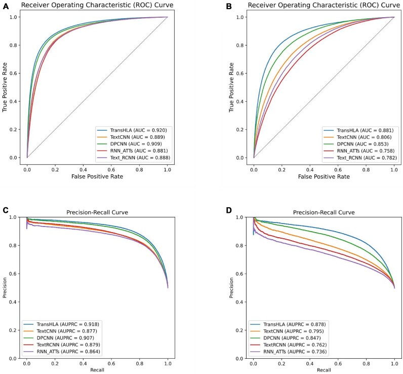
Background: Precise prediction of epitope presentation on human leukocyte antigen (HLA) molecules is crucial for advancing vaccine development and immunotherapy. Conventional HLA–peptide binding affinity prediction tools often focus on specific alleles and lack a universal approach for comprehensive HLA site analysis. This limitation hinders efficient filtering of invalid peptide segments. Results: We introduce TransHLA, a pioneering tool designed for epitope prediction across all HLA alleles, integrating Transformer and Residue CNN architectures. TransHLA utilizes the ESM2 large language model for sequence and structure embeddings, achieving high predictive accuracy. For HLA class I, it reaches an accuracy of 84.72% and an area under the curve (AUC) of 91.95% on IEDB test data. For HLA class II, it achieves 79.94% accuracy and an AUC of 88.14%. Our case studies using datasets like CEDAR and VDJdb demonstrate that TransHLA surpasses existing models in specificity and sensitivity for identifying immunogenic epitopes and neoepitopes. Conclusions: TransHLA significantly enhances vaccine design and immunotherapy by efficiently identifying broadly reactive peptides. Our resources, including data and code, are publicly accessible at https://github.com/SkywalkerLuke/TransHLA
Overview
The claims and statistics of this paper haven’t been checked yet.
Run the check — free, no account needed. It reads the open-access text and checks the sources it cites.
Integrity signals
Registry
Cites
Statistics
Figures
Verification
Claims the paper makes
The claims and statistics of this paper haven’t been checked yet.
Run the check — free, no account needed. It reads the open-access text and checks the sources it cites.
Self-check
Percentages, sums and reported differences re-computed from the paper’s own numbers.
The self-check needs the paper’s full text.
Citations
Cites
Deep Residual Learning for Image Recognition
He, K., Zhang, X., Ren, S., & Sun, J. (2016) · 2016 IEEE Conference on Computer Vision and Pattern Recognition (CVPR) · doi:10.1109/cvpr.2016.90 · Cited by 235,474
BERT: Pre-training of Deep Bidirectional Transformers for Language Understanding
Devlin, J., Chang, M.-W., Lee, K., & Toutanova, K. (2018) · arXiv · doi:10.48550/arxiv.1810.04805 · Cited by 73,173
BERT: Pre-training of Deep Bidirectional Transformers for Language Understanding
Devlin, J., Chang, M.-W., Lee, K., & Toutanova, K. (2019) · Proceedings of the 2019 Conference of the North American Chapter of the Association for Computational Linguistics: Human Language Technologies, Volume 1 (Long and Short Papers) · doi:10.18653/v1/n19-1423 · Cited by 54,325
Highly accurate protein structure prediction with AlphaFold
Jumper, J., Evans, R., Pritzel, A., Green, T., Figurnov, M., Ronneberger, O., Tunyasuvunakool, K., Bates, R., Žídek, A., Potapenko, A., et al. (2021) · Nature · doi:10.1038/s41586-021-03819-2 · Cited by 43,764
Language Models are Few-Shot Learners
Brown, T. B., Mann, B., Ryder, N., Subbiah, M., Kaplan, J., Dhariwal, P., Neelakantan, A., Shyam, P., Sastry, G., Askell, A., et al. (2020) · arXiv · doi:10.48550/arxiv.2005.14165 · Cited by 23,372
Evolutionary-scale prediction of atomic level protein structure with a language model
Lin, Z., Akin, H., Rao, R., Hie, B., Zhu, Z., Lu, W., Smetanin, N., Verkuil, R., Kabeli, O., Shmueli, Y., et al. (2022) · doi:10.1101/2022.07.20.500902 · Cited by 5,990
ProtTrans: Toward Understanding the Language of Life Through Self-Supervised Learning
Elnaggar, A., Heinzinger, M., Dallago, C., Rehawi, G., Wang, Y., Jones, L., Gibbs, T., Feher, T., Angerer, C., Steinegger, M., et al. (2022) · IEEE Transactions on Pattern Analysis and Machine Intelligence · doi:10.1109/tpami.2021.3095381 · Cited by 2,329
Research works printed but not linked: 48
- Chaffey, 2003, Molecular biology of the cell
- Murphy, 2016, Janeway’s immunobiology, 10.1201/9781315533247 · DOI
- Abbas, 2014, Cellular and molecular immunology E-book
- Shao, 2020, High-throughput prediction of MHC class I and II neoantigens with MHCnuggets, Cancer Immunol Res, 8, 396, 10.1158/2326-6066.CIR-19-0464 · DOI
- O’Donnell, 2020, MHCflurry 2.0: improved pan-allele prediction of MHC class I-presented peptides by incorporating antigen processing, Cell Syst, 11, 42, 10.1016/j.cels.2020.06.010 · DOI · Paper page
- Liu, 2021, DeepSeqPanII: an interpretable recurrent neural network model with attention mechanism for peptide-HLA class II binding prediction, IEEE/ACM T Comput Biol Bioinform, 19, 2188, 10.1109/TCBB.2021.3074927 · DOI
- Nguyen, 2021, The pockets guide to HLA class I molecules, Biochem Soc Trans, 49, 2319, 10.1042/BST20210410 · DOI
- Mei, 2021, Anthem: a user customised tool for fast and accurate prediction of binding between peptides and HLA class I molecules, Briefings Bioinf, 22, bbaa415, 10.1093/bib/bbaa415 · DOI
- Reynisson, 2020, NetMHCpan-4.1 and NetMHCIIpan-4.0: improved predictions of MHC antigen presentation by concurrent motif deconvolution and integration of MS MHC eluted ligand data, Nucleic Acids Res, 48, W449, 10.1093/nar/gkaa379 · DOI · Paper page
- Chu, 2022, A transformer-based model to predict peptide–HLA class I binding and optimize mutated peptides for vaccine design, Nat Mach Intel, 4, 300, 10.1038/s42256-022-00459-7 · DOI
- Tadros, 2024, Predicting MHC-I ligands across alleles and species: how far can we go?, bioRxiv, 2024
- Racle, 2023, Machine learning predictions of MHC-II specificities reveal alternative binding mode of class II epitopes, Immunity, 56, 1359, 10.1016/j.immuni.2023.03.009 · DOI
- Vaswani, 2017, Attention is all you need, Adv Neur Inf Proc Syst, 30, 10.48550/arXiv.1706.03762 · DOI · Paper page
- Kim, 2014, Convolutional neural networks for sentence classification, 10.3115/v1/D14-1181 · DOI · Paper page
- Lai, 2015, Recurrent convolutional neural networks for text classification, 10.1609/aaai.v29i1.9513 · DOI
- Johnson, 2017, Deep pyramid convolutional neural networks for text categorization, 562, 10.18653/v1/P17-1052 · DOI
- Zhou, 2016, Attention-based bidirectional long short-term memory networks for relation classification, 207, 10.18653/v1/P16-2034 · DOI
- Vita, 2019, The immune epitope database (IEDB): 2018 update, Nucleic Acids Res, 47, D339, 10.1093/nar/gky1006 · DOI
- Koşaloğlu-Yalçın, 2023, The cancer epitope database and analysis resource (CEDAR), Nucleic Acids Res, 51, D845, 10.1093/nar/gkac902 · DOI
- Shugay, 2018, VDJdb: a curated database of T-cell receptor sequences with known antigen specificity, Nucleic Acids Res, 46, D419, 10.1093/nar/gkx760 · DOI
- Nolan, 2025, A large-scale database of T-cell receptor beta (TCR$\beta$) sequences and binding associations from natural and synthetic exposure to SARS-CoV-2, Front Immunol, 10.3389/fimmu.2025.1488851 · DOI
- Lu, 2022, dbPepNeo2. 0: a database for human tumor neoantigen peptides from mass spectrometry and TCR recognition, Front Immunol, 13, 855976, 10.3389/fimmu.2022.855976 · DOI
- Xia, 2021, NEPdb: a database of T-cell experimentally-validated neoantigens and pan-cancer predicted neoepitopes for cancer immunotherapy, Front Immunol, 12, 644637, 10.3389/fimmu.2021.644637 · DOI
- Buchfink, 2015, Fast and sensitive protein alignment using DIAMOND, Nat Methods, 12, 59, 10.1038/nmeth.3176 · DOI · Paper page
- Pruitt, 2007, NCBI reference sequences (RefSeq): a curated non-redundant sequence database of genomes, transcripts and proteins, Nucleic Acids Res, 35, D61, 10.1093/nar/gkl842 · DOI
- Fu, 2012, CD-HIT: accelerated for clustering the next-generation sequencing data, Bioinformatics, 28, 3150, 10.1093/bioinformatics/bts565 · DOI · Paper page
- Chen, 2024, xTrimoPGLM: unified 100B-scale pre-trained transformer for deciphering the language of protein, 10.48550/arXiv.2401.06199 · DOI
- Du, 2023, UniDL4BioPep: a universal deep learning architecture for binary classification in peptide bioactivity, Briefings Bioinf, 24, bbad135, 10.1093/bib/bbad135 · DOI
- Xu, 2024, PTransIPs: identification of phosphorylation sites enhanced by protein PLM embeddings, IEEE J Biom Health Inf, 28, 3762, 10.1109/JBHI.2024.3377362 · DOI
- Mirdita, 2022, ColabFold: making protein folding accessible to all, Nat Methods, 19, 679, 10.1038/s41592-022-01488-1 · DOI · Paper page
- Racle, 2019, Robust prediction of HLA class II epitopes by deep motif deconvolution of immunopeptidomes, Nat Biotechnol, 37, 1283, 10.1038/s41587-019-0289-6 · DOI
- Nagler, 2021, Identification of presented SARS-CoV-2 HLA class I and HLA class II peptides using HLA peptidomics, Cell Rep, 35, 109305, 10.1016/j.celrep.2021.109305 · DOI
- Yang, 2024, MHCII-peptide presentation: an assessment of the state-of-the-art prediction methods, Front Immunol, 15, 1293706, 10.3389/fimmu.2024.1293706 · DOI
- Maćkiewicz, 1993, Principal components analysis (PCA), Comput Geosci, 19, 303, 10.1016/0098-3004(93)90090-R · DOI
- Westhof, 1984, Correlation between segmental mobility and the location of antigenic determinants in proteins, Nature, 311, 123, 10.1038/311123a0 · DOI
- Kim, 2021, Epitopes of protein binders are related to the structural flexibility of a target protein surface, J Chem Inf Model, 61, 2099, 10.1021/acs.jcim.0c01397 · DOI
- Klatt, 2020, Solving an MHC allele–specific bias in the reported immunopeptidome, JCI Insight, 5, 10.1172/jci.insight.141264 · DOI
- Cock, 2009, Biopython: freely available Python tools for computational molecular biology and bioinformatics, Bioinformatics, 25, 1422, 10.1093/bioinformatics/btp163 · DOI · Paper page
- Vihinen, 1994, Accuracy of protein flexibility predictions, Proteins Struct Funct Bioinf, 19, 141, 10.1002/prot.340190207 · DOI
- Chen, 2016, Xgboost: A scalable tree boosting system, 785, 10.1145/2939672.2939785 · DOI · Paper page
- Mariño Pérez, 2022, Visualizing protein breathing motions associated with aromatic ring flipping, Nature, 602, 695, 10.1038/s41586-022-04417-6 · DOI
- Anjana, 2012, Aromatic-aromatic interactions in structures of proteins and protein-DNA complexes: a study based on orientation and distance, Bioinformation, 8, 1220, 10.6026/97320630081220 · DOI
- Baker, 2012, Hydrogen bonding in biological macromolecules, 10.1107/97809553602060000887 · DOI
- Grimaldi, 2015, Stability of proteins on hydrophilic surfaces, Langmuir, 31, 1005, 10.1021/la503865b · DOI
- Drelich, 2011, Hydrophilic and superhydrophilic surfaces and materials, Soft Matter, 7, 9804, 10.1039/c1sm05849e · DOI
- Perticaroli, 2013, Secondary structure and rigidity in model proteins, Soft Matt, 9, 9548, 10.1039/c3sm50807b · DOI
- Mamonova, 2013, Stability and rigidity/flexibility—two sides of the same coin?, Biochim Biophys Acta, 1834, 854, 10.1016/j.bbapap.2013.02.011 · DOI
- Tianchi L, Xueying W, Wan N, et al. Supporting data for “TransHLA: A Hybrid Transformer Model for HLA-Presented Epitope Detection.”GigaScience Database. 2024. 10.5524/102633. · DOI
Metadata
- Authors
- Lu, T., Wang, X., Nie, W., Huo, M., & Li, S.
- Affiliations
- City University of Hong Kong (HK)
- Department of Computer Science, City University of Hong Kong , Kowloon 999077 ,
- Venue
- GigaScience
- Publisher
- Oxford University Press (OUP)
- Published
- 2025
- Volume
- 14
- ISSN
- 2047-217X (ISSN-L)
- PMC
- PMC11878767
- OpenAlex
- W4408141849
- Cited by
- 10
- References
- 7
- Citations by year
- 2026: 5 · 2025: 5
- FWCI
- 2.99
- Type
- journal article
- Language
- en
- Open access
- gold
- doi.org
- Licence
- Locations
- GigaScience (publishedVersion, cc-by)
- bioRxiv (Cold Spring Harbor Laboratory) (acceptedVersion, cc-by)
- Europe PMC (PubMed Central) (publishedVersion, cc-by)
- PubMed Central (publishedVersion, cc-by)
- PubMed (publishedVersion)
- CityU Scholars (submittedVersion, cc-by)
- Topics
- Life Sciences › Biochemistry, Genetics and Molecular Biology › Molecular Biology › vaccines and immunoinformatics approaches
- Life Sciences › Biochemistry, Genetics and Molecular Biology › Molecular Biology › Machine Learning in Bioinformatics
- Life Sciences › Immunology and Microbiology › Immunology › Immunotherapy and Immune Responses
- Keywords
- HLA class I, HLA class II, ESM-2 embeddings, immunogenic epitopes, neoantigens, vaccine design, immunotherapy, IEDB, cedar
- MeSH terms
- Alleles, Epitopes, HLA Antigens, Humans, Software, Antigen Presentation, Epitope Mapping, Computational Biology
About these checks
Registry, retraction, Cites and statistics results are computed, not judged, with one exception: a printed reference that no index lists is read once by a model before it counts as a citation error. Claims are extracted by a model and kept only when their quote appears verbatim in the text; each cited source is then checked, with a model’s help, for whether it says what the claim relies on. Those verdicts can be wrong — read the claim’s quote and the cited source before relying on one.
Statistics and arithmetic are recomputed, not judged.
Confidence bands are shown only for fields calibrated on our annotated test set. None is calibrated yet, so no band is shown.
What counts as a claim ›How we measure confidence ›How we check this ›
Are you an author, or do you see a problem with this page? Request a review
How to cite
Cite CoChat for the checks on this page, and the paper for its own findings.
- This page
- CoChat. (2026). Citation check: TransHLA: a Hybrid Transformer model for HLA-presented epitope detection. https://app.cochat.ai/papers/10.1093/gigascience/giaf008
- The paper
- Lu, T., Wang, X., Nie, W., Huo, M., & Li, S. (2025). TransHLA: a Hybrid Transformer model for HLA-presented epitope detection. GigaScience. https://doi.org/10.1093/gigascience/giaf008
