

The DOI matches its Crossref record; 44 of the 44 references it cites were found in a registry or an index.
Defective autophagy in neurons and astrocytes from mice deficient in PI(3,5)P2
Open accessAbstract
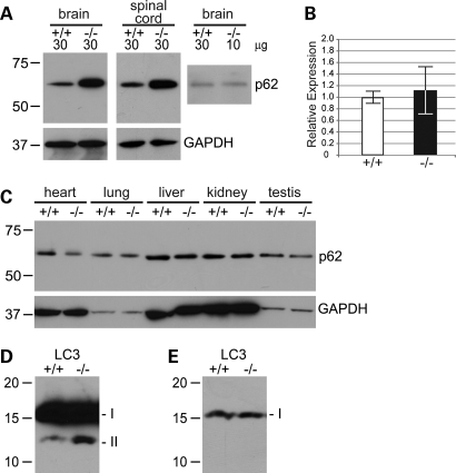
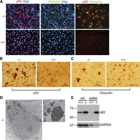
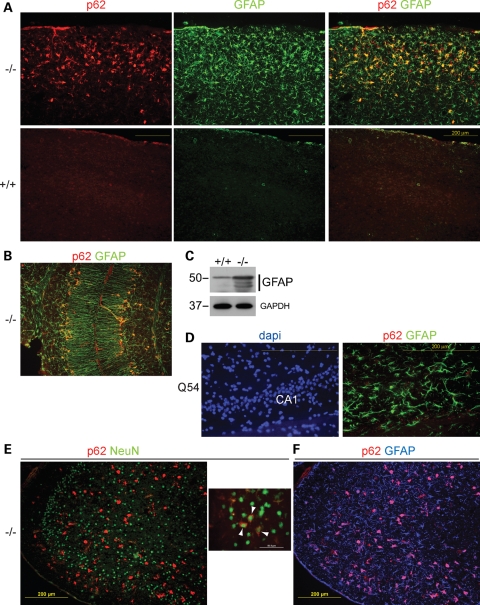
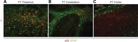
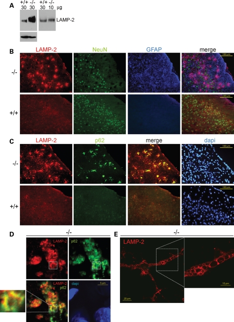
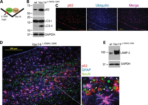
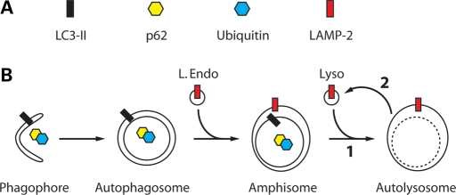
Mutations affecting the conversion of PI3P to the signaling lipid PI(3,5)P(2) result in spongiform degeneration of mouse brain and are associated with the human disorders Charcot-Marie-Tooth disease and amyotrophic lateral sclerosis (ALS). We now report accumulation of the proteins LC3-II, p62 and LAMP-2 in neurons and astrocytes of mice with mutations in two components of the PI(3,5)P(2) regulatory complex, Fig4 and Vac14. Cytoplasmic inclusion bodies containing p62 and ubiquinated proteins are present in regions of the mutant brain that undergo degeneration. Co-localization of p62 and LAMP-2 in affected cells indicates that formation or recycling of the autolysosome is impaired. These results establish a role for PI(3,5)P(2) in autophagy in the mammalian central nervous system (CNS) and demonstrate that mutations affecting PI(3,5)P(2) can contribute to inclusion body disease.
Overview
The claims and statistics of this paper haven’t been checked yet.
Run the check — free, no account needed. It reads the open-access text and checks the sources it cites.
Integrity signals
Registry
Cites
Statistics
Figures

Verification
Claims the paper makes
The claims and statistics of this paper haven’t been checked yet.
Run the check — free, no account needed. It reads the open-access text and checks the sources it cites.
Claims other papers make about this one
Each is checked against this paper’s own text: does it say what the citing paper says it does?
In the citing paper
“Indeed, accumulation of LC3-II and p62 proteins is a well-known feature of autophagy-deficient tissues [32, 64, 80].”
From Senescence-related impairment of autophagy induces toxic intraneuronal amyloid-β accumulation in a mouse model of amyloid pathology (2023) · cites this paper as [32]
Self-check
Percentages, sums and reported differences re-computed from the paper’s own numbers.
The self-check needs the paper’s full text.
Citations
Cites
Suppression of basal autophagy in neural cells causes neurodegenerative disease in mice
Hara, T., Nakamura, K., Matsui, M., Yamamoto, A., Nakahara, Y., Suzuki-Migishima, R., Yokoyama, M., Mishima, K., Saito, I., Okano, H., et al. (2006) · Nature · doi:10.1038/nature04724 · Cited by 3,882
Rapamycin fed late in life extends lifespan in genetically heterogeneous mice
Harrison, D. E., Strong, R., Sharp, Z. D., Nelson, J. F., Astle, C. M., Flurkey, K., Nadon, N. L., Wilkinson, J. E., Frenkel, K., Carter, C. S., et al. (2009) · Nature · doi:10.1038/nature08221 · Cited by 3,766
Loss of autophagy in the central nervous system causes neurodegeneration in mice
Komatsu, M., Waguri, S., Chiba, T., Murata, S., Iwata, J.-I., Tanida, I., Ueno, T., Koike, M., Uchiyama, Y., Kominami, E., et al. (2006) · Nature · doi:10.1038/nature04723 · Cited by 3,500
Protein aggregation and neurodegenerative disease
Ross, C. A., & Poirier, M. A. (2004) · Nature Medicine · doi:10.1038/nm1066 · Cited by 3,454
TDP-43 Mutations in Familial and Sporadic Amyotrophic Lateral Sclerosis
Sreedharan, J., Blair, I. P., Tripathi, V. B., Hu, X., Vance, C., Rogelj, B., Ackerley, S., Durnall, J. C., Williams, K. L., Buratti, E., et al. (2008) · Science · doi:10.1126/science.1154584 · Cited by 2,630
How to Interpret LC3 Immunoblotting
Mizushima, N., & Yoshimori, T. (2007) · Autophagy · doi:10.4161/auto.4600 · Cited by 2,595
Guidelines for the use and interpretation of assays for monitoring autophagy in higher eukaryotes
Klionsky, D. J., Abeliovich, H., Agostinis, P., Agrawal, D. K., Aliev, G., Askew, D. S., Baba, M., Baehrecke, E. H., Bahr, B. A., Ballabio, A., et al. (2008) · Autophagy · doi:10.4161/auto.5338 · Cited by 2,230
Autophagy: from phenomenology to molecular understanding in less than a decade
Klionsky, D. J. (2007) · Nature Reviews Molecular Cell Biology · doi:10.1038/nrm2245 · Cited by 1,977
Axe, E. L., Walker, S. A., Manifava, M., Chandra, P., Roderick, H. L., Habermann, A., Griffiths, G., & Ktistakis, N. T. (2008) · The Journal of Cell Biology · doi:10.1083/jcb.200803137 · Cited by 1,885
Astrocytes as determinants of disease progression in inherited amyotrophic lateral sclerosis
Yamanaka, K., Chun, S. J., Boillee, S., Fujimori-Tonou, N., Yamashita, H., Gutmann, D. H., Takahashi, R., Misawa, H., & Cleveland, D. W. (2008) · Nature Neuroscience · doi:10.1038/nn2047 · Cited by 1,140 · cited 1×
“Using conditional knock-out technology, tissue-specific knock-out of mutant SOD1 expression in astrocytes was shown to delay disease progression (31).”
Cited by
Sorted by citations
Hardiman, O. M., Al‐Chalabi, A., Chiò, A., Corr, E. M., Logroscino, G., Robberecht, W., Shaw, P. J., Simmons, Z., & van den Berg, L. H. (2017) · Nature Reviews Disease Primers · doi:10.1038/nrdp.2017.71 · Cited by 2,262
Dong, X., Shen, D., Wang, X., Dawson, T., Li, X., Zhang, Q., Cheng, X., Zhang, Y., Weisman, L. S., Delling, M., et al. (2010) · Nature Communications · doi:10.1038/ncomms1037 · Cited by 645
Canonical and non-canonical autophagy: variations on a common theme of self-eating?
Codogno, P., Mehrpour, M., & Proikas‐Cezanne, T. (2011) · Nature Reviews Molecular Cell Biology · doi:10.1038/nrm3249 · Cited by 565
The different autophagy degradation pathways and neurodegeneration
Fleming, A., Bourdenx, M., Fujimaki, M., Karabiyik, C., Krause, G. J., Lopez, A., Martín‐Segura, A., Puri, C., Scrivo, A., Skidmore, J., et al. (2022) · Neuron · doi:10.1016/j.neuron.2022.01.017 · Cited by 500
Gómez-Virgilio, L., Silva-Lucero, M.-D.-C., Flores-Morelos, D.-S., Gallardo-Nieto, J., Lopez-Toledo, G., Abarca-Fernandez, A.-M., Zacapala-Gómez, A.-E., Luna-Muñoz, J., Montiel-Sosa, F., Soto-Rojas, L. O., et al. (2022) · Cells · doi:10.3390/cells11152262 · Cited by 431
Metadata
- Authors
- Ferguson, C. J., Lenk, G. M., & Meisler, M. H.
- Affiliations
- University of Michigan (US)
- Corresponding author
- Meisler, Miriam H.
- Venue
- Human Molecular Genetics
- Publisher
- Oxford University Press (OUP)
- Published
- 2009-09-29
- Volume
- 18
- Issue
- 24
- Pages
- 4868-4878
- ISSN
- 0964-6906 (ISSN-L)
- 1460-2083
- PMC
- PMC2778378
- OpenAlex
- W2135384508
- Cited by
- 202
- References
- 44
- Citations by year
- 2026: 5 · 2025: 5 · 2024: 4 · 2023: 14 · 2022: 12 · 2021: 13 · 2020: 13 · 2019: 14 · 2018: 9 · 2017: 15 · 2016: 7 · 2015: 11 · 2014: 20 · 2013: 15 · 2012: 21
- FWCI
- 10.07
- Type
- journal article
- Language
- en
- Open access
- hybrid
- doi.org
- Licence
- cc-by-nc
- Locations
- Human Molecular Genetics (publishedVersion, cc-by-nc)
- PubMed Central (publishedVersion, cc-by-nc)
- PubMed (publishedVersion)
- Funders
- Hartwell Foundation
- National Institutes of Health
- National Institute of General Medical Sciences (T 32 GM07863, T32GM007863, R01GM024872, R01-GM24872, T32GM008322)
- Topics
- Life Sciences › Biochemistry, Genetics and Molecular Biology › Cell Biology › Autophagy in Disease and Therapy
- Life Sciences › Neuroscience › Cellular and Molecular Neuroscience › Hereditary Neurological Disorders
- Life Sciences › Biochemistry, Genetics and Molecular Biology › Biochemistry › Glycogen Storage Diseases and Myoclonus
- Keywords
- PI(3,5)P2, autophagy, neurons, astrocytes, FIG4, VAC14, LC3-II, p62, LAMP2, autolysosomes, spongiform degeneration, Charcot-Marie-Tooth disease, amyotrophic lateral sclerosis, inclusion bodies, ubiquitinated proteins, central nervous system
- MeSH terms
- Phosphoinositide Phosphatases, Amyotrophic Lateral Sclerosis, Animals, Astrocytes, Autophagy, Brain, Inclusion Bodies, Charcot-Marie-Tooth Disease, DNA-Binding Proteins, Flavoproteins, Humans, Mice, Mutant Strains, Microtubule-Associated Proteins, Neurons, Transcription Factors, Up-Regulation, Phosphatidylinositol Phosphates, Ubiquitin, Mice, Transcription Factor TFIIH, Lysosomal-Associated Membrane Protein 2
About these checks
Registry, retraction, Cites and statistics results are computed, not judged, with one exception: a printed reference that no index lists is read once by a model before it counts as a citation error. Claims are extracted by a model and kept only when their quote appears verbatim in the text; each cited source is then checked, with a model’s help, for whether it says what the claim relies on. Those verdicts can be wrong — read the claim’s quote and the cited source before relying on one.
Statistics and arithmetic are recomputed, not judged.
Confidence bands are shown only for fields calibrated on our annotated test set. None is calibrated yet, so no band is shown.
What counts as a claim ›How we measure confidence ›How we check this ›
Are you an author, or do you see a problem with this page? Request a review
