Cited by 102Photosynthetic Processes and MechanismsPlant Water Relations and Carbon DynamicsPlant biochemistry and biosynthesis

The DOI matches its Crossref record.

Improved analysis of C4and C3photosynthesis via refined in vitro assays of their carbon fixation biochemistry

Open access

Abstract

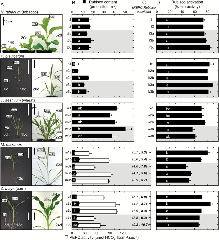
Plants operating C3 and C4 photosynthetic pathways exhibit differences in leaf anatomy and photosynthetic carbon fixation biochemistry. Fully understanding this underpinning biochemical variation is requisite to identifying solutions for improving photosynthetic efficiency and growth. Here we refine assay methods for accurately measuring the carboxylase and decarboxylase activities in C3 and C4 plant soluble protein. We show that differences in plant extract preparation and assay conditions are required to measure NADP-malic enzyme and phosphoenolpyruvate carboxylase (pH 8, Mg(2+), 22 °C) and phosphoenolpyruvate carboxykinase (pH 7, >2mM Mn(2+), no Mg(2+)) maximal activities accurately. We validate how the omission of MgCl2 during leaf protein extraction, lengthy (>1min) centrifugation times, and the use of non-pure ribulose-1,5-bisphosphate (RuBP) significantly underestimate Rubisco activation status. We show how Rubisco activation status varies with leaf ontogeny and is generally lower in mature C4 monocot leaves (45-60% activation) relative to C3 monocots (55-90% activation). Consistent with their >3-fold lower Rubisco contents, full Rubisco activation in soluble protein from C4 leaves (<5min) was faster than in C3 plant samples (<10min), with addition of Rubisco activase not required for full activation. We conclude that Rubisco inactivation in illuminated leaves primarily stems from RuBP binding to non-carbamylated enzyme, a state readily reversible by dilution during cellular protein extraction.

Overview

The claims and statistics of this paper haven’t been checked yet.

Run the check — free, no account needed. It reads the open-access text and checks the sources it cites.

A few free checks a day · papers already verified open instantly · results are public

Integrity signals

Registry

Crossref · 10.1093/jxb/erw154

Cites

Statistics

Figures

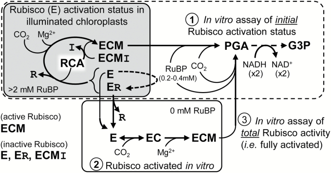
Fig. 1. Summary of Rubisco activation status in vivo and modulation in vitro. Summary of the NADH-linked assay used to determine Rubisco activation status using rapidly extracted soluble protein from young, non-stressed leaves sampled during illumination [where inactive ECMI complexes containing the Rubisco inhibitors (I) CA1P or XuBP would be negligible]. See text for details of the three assay stages indicated. R, RuBP; RCA, Rubisco activase. Details of the NADH-linked assay are summarized in Supplementary Fig. S1 and Supplementary Table S1.
Fig. 1. Summary of Rubisco activation status in vivo and modulation in vitro. Summary of the NADH-linked assay used to determine Rubisco activation status using rapidly extracted soluble protein from young, non-stressed leaves sampled during illumination [where inactive ECMI complexes containing the Rubisco inhibitors (I) CA1P or XuBP would be negligible]. See text for details of the three assay stages indicated. R, RuBP; RCA, Rubisco activase. Details of the NADH-linked assay are summarized in Supplementary Fig. S1 and Supplementary Table S1.CC BY · PubMed Central
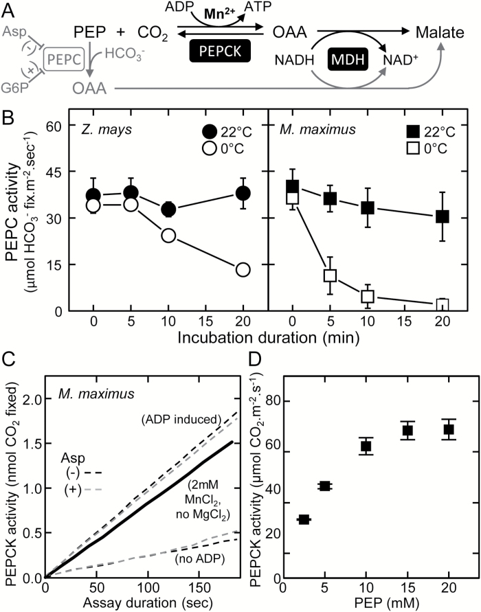
Fig. 2. Optimizing the measurement of PEPC and PEPCK activities in leaf extract. (A) Summarizing the commonality of the malate dehydrogenase- (MDH) coupled, NADH oxidation-linked assay to quantify the carboxylation of phosphoenolpyruvate (PEP) into oxaloacetate (OAA) by PEP carboxykinase (PEPCK; in black) and PEP carboxylase (PEPC; in gray), an enzyme inhibited (–) by aspartate (Asp; Huber and Edwards, 1975) and activated (+) by glucose-6-phosphate (G6P). (B) Effect of storage temperature (room temperature, 22 °C, or ice, 0 °C) and time on PEPC activity in the soluble protein from young leaves from mature Z. mays and M. maximus plants (n=3 biological replicates ±SD; see leaves m3b and c3b in Fig. 5A for examples). (C) Representative assay of PEP carboxylation by PEPCK measured in leaf soluble protein extract using the no MgCl2 method of Sharwood et al. (2014) ( black line) and the modified ADP method of this study in assays with (gray dashed lines) or without (black dashed lines) 5mM aspartate (a PEPC inhibitor, A). (D) Response of PEPCK activity to [PEP] in M. maximus leaf soluble protein. To prevent PEPC interference ensure: 1) No MgCl2 in assay and extraction buffers, 2) MnCl2 up to 5mM in extraction and assay buffers, 3) pH of extraction and assay buffers <7.0, 4) No glucose-6-phosphate, and 5) include aspartate in assay buffer.
Fig. 2. Optimizing the measurement of PEPC and PEPCK activities in leaf extract. (A) Summarizing the commonality of the malate dehydrogenase- (MDH) coupled, NADH oxidation-linked assay to quantify the carboxylation of phosphoenolpyruvate (PEP) into oxaloacetate (OAA) by PEP carboxykinase (PEPCK; in black) and PEP carboxylase (PEPC; in gray), an enzyme inhibited (–) by aspartate (Asp; Huber and Edwards, 1975) and activated (+) by glucose-6-phosphate (G6P). (B) Effect of storage temperature (room temperature, 22 °C, or ice, 0 °C) and time on PEPC activity in the soluble protein from young leaves from mature Z. mays and M. maximus plants (n=3 biological replicates ±SD; see leaves m3b and c3b in Fig. 5A for examples). (C) Representative assay of PEP carboxylation by PEPCK measured in leaf soluble protein extract using the no MgCl2 method of Sharwood et al. (2014) ( black line) and the modified ADP method of this study in assays with (gray dashed lines) or without (black dashed lines) 5mM aspartate (a PEPC inhibitor, A). (D) Response of PEPCK activity to [PEP] in M. maximus leaf soluble protein. To prevent PEPC interference ensure: 1) No MgCl2 in assay and extraction buffers, 2) MnCl2 up to 5mM in extraction and assay buffers, 3) pH of extraction and assay buffers <7.0, 4) No glucose-6-phosphate, and 5) include aspartate in assay buffer.CC BY · PubMed Central
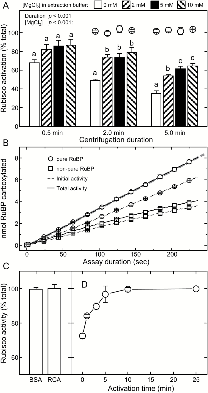
Fig. 3. Evaluating the experimental methodology for measuring leaf Rubisco activation status. (A) Appraising how MgCl2 inclusion and quickness of soluble leaf protein extraction influences Rubisco activation quantification. NADH-linked assays were performed on N2-frozen replica (n=5) tobacco leaf discs (0.5cm2) taken from a young, nearly fully expanded upper canopy leaf (15cm in diameter) and stored at –80 °C for up to 3 months without effect on recoverable activity. Circles indicate the total activities measured after 10min activation relative to the 0.5min centrifuged sample (B) Representative NADH-linked spectrophotometric measures of initial (dashed lines) and total (solid lines) Rubisco activities made using low purity commercial RuBP (squares) or that purified according to Kane et al. (1998) (circles). Rates correspond to protein from 0.9mm2 of leaf with a Rubisco active site concentration in each assay of ~34.4nM (i.e. a k cat of 2.2s−1). (C) Incubation of leaf protein extract for 2min at 25 °C with 80 μg ml–1 of either BSA or purified tobacco Rubisco activase (RCA) had no effect on the measured rates of fully activated Rubisco, while the same treatment re-activated >80% of inhibited ER complexes formed using a comparable concentration of purified tobacco Rubisco (Supplementary Fig. S2). (D) Change in the activity status of Rubisco in tobacco soluble protein activated at 25 °C for up to 25min. Data in (C) and (D) are the averages (±SE) from analyses with three separate leaf samples expressed as a percentage of the total activities measured after 10min (C) and 25min (D) activation. For (A), the significance level (P) for the [MgCl2] and centrifugation duration factors are shown. Letters indicate the ranking (lowest=a) of means within each centrifugation duration using a post-hoc Tukey test. Values followed by the same letter are not significantly different at the 5% level (P>0.05).
Fig. 3. Evaluating the experimental methodology for measuring leaf Rubisco activation status. (A) Appraising how MgCl2 inclusion and quickness of soluble leaf protein extraction influences Rubisco activation quantification. NADH-linked assays were performed on N2-frozen replica (n=5) tobacco leaf discs (0.5cm2) taken from a young, nearly fully expanded upper canopy leaf (15cm in diameter) and stored at –80 °C for up to 3 months without effect on recoverable activity. Circles indicate the total activities measured after 10min activation relative to the 0.5min centrifuged sample (B) Representative NADH-linked spectrophotometric measures of initial (dashed lines) and total (solid lines) Rubisco activities made using low purity commercial RuBP (squares) or that purified according to Kane et al. (1998) (circles). Rates correspond to protein from 0.9mm2 of leaf with a Rubisco active site concentration in each assay of ~34.4nM (i.e. a k cat of 2.2s−1). (C) Incubation of leaf protein extract for 2min at 25 °C with 80 μg ml–1 of either BSA or purified tobacco Rubisco activase (RCA) had no effect on the measured rates of fully activated Rubisco, while the same treatment re-activated >80% of inhibited ER complexes formed using a comparable concentration of purified tobacco Rubisco (Supplementary Fig. S2). (D) Change in the activity status of Rubisco in tobacco soluble protein activated at 25 °C for up to 25min. Data in (C) and (D) are the averages (±SE) from analyses with three separate leaf samples expressed as a percentage of the total activities measured after 10min (C) and 25min (D) activation. For (A), the significance level (P) for the [MgCl2] and centrifugation duration factors are shown. Letters indicate the ranking (lowest=a) of means within each centrifugation duration using a post-hoc Tukey test. Values followed by the same letter are not significantly different at the 5% level (P>0.05).CC BY · PubMed Central
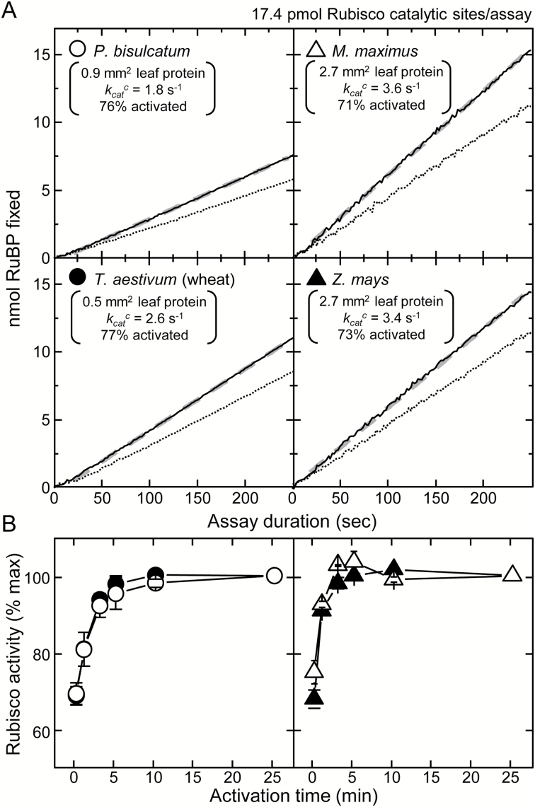
Fig. 4. Variations in Rubisco content and activation status during leaf ontogeny can account for variations in the in vivo estimates of V c max. (A) Representative NADH-linked assay data showing the linear RuBP carboxylation rates in assays of initial (dashed lines) and total (solid lines, after 10–15min activation) Rubsico activities at 25 °C using soluble leaf protein from both C3 (P. bisulcatum, T. aestivum) and C4 (M. maximus, Z. mays) monocotyledon species. Shown are details of the calculated Rubisco activation status (% of maximum), the derived carboxylation rates (k cat c, quantified from the slope of the fitted linear regression, gray dashed line, divided by Rubisco content quantified by [14C]CABP binding) and the area of leaf protein required to attain the 17.4 pmol Rubsico catalytic sites used to normalize the plotted data to highlight the variations in k cat c between each species (see also Table 1). (B) Response of Rubisco activation and activity in the soluble leaf protein of each species following incubation at 25 °C. Shown is the average (±SE) of analyses from three separate leaf samples for each species expressed as a percentage of the maximum activities measured after 25min activation.
Fig. 4. Variations in Rubisco content and activation status during leaf ontogeny can account for variations in the in vivo estimates of V c max. (A) Representative NADH-linked assay data showing the linear RuBP carboxylation rates in assays of initial (dashed lines) and total (solid lines, after 10–15min activation) Rubsico activities at 25 °C using soluble leaf protein from both C3 (P. bisulcatum, T. aestivum) and C4 (M. maximus, Z. mays) monocotyledon species. Shown are details of the calculated Rubisco activation status (% of maximum), the derived carboxylation rates (k cat c, quantified from the slope of the fitted linear regression, gray dashed line, divided by Rubisco content quantified by [14C]CABP binding) and the area of leaf protein required to attain the 17.4 pmol Rubsico catalytic sites used to normalize the plotted data to highlight the variations in k cat c between each species (see also Table 1). (B) Response of Rubisco activation and activity in the soluble leaf protein of each species following incubation at 25 °C. Shown is the average (±SE) of analyses from three separate leaf samples for each species expressed as a percentage of the maximum activities measured after 25min activation.CC BY · PubMed Central
Fig. 5. Variation in PEPC activity, Rubisco content, and Rubisco activation status with leaf ontogeny and development in C3 and C4 plants. (A) Pictures showing the leaves at differing stages of ontogeny and plant development (as labeled) that were analyzed for (B) Rubisco content (black bars, determined by [14C]CABP binding) and PEPC activity (white bars). (C) For both C4 species, the corresponding PEPC:Rubisco activity ratios are shown in parenthess; the first value is determined from the rates measured using the NADH-linked assays (in italics) and the second value (in bold) takes into account the higher Rubisco k cat c values quantified using 14CO2 assays (Table 1) and (D) the activation status of Rubisco in each leaf analyzed. The age of each plant (days, d) post-cotyledon emergence is indicated with the scale bar=10cm. All data are averages (±SD) of n=3 leaf discs taken from each leaf (or for the juvenile samples b1, c1, and w1, from replica plantlets). Regions shaded gray in (B) and (D) indicates data for leaves sampled from more mature plants. For (B) and (D), letters indicate the ranking (lowest=a) of means within each species using a post-hoc Tukey test. Values followed by the same letter are not significantly different at the 5% level (P>0.05). The levels of Rubisco measured correlate with those previously measured in the leaves of tobacco (Whitney et al., 2011), P. bisulcatum (Pinto et al., 2014), M. maximus (Pinto et al., 2016), and maize (Sharwood et al., 2014).
Fig. 5. Variation in PEPC activity, Rubisco content, and Rubisco activation status with leaf ontogeny and development in C3 and C4 plants. (A) Pictures showing the leaves at differing stages of ontogeny and plant development (as labeled) that were analyzed for (B) Rubisco content (black bars, determined by [14C]CABP binding) and PEPC activity (white bars). (C) For both C4 species, the corresponding PEPC:Rubisco activity ratios are shown in parenthess; the first value is determined from the rates measured using the NADH-linked assays (in italics) and the second value (in bold) takes into account the higher Rubisco k cat c values quantified using 14CO2 assays (Table 1) and (D) the activation status of Rubisco in each leaf analyzed. The age of each plant (days, d) post-cotyledon emergence is indicated with the scale bar=10cm. All data are averages (±SD) of n=3 leaf discs taken from each leaf (or for the juvenile samples b1, c1, and w1, from replica plantlets). Regions shaded gray in (B) and (D) indicates data for leaves sampled from more mature plants. For (B) and (D), letters indicate the ranking (lowest=a) of means within each species using a post-hoc Tukey test. Values followed by the same letter are not significantly different at the 5% level (P>0.05). The levels of Rubisco measured correlate with those previously measured in the leaves of tobacco (Whitney et al., 2011), P. bisulcatum (Pinto et al., 2014), M. maximus (Pinto et al., 2016), and maize (Sharwood et al., 2014).CC BY · PubMed Central
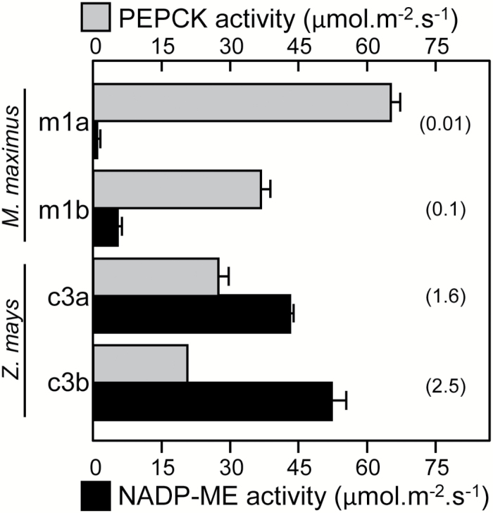
Fig. 6. Maximal activity of the decarboxylases in C4 grasses. Comparison of PEPCK (gray) and NADP-ME (black) activities in differing aged Z. mays and M. maximus leaves (n=3, ± SE). The leaves analyzed are shown in Fig. 4A. The PEPCK:NADP-ME ratio activities are indicated in parentheses.
Fig. 6. Maximal activity of the decarboxylases in C4 grasses. Comparison of PEPCK (gray) and NADP-ME (black) activities in differing aged Z. mays and M. maximus leaves (n=3, ± SE). The leaves analyzed are shown in Fig. 4A. The PEPCK:NADP-ME ratio activities are indicated in parentheses.CC BY · PubMed Central

Verification

Claims the paper makes

The claims and statistics of this paper haven’t been checked yet.

Run the check — free, no account needed. It reads the open-access text and checks the sources it cites.

A few free checks a day · papers already verified open instantly · results are public

Claims other papers make about this one

Each is checked against this paper’s own text: does it say what the citing paper says it does?

Self-check

Percentages, sums and reported differences re-computed from the paper’s own numbers.

The self-check needs the paper’s full text.

Citations

Cites

No references could be resolved yet.

Research works printed but not linked: 81

  1. 10.1093/jxb/ern091 · DOI
  2. 10.1016/j.plaphy.2008.01.001 · DOI
  3. 10.1042/BJ20111443 · DOI
  4. 10.1016/S0003-9861(03)00100-0 · DOI
  5. 10.1016/0014-5793(96)00555-8 · DOI
  6. 10.1016/B978-0-12-461013-2.50010-1 · DOI
  7. 10.1016/S0076-6879(05)98044-0 · DOI
  8. 10.1007/978-1-60761-925-3_29 · DOI
  9. 10.1016/j.tplants.2010.03.006 · DOI · Paper page
  10. 10.1104/pp.113.228221 · DOI
  11. 10.1111/j.1365-3040.2001.00668.x · DOI
  12. 10.1104/pp.15.00586 · DOI
  13. 10.1038/nplants.2014.2 · DOI
  14. 10.1093/jxb/erq071 · DOI
  15. 10.1104/pp.128.1.160 · DOI
  16. 10.1073/pnas.230451497 · DOI · Paper page
  17. 10.1104/pp.93.4.1390 · DOI
  18. 10.1126/science.1177216 · DOI
  19. 10.1093/pcp/pcl028 · DOI
  20. 10.1007/BF00377192 · DOI
  21. 10.1007/BF00386231 · DOI · Paper page
  22. 10.1093/jxb/err080 · DOI
  23. 10.1093/jxb/erq303 · DOI · Paper page
  24. 10.1104/pp.104.054759 · DOI
  25. 10.1093/jxb/29.3.547 · DOI
  26. 10.1038/324274a0 · DOI
  27. 10.1016/S0304-4173(87)80009-5 · DOI
  28. 10.1071/PP9780571 · DOI
  29. Jordan, 1983, Inhibition of ribulose bisphosphate carboxylase by substrate ribulose 1,5-bisphosphate, Journal of BIological Chemistry, 258, 13752, 10.1016/S0021-9258(17)43982-2 · DOI
  30. Kanai R Edwards GE . 1999. The biochemistry of C4 photosynthesis. In: Rowan FS Russell KM , eds. C4 plant biology . San Diego: Academic Press, 49–87. · DOI
  31. 10.1104/pp.117.3.1059 · DOI
  32. 10.1093/pcp/pci052 · DOI
  33. 10.1016/j.plantsci.2015.03.004 · DOI
  34. 10.1104/pp.108.129890 · DOI
  35. 10.1007/978-1-60761-925-3_27 · DOI
  36. 10.1042/bj1590563 · DOI
  37. Lilley, 1974, An improved spectrophotometric assay for ribulosebisphosphate carboxylase, Biochimica et Biophysica Acta, 358, 226, 10.1016/0005-2744(74)90274-5 · DOI
  38. 10.1016/j.cell.2015.03.019 · DOI
  39. 10.1093/pcp/pcg113 · DOI
  40. 10.1104/pp.99.4.1551 · DOI
  41. 10.1007/s11120-013-9819-0 · DOI
  42. 10.1042/BJ20080668 · DOI
  43. 10.1093/aob/mct017 · DOI
  44. 10.1093/aob/mcf103 · DOI · Paper page
  45. 10.1046/j.1365-3040.1997.d01-85.x · DOI
  46. 10.1093/jxb/ers336 · DOI
  47. 10.1093/jxb/ern084 · DOI
  48. 10.1042/BJ20060430 · DOI
  49. 10.1074/jbc.M305493200 · DOI
  50. 10.1104/pp.112.203240 · DOI
  51. 10.1007/s11120-015-0092-2 · DOI
  52. 10.1023/B:PRES.0000017179.31351.f0 · DOI
  53. 10.1111/pce.12636 · DOI
  54. 10.1093/jxb/eru155 · DOI
  55. 10.1093/jxb/ers257 · DOI
  56. 10.1023/A:1022421515027 · DOI
  57. 10.1104/pp.58.5.603 · DOI
  58. 10.1111/j.1399-3054.1988.tb00638.x · DOI
  59. 10.1111/j.1365-3040.1991.tb01528.x · DOI
  60. Ruuska, 1998, The interplay between limiting processes in C3 photosynthesis studied by rapid-response gas exchange using transgenic tobacco impaired in photosynthesis, Functional Plant Biology, 25, 859, 10.1071/PP98079 · DOI
  61. 10.1104/pp.104.047977 · DOI
  62. 10.1111/j.1469-8137.2004.00974.x · DOI
  63. 10.1146/annurev-arplant-042811-105511 · DOI
  64. 10.1111/j.0031-9317.2004.0173.x · DOI
  65. 10.1104/pp.90.2.679 · DOI
  66. 10.1007/s11120-013-9964-5 · DOI
  67. 10.1111/j.1365-3040.2007.01710.x · DOI
  68. 10.1104/pp.107.109058 · DOI
  69. 10.1093/jxb/eru130 · DOI
  70. 10.1111/pce.12386 · DOI
  71. 10.1111/j.1365-3040.2007.01679.x · DOI
  72. 10.1093/oxfordjournals.jbchem.a022791 · DOI
  73. von Caemmerer S . 2000. Biochemical models of leaf photosynthesis. Melbourne: CSIRO Publishing. · DOI
  74. 10.1093/jxb/eru127 · DOI
  75. 10.1104/pp.104.056077 · DOI
  76. 10.1111/pce.12166 · DOI · Paper page
  77. 10.1104/pp.128.1.165 · DOI
  78. 10.1046/j.1365-313x.2001.01056.x · DOI
  79. 10.1007/978-1-62703-995-6_15 · DOI
  80. 10.1073/pnas.1109503108 · DOI
  81. 10.1104/pp.121.2.579 · DOI

Cited by

Sorted by citations

See all 102

Metadata

Authors
Sharwood, R. E., Sonawane, B. V., Ghannoum, O., & Whitney, S. M.
Affiliations
  • Australian National University (AU)
  • Western Sydney University (AU)
Corresponding author
Sharwood, Robert E.
Venue
Journal of Experimental Botany
Published
2016-04-27
Volume
67
Issue
10
Pages
3137-3148
ISSN
  • 0022-0957 (ISSN-L)
  • 1460-2431
OpenAlex
W2343690410
Cited by
102
References
0
Citations by year
2026: 5 · 2025: 10 · 2024: 8 · 2023: 9 · 2022: 8 · 2021: 11 · 2020: 17 · 2019: 10 · 2018: 11 · 2017: 8 · 2016: 5
FWCI
5.89
Type
journal article
Language
en
Open access
Licence
cc-by
Locations
  • Journal of Experimental Botany (publishedVersion, cc-by)
  • PubMed Central (publishedVersion, cc-by)
  • PubMed (publishedVersion)
  • HighWire Press Open Archive (submittedVersion)
  • ANU Open Research (Australian National University) (submittedVersion, cc-by)
Funders
Australian Research Council (DE130101760, DP120101603, CE140100015)
Topics
  • Life Sciences › Biochemistry, Genetics and Molecular Biology › Molecular Biology › Photosynthetic Processes and Mechanisms
  • Physical Sciences › Environmental Science › Global and Planetary Change › Plant Water Relations and Carbon Dynamics
  • Life Sciences › Biochemistry, Genetics and Molecular Biology › Molecular Biology › Plant biochemistry and biosynthesis
Keywords
C3 photosynthesis, C4 photosynthesis, Rubisco activation, NADP-malic enzyme, phosphoenolpyruvate carboxylase, phosphoenolpyruvate carboxykinase, leaf ontogeny, Rubisco activase, photosynthetic efficiency
MeSH terms
Carbon Dioxide, Zea mays, Poaceae, Panicum, Photosynthesis, Ribulose-Bisphosphate Carboxylase, Nicotiana, Triticum, Plant Leaves, Phosphoenolpyruvate Carboxykinase (ATP), Malate Dehydrogenase (NADP+), In Vitro Techniques

About these checks

Registry, retraction, Cites and statistics results are computed, not judged, with one exception: a printed reference that no index lists is read once by a model before it counts as a citation error. Claims are extracted by a model and kept only when their quote appears verbatim in the text; each cited source is then checked, with a model’s help, for whether it says what the claim relies on. Those verdicts can be wrong — read the claim’s quote and the cited source before relying on one.

Statistics and arithmetic are recomputed, not judged.

Confidence bands are shown only for fields calibrated on our annotated test set. None is calibrated yet, so no band is shown.

What counts as a claim ›How we measure confidence ›How we check this ›

Are you an author, or do you see a problem with this page? Request a review

How to cite

Cite CoChat for the checks on this page, and the paper for its own findings.

This page
CoChat. (2026). Citation check: Improved analysis of C4and C3photosynthesis via refined in vitro assays of their carbon fixation biochemistry. https://app.cochat.ai/papers/10.1093/jxb/erw154
The paper
Sharwood, R. E., Sonawane, B. V., Ghannoum, O., & Whitney, S. M. (2016). Improved analysis of C4and C3photosynthesis via refined in vitro assays of their carbon fixation biochemistry. Journal of Experimental Botany. https://doi.org/10.1093/jxb/erw154