

The DOI matches its Crossref record; 20 of the 20 references it cites were found in a registry or an index.
Reshaping of the Anterior Nasal Spine: An Important Step in Rhinoplasty
Open accessAbstract
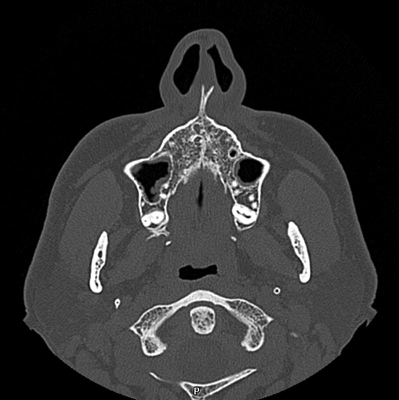
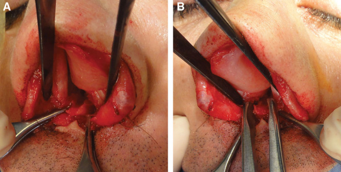
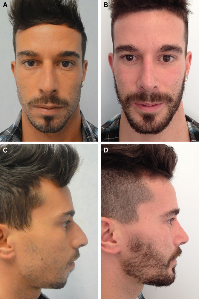
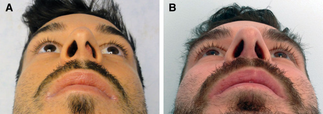
Background: The importance of analysis of the nasal spine should not be underestimated in the correct planning of rhinoplasty. Deformations in position with respect to the midline and/or in size are often present, and their correction to ensure harmony between the spine and the other components of the nasal pyramid constitutes a key step in rhinoplasty that can lead to excellent results. Methods: The study includes 160 patients who underwent surgical treatment of the anterior nasal spine with or without other techniques of nasal reshaping. Eighty-seven of these patients presented with hyperplasia of the nasal spine, 43 with hypoplasia, and 30 with deviation. A combination of deviation and hyperplasia was present in 15 cases. Results: No patients developed postoperative complications. Five patients who underwent anterior nasal spine reduction reported postoperative numbness in the premaxillary area, but sensitivity was fully regained within 4 months after surgery in these patients. All the patients reported postoperative improvement of nasal airflow. A total of 142 patients considered their postoperative aesthetic result as excellent and 18 as good. Conclusion: The simplicity of the surgical procedures performed on the nasal spine and the marked aesthetic improvements thus achieved suggest that greater attention should be paid to this anatomical region.
Overview
The claims and statistics of this paper haven’t been checked yet.
Run the check — free, no account needed. It reads the open-access text and checks the sources it cites.
Integrity signals
Registry
Cites
Statistics
Figures
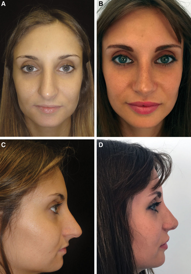
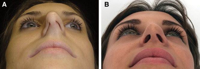
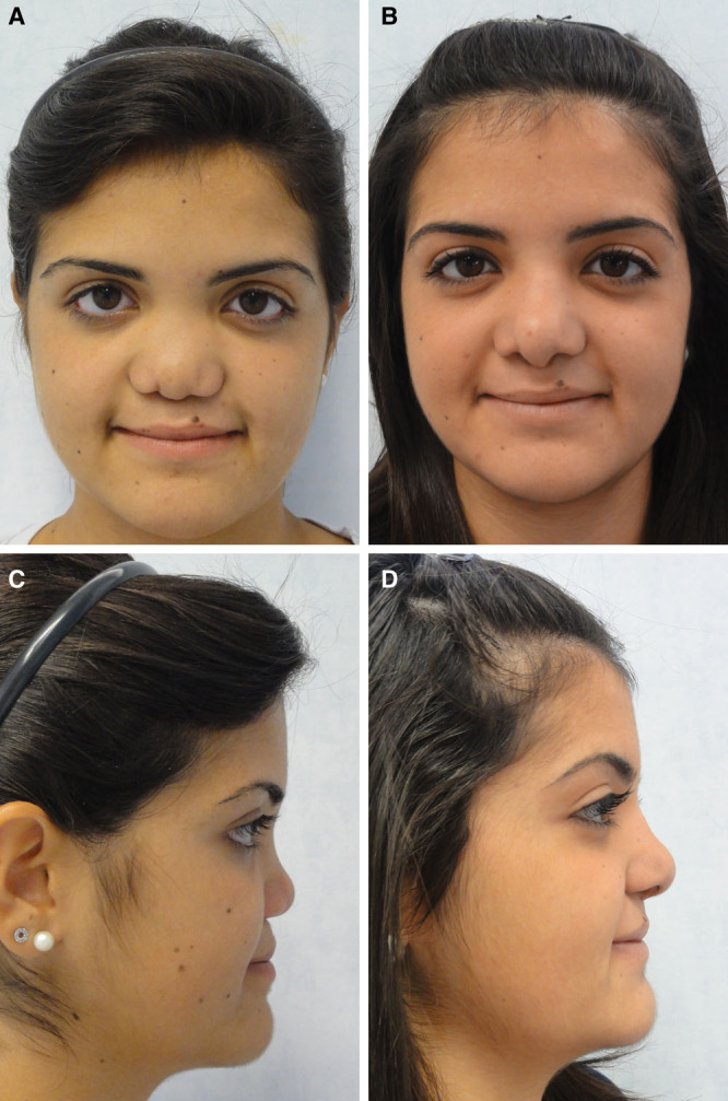
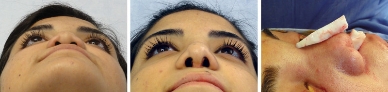
Verification
Claims the paper makes
The claims and statistics of this paper haven’t been checked yet.
Run the check — free, no account needed. It reads the open-access text and checks the sources it cites.
Claims other papers make about this one
Each is checked against this paper’s own text: does it say what the citing paper says it does?
The cited source says what the claim attributes to it.
In the citing paper
“It holds the attachment for the caudal nasal septum providing support to the nasal tip and projection to both the nose and the upper lip.7,8”
From Sliding Spine Relocation Surgery with Anterior Septal Reconstruction (2024) · cites this paper as [8]
Self-check
Percentages, sums and reported differences re-computed from the paper’s own numbers.
The self-check needs the paper’s full text.
Citations
Cites
Arnett, G. W., Jelic, J. S., Kim, J., et al. (1999) · American Journal of Orthodontics and Dentofacial Orthopedics · doi:10.1016/s0889-5406(99)70234-9 · Cited by 393
Constantian, M. B., & Clardy, B. R. (1996) · Plastic & Reconstructive Surgery · doi:10.1097/00006534-199607000-00007 · Cited by 292
Byrd, H. S., & Hobar, P. C. (1993) · Plastic & Reconstructive Surgery · doi:10.1097/00006534-199304000-00011 · Cited by 178
Quantitative analysis of nasal tip projection
Crumley, R. L., & Lanser, M. J. (1988) · The Laryngoscope · doi:10.1288/00005537-198802000-00017 · Cited by 170
Rhinoplasty: a practical guide for surgical planning.
Byrd, H. S., & Hobar, P. C. (1993) · PubMed · Cited by 155
Guyuron, B., & Behmand, R. A. (2003) · Plastic and Reconstructive Surgery · doi:10.1097/01.prs.0000060802.70218.fe · Cited by 87
Johnson, C. M., & Godin, M. S. (1995) · Plastic & Reconstructive Surgery · doi:10.1097/00006534-199501000-00006 · Cited by 50
Soliemanzadeh, P., & Kridel, R. (2005) · Archives of Facial Plastic Surgery · doi:10.1001/archfaci.7.6.374 · Cited by 50
Simplifying the Management of Caudal Septal Deviation in Rhinoplasty
Constantine, F. C., Ahmad, J., Geissler, P., & Rohrich, R. J. (2014) · Plastic and Reconstructive Surgery · doi:10.1097/prs.0000000000000236 · Cited by 49
An Algorithm for Treatment of the Drooping Nose
Sajjadian, A., & Guyuron, B. (2009) · Aesthetic Surgery Journal · doi:10.1016/j.asj.2009.02.006 · Cited by 48
Refinements in Extracorporal Septoplasty
Gubisch, W., & Constantinescu, M. A. (1999) · Plastic & Reconstructive Surgery · doi:10.1097/00006534-199909020-00041 · Cited by 48
Refinements in Extracorporal Septoplasty
Gubisch, W., & Constantinescu, M. A. (1999) · Plastic & Reconstructive Surgery · doi:10.1097/00006534-199909040-00038 · Cited by 34
Keppler‐Noreuil, K. M., & Wenzel, T. J. (2010) · Journal of Craniofacial Surgery · doi:10.1097/scs.0b013e3181ef2b71 · Cited by 24
Tanaka, Y. (2014) · Plastic & Reconstructive Surgery Global Open · doi:10.1097/gox.0000000000000161 · Cited by 23
A Multivariate Analysis of Nasal Tip Deprojection
Unger, J. G., Lee, M. R., Kwon, R. K., et al. (2012) · Plastic & Reconstructive Surgery · doi:10.1097/prs.0b013e31824a2e05 · Cited by 23
Fracture of the anterior nasal spine.
Escada, P., & Penha, R. (1999) · PubMed · Cited by 11
Fracture of the Anterior Nasal Spine
Kim, H., Kim, Y. J., Nam, S. H., & Choi, Y. W. (2012) · Journal of Craniofacial Surgery · doi:10.1097/scs.0b013e31824cdc8c · Cited by 10
Functional anatomy of the premaxillary area.
Hafkamp, H. C., Bruintjes, T. D., & Huizing, E. H. (1999) · PubMed · Cited by 7
Case report and surgical solution for nasal spine agenesis in a woman with Binder syndrome
Kansu, L., Akkuzu, B., & Avcı, S. (2007) · European Archives of Oto-Rhino-Laryngology · doi:10.1007/s00405-007-0532-5 · Cited by 6
Deprojection of the Nasal Tip in Revision Rhinoplasty
Kridel, R., & Undavia, S. S. (2012) · Facial Plastic Surgery · doi:10.1055/s-0032-1319843 · Cited by 6
Cited by
Sorted by citations
Ng, C. L., Rival, R., & Solomon, P. (2020) · Aesthetic Plastic Surgery · doi:10.1007/s00266-019-01568-2 · Cited by 5
Gubisch, W. (2020) · Aesthetic Plastic Surgery · doi:10.1007/s00266-019-01601-4 · Cited by 3
Sliding Spine Relocation Surgery with Anterior Septal Reconstruction
Fleury Curado, T., El Abany, A., & Most, S. P. (2024) · Craniomaxillofacial Trauma & Reconstruction · doi:10.1177/19433875231152947
Metadata
- Authors
- Marianetti, T. M., Boccieri, A., & Pascali, M.
- Affiliations
- University of Rome Tor Vergata (IT)
- Corresponding author
- Marianetti, Tito M.
- Venue
- Plastic and Reconstructive Surgery - Global Open
- Published
- 2016-09
- Volume
- 4
- Issue
- 9
- Pages
- e1026
- ISSN
- 2169-7574 (ISSN-L)
- PMC
- PMC5055009
- OpenAlex
- W2521752508
- Cited by
- 22
- References
- 20
- Citations by year
- 2026: 2 · 2025: 3 · 2023: 4 · 2022: 1 · 2021: 1 · 2020: 5 · 2019: 3 · 2018: 1 · 2017: 2
- FWCI
- 2.22
- Type
- journal article
- Language
- en
- Open access
- gold
- doi.org
- Licence
- Locations
- Plastic & Reconstructive Surgery Global Open (publishedVersion, cc-by-nc-nd)
- PubMed Central (publishedVersion, cc-by-nc-nd)
- PubMed (publishedVersion)
- Topics
- Health Sciences › Medicine › Surgery › Nasal Surgery and Airway Studies
- Health Sciences › Medicine › Surgery › Reconstructive Facial Surgery Techniques
- Health Sciences › Dentistry › Oral Surgery › Cleft Lip and Palate Research
- Keywords
- anterior nasal spine, rhinoplasty, nasal pyramid, nasal airflow, cosmetic outcome, nasal deformity
About these checks
Registry, retraction, Cites and statistics results are computed, not judged, with one exception: a printed reference that no index lists is read once by a model before it counts as a citation error. Claims are extracted by a model and kept only when their quote appears verbatim in the text; each cited source is then checked, with a model’s help, for whether it says what the claim relies on. Those verdicts can be wrong — read the claim’s quote and the cited source before relying on one.
Statistics and arithmetic are recomputed, not judged.
Confidence bands are shown only for fields calibrated on our annotated test set. None is calibrated yet, so no band is shown.
What counts as a claim ›How we measure confidence ›How we check this ›
Are you an author, or do you see a problem with this page? Request a review
