

The DOI matches its Crossref record; 5 of the 5 references it cites were found in a registry or an index.
Anterior Ear Reconstruction with the Posterior Pull-through Flap
Open accessAbstract
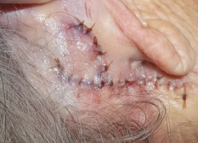
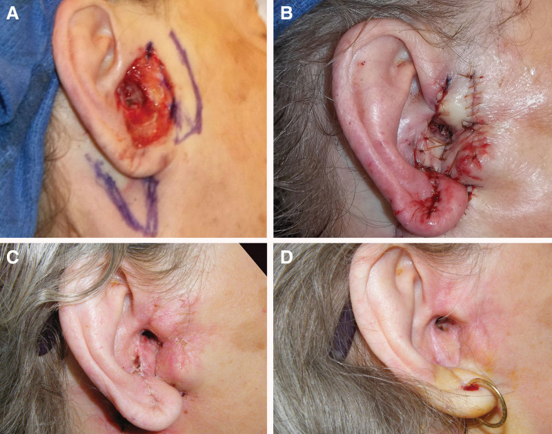
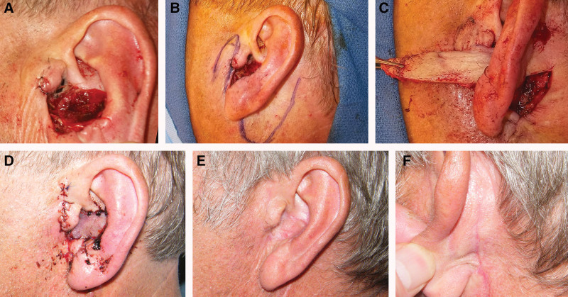
Background: Defects of the central ear after skin cancer removal remain a common problem for the reconstructive surgeon. The experience with a one-stage, postauricular, skin-island flap passed through the cartilage to reconstruct ear defects is reported. Methods: Patients with an intact ear helix and an anterior full-thickness defect (cartilage defects included) were reconstructed with a skin-island flap based on posterior subcutaneous tissue deep to the flap base. The flap was passed through a generous aperture created in the ear cartilage. The skin at the base was incised superficially after the flap was passed through the cartilage to create a skin-island and avoid burial of epithelium. The flap periphery and postauricular harvest site were sutured with absorbable chromic sutures. Results: Twenty-two patients (six women, 16 men) with ear defects underwent single-stage reconstruction over a 9-year period. The defects reconstructed measured in diameter from 2 cm to 4.5 cm. Six patients required a second skin flap from the preauricular area to close ear canal defects. The pull-through flap added structural support, and prevented ear distortion. No flap necrosis occurred. Venous congestion was common and self-limiting. No epithelial cysts developed. Conclusions: A one-stage postauricular skin-island flap can reliably reconstruct anterior ear defects without distorting shape or position of the ear. Care is needed to provide a generous aperture through the ear cartilage to accommodate the flap. Healing proceeds predictably, and minimal complications are associated with this posteriorly-based, pass-through, skin-island flap in ear reconstruction.
Overview
The claims and statistics of this paper haven’t been checked yet.
Run the check — free, no account needed. It reads the open-access text and checks the sources it cites.
Integrity signals
Registry
Cites
Statistics
Figures
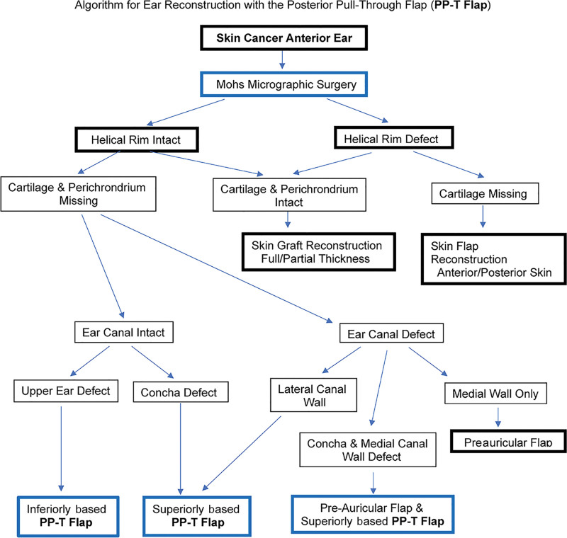
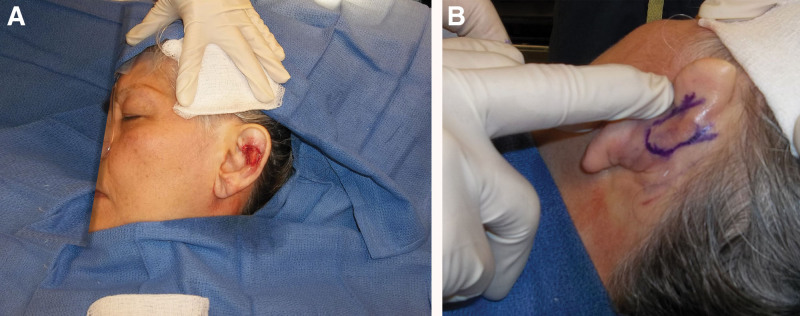
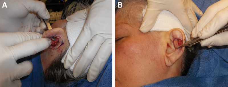
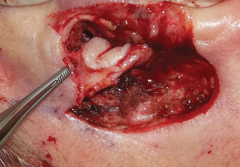
Verification
Claims the paper makes
The claims and statistics of this paper haven’t been checked yet.
Run the check — free, no account needed. It reads the open-access text and checks the sources it cites.
Claims other papers make about this one
Each is checked against this paper’s own text: does it say what the citing paper says it does?
The source backs part of the claim, not all of it — or backs it for a different population, condition or comparison.
In the citing paper
“Additionally, scars tend to be well hidden, and the donor site heals well secondarily.19,32,33”
From Ear Reconstruction following Mohs Micrographic Surgery (2024) · cites this paper as [33]
Experimental — accuracy not yet measured
Self-check
Percentages, sums and reported differences re-computed from the paper’s own numbers.
The self-check needs the paper’s full text.
Citations
Cites
A simple island flap for reconstruction of concha-helix defects
Masson, J. K. (1972) · British Journal of Plastic Surgery · doi:10.1016/s0007-1226(72)80083-3 · Cited by 134
Pull-through subcutaneous pedicle flap for an anterior auricular defect
NGUYEN, D. H., & BORDEAUX, J. S. (2010) · Dermatologic Surgery · doi:10.1111/j.1524-4725.2010.01575.x · Cited by 17
The trap door flap: A reliable, reproducible method of anterior pinna reconstruction
McInerney, N. M., Piggott, R. P., & Regan, P. J. (2013) · Journal of Plastic Reconstructive & Aesthetic Surgery · doi:10.1016/j.bjps.2013.05.037 · Cited by 15
The trap door flap for reconstructing defects of the concha
Lynch, J., Mahajan, A., & Regan, P. J. (2003) · British Journal of Plastic Surgery · doi:10.1016/s0007-1226(03)00307-2 · Cited by 13
The Revolving Door Flap: Revisiting an Elegant but Forgotten Flap for Ear Defect Reconstruction
Golash, A., Bera, S., Kanoi, A. V., & Golash, A. (2020) · Indian Journal of Plastic Surgery · doi:10.1055/s-0040-1709531 · Cited by 12
Cited by
Sorted by citations
Ear Reconstruction following Mohs Micrographic Surgery
Mohan, V. C., Owens, W. R., Schmidt, J. L., Burns, H. R., & Buchanan, E. P. (2024) · Seminars in Plastic Surgery · doi:10.1055/s-0044-1791830
Metadata
- Authors
- Gatti, J. E., Charipova, K., & Sollitto, R. B.
- Affiliations
- Virtua Voorhees Hospital (US)
- Georgetown University Medical Center (US)
- Corresponding author
- Gatti, John E.
- Venue
- Plastic and Reconstructive Surgery - Global Open
- Published
- 2023-10
- Volume
- 11
- Issue
- 10
- Pages
- e5338
- ISSN
- 2169-7574 (ISSN-L)
- PMC
- PMC10569762
- OpenAlex
- W4387571468
- Cited by
- 1
- References
- 5
- Citations by year
- 2024: 1
- FWCI
- 0.27
- Type
- journal article
- Language
- en
- Open access
- gold
- dx.doi.org
- Licence
- Locations
- Plastic & Reconstructive Surgery Global Open (publishedVersion, cc-by-nc-nd)
- PubMed Central (publishedVersion, cc-by-nc-nd)
- PubMed (publishedVersion)
- DOAJ (DOAJ: Directory of Open Access Journals) (submittedVersion, cc-by-sa)
- Topics
- Health Sciences › Medicine › Surgery › Reconstructive Facial Surgery Techniques
- Health Sciences › Medicine › Surgery › Reconstructive Surgery and Microvascular Techniques
- Health Sciences › Medicine › Oncology › Ear and Head Tumors
- Keywords
- skin cancer excision, one-stage reconstruction, venous congestion
About these checks
Registry, retraction, Cites and statistics results are computed, not judged, with one exception: a printed reference that no index lists is read once by a model before it counts as a citation error. Claims are extracted by a model and kept only when their quote appears verbatim in the text; each cited source is then checked, with a model’s help, for whether it says what the claim relies on. Those verdicts can be wrong — read the claim’s quote and the cited source before relying on one.
Statistics and arithmetic are recomputed, not judged.
Confidence bands are shown only for fields calibrated on our annotated test set. None is calibrated yet, so no band is shown.
What counts as a claim ›How we measure confidence ›How we check this ›
Are you an author, or do you see a problem with this page? Request a review
