

The DOI matches its Crossref record; 52 of the 54 references it cites were found in a registry or an index.
The Basic Helix-Loop-Helix Transcription Factor E47 Reprograms Human Pancreatic Cancer Cells to a Quiescent Acinar State With Reduced Tumorigenic Potential
Open accessAbstract
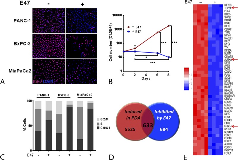
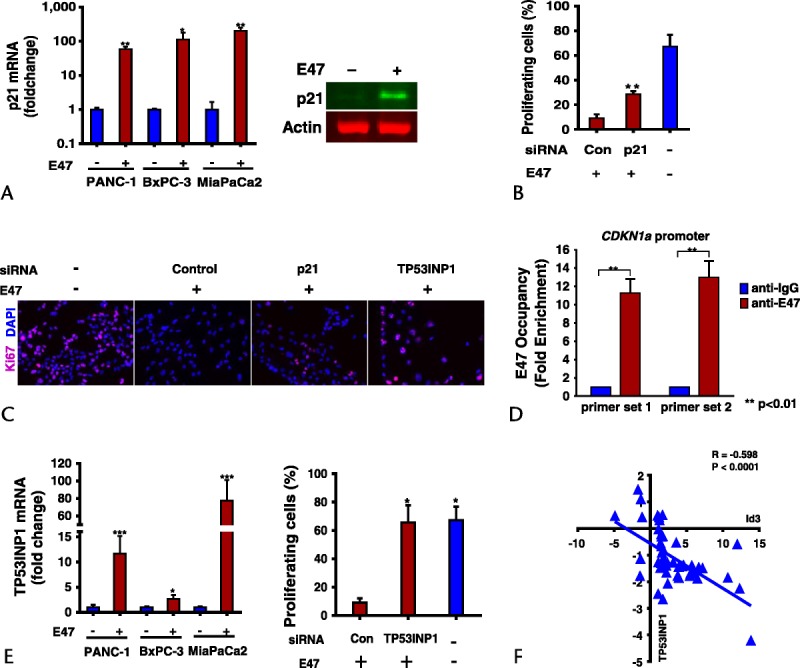
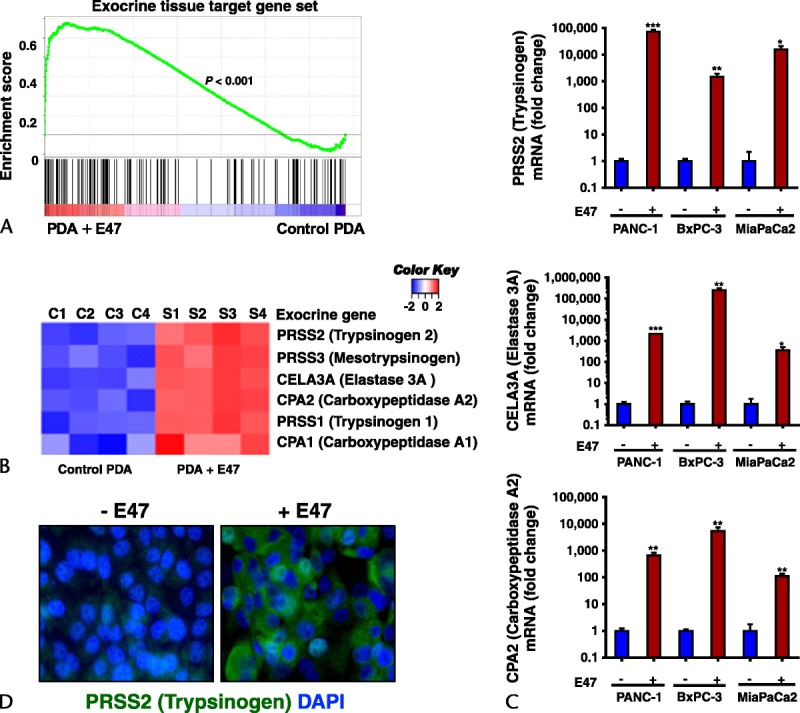
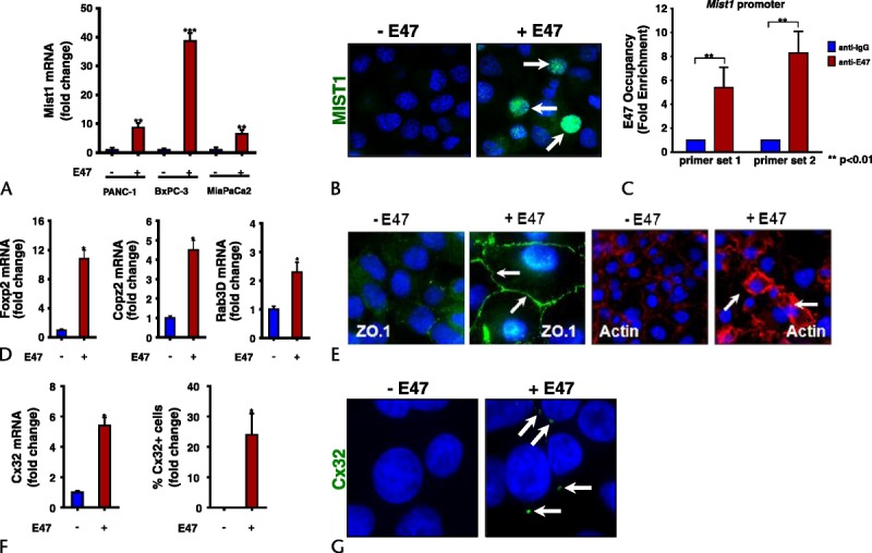
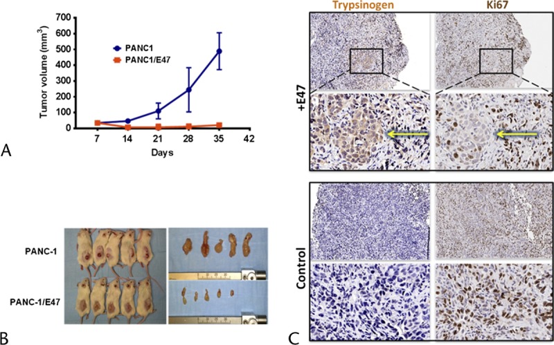
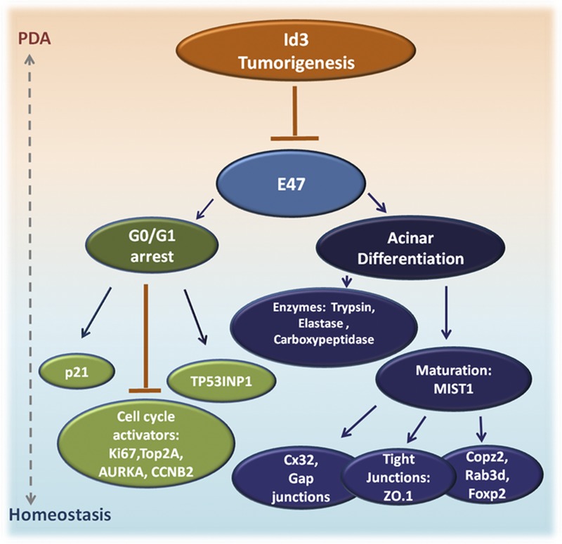
OBJECTIVES: Pancreatic ductal adenocarcinoma (PDA) initiates from quiescent acinar cells that attain a Kras mutation, lose signaling from basic helix-loop-helix (bHLH) transcription factors, undergo acinar-ductal metaplasia, and rapidly acquire increased growth potential. We queried whether PDA cells can be reprogrammed to revert to their original quiescent acinar cell state by shifting key transcription programs. METHODS: Human PDA cell lines were engineered to express an inducible form of the bHLH protein E47. Gene expression, growth, and functional studies were investigated using microarray, quantitative polymerase chain reaction, immunoblots, immunohistochemistry, small interfering RNA, chromatin immunoprecipitation analyses, and cell transplantation into mice. RESULTS: In human PDA cells, E47 activity triggers stable G0/G1 arrest, which requires the cyclin-dependent kinase inhibitor p21 and the stress response protein TP53INP1. Concurrently, E47 induces high level expression of acinar digestive enzymes and feed forward activation of the acinar maturation network regulated by the bHLH factor MIST1. Moreover, induction of E47 in human PDA cells in vitro is sufficient to inhibit tumorigenesis. CONCLUSIONS: Human PDA cells retain a high degree of plasticity, which can be exploited to induce a quiescent acinar cell state with reduced tumorigenic potential. Moreover, bHLH activity is a critical node coordinately regulating human PDA cell growth versus cell fate.
Overview
The claims and statistics of this paper haven’t been checked yet.
Free: it reads the open-access text and checks the first 5 claims that cite a source we can check. Create a free account to check every claim.
Integrity signals
Registry
Cites
Statistics
Figures
Verification
Claims the paper makes
The claims and statistics of this paper haven’t been checked yet.
Free: it reads the open-access text and checks the first 5 claims that cite a source we can check. Create a free account to check every claim.
Claims other papers make about this one
Each is checked against this paper’s own text: does it say what the citing paper says it does?
The cited source doesn’t support the claim as written.
In the citing paper
“During acinar-to-ductal metaplasia, dedifferentiated acinar cells acquire a senescent state characterized by cell cycle arrest, induction of p53, CDKN1A, CDKN2A and SA-β-gal activity190–193.”
From SenNet recommendations for detecting senescent cells in different tissues (2024) · cites this paper as [192]
Checked by a reasoning model — may be wrong.
Experimental — accuracy not yet measured
Self-check
Percentages, sums and reported differences re-computed from the paper’s own numbers.
The self-check needs the paper’s full text.
Citations
Cites
Moore, M. J., Goldstein, D., Hamm, J., Figer, A., Hecht, J. R., Gallinger, S., Au, H. J., Murawa, P., Walde, D., Wolff, R. A., et al. (2007) · Journal of Clinical Oncology · doi:10.1200/jco.2006.07.9525 · Cited by 3,848
A gene atlas of the mouse and human protein-encoding transcriptomes
Su, A. I., Wiltshire, T., Batalov, S., Lapp, H., Ching, K. A., Block, D., Zhang, J., Soden, R., Hayakawa, M., Kreiman, G., et al. (2004) · Proceedings of the National Academy of Sciences · doi:10.1073/pnas.0400782101 · Cited by 3,475 · cited 1×
“The first evidence that PDA cells could indeed revert to an acinar identity was obtained through computational GSEA comparison of our microarray data with a set of human pancreas-enriched genes (GSE1133).(30) As shown in Figure 3A, E47 significantly induced an acinar gene signature profile, achieving an enrichment sco…”
ResultPreinvasive and invasive ductal pancreatic cancer and its early detection in the mouse
Hingorani, S. R., Petricoin, E. F., Maitra, A., Rajapakse, V., King, C., Jacobetz, M. A., Ross, S., Conrads, T. P., Veenstra, T. D., Hitt, B. A., et al. (2003) · Cancer Cell · doi:10.1016/s1535-6108(03)00309-x · Cited by 2,471
Subtypes of pancreatic ductal adenocarcinoma and their differing responses to therapy
Collisson, E. A., Sadanandam, A., Olson, P., Gibb, W. J., Truitt, M., Gu, S., Cooc, J., Weinkle, J., Kim, G. E., Jakkula, L., et al. (2011) · Nature Medicine · doi:10.1038/nm.2344 · Cited by 1,967 · cited 1×
“GSE16515 and GSE15471 datasets were used to identify genes, which are highly expressed in human PDA tumors relative to normal pancreas tissue, and to determine the correlation between Id3 and TP53INP1 expression in PDA-tumor samples relative to control tissue (statistically analyzed by Pearson coefficient).(28,29) Gen…”
MethodHelix-Loop-Helix Proteins: Regulators of Transcription in Eucaryotic Organisms
Massari, M. E., & Murre, C. (2000) · Molecular and Cellular Biology · doi:10.1128/mcb.20.2.429-440.2000 · Cited by 1,816
Guerra, C., Schuhmacher, A. J., Cañamero, M., Grippo, P. J., Verdaguer, L., Pérez-Gallego, L., Dubus, P., Sandgren, E. P., & Barbacid, M. (2007) · Cancer Cell · doi:10.1016/j.ccr.2007.01.012 · Cited by 1,224
Oncogenic Kras is required for both the initiation and maintenance of pancreatic cancer in mice
Collins, M. A., Bednar, F., Zhang, Y., Brisset, J.-C., Galbán, S., Galbán, C. J., Rakshit, S., Flannagan, K. S., Adsay, N. V., & Pasca di Magliano, M. (2012) · Journal of Clinical Investigation · doi:10.1172/jci59227 · Cited by 770
FKBP51 Affects Cancer Cell Response to Chemotherapy by Negatively Regulating Akt
Pei, H., Li, L., Fridley, B. L., Jenkins, G. D., Kalari, K. R., Lingle, W., Petersen, G., Lou, Z., & Wang, L. (2009) · Cancer Cell · doi:10.1016/j.ccr.2009.07.016 · Cited by 709 · cited 1×
“GSE16515 and GSE15471 datasets were used to identify genes, which are highly expressed in human PDA tumors relative to normal pancreas tissue, and to determine the correlation between Id3 and TP53INP1 expression in PDA-tumor samples relative to control tissue (statistically analyzed by Pearson coefficient).(28,29) Gen…”
MethodKopp, J. L., von Figura, G., Mayes, E., Liu, F.-F., Dubois, C. L., Morris, J. P., Pan, F. C., Akiyama, H., Wright, C. V. E., Jensen, K., et al. (2012) · Cancer Cell · doi:10.1016/j.ccr.2012.10.025 · Cited by 697 · cited 1×
“The cells acquire a duct-like transcriptome that drives the formation of precancerous pancreatic intraepithelial neoplasia (PanIN) lesions.(5,6,9) Subsequent mutations in additional oncogenes or tumor-suppressor genes fuel disease progression, leading to aggressive PDA.”
Gironella, M., Seux, M., Xie, M.-J., Cano, C., Tomasini, R., Gommeaux, J., Garcia, S., Nowak, J., Yeung, M. L., Jeang, K.-T., et al. (2007) · Proceedings of the National Academy of Sciences · doi:10.1073/pnas.0703942104 · Cited by 551
Cited by
SenNet recommendations for detecting senescent cells in different tissues
Suryadevara, V., Hudgins, A. D., Rajesh, A., Pappalardo, A., Karpova, A., Dey, A. K., Hertzel, A., Agudelo, A., Rocha, A., Soygur, B., et al. (2024) · Nature Reviews Molecular Cell Biology · doi:10.1038/s41580-024-00738-8 · Cited by 408
Acinar cell plasticity and development of pancreatic ductal adenocarcinoma
Storz, P. (2017) · Nature Reviews Gastroenterology & Hepatology · doi:10.1038/nrgastro.2017.12 · Cited by 389
Defining the molecular pathology of pancreatic body and tail adenocarcinoma
Initiative, A. P. C. G., Dreyer, S. B., Jamieson, N. B., Upstill‐Goddard, R., Bailey, P. J., McKay, C. J., Biankin, A. V., & Chang, D. K.-F. (2018) · British journal of surgery · doi:10.1002/bjs.10772 · Cited by 132
Regulation of Cellular Identity in Cancer
Roy, N., & Hebrok, M. (2015) · Developmental Cell · doi:10.1016/j.devcel.2015.12.001 · Cited by 104
“In support of the broader use of such an approach, a recent report showed that by overexpressing the bHLH protein E47, neoplastic cells reverted to an acinar state with reduced PanIN tumorigenicity (Kim et al., 2015).”
ResultCrawford, H. C., di Magliano, M. P., & Banerjee, S. (2019) · Gastroenterology · doi:10.1053/j.gastro.2018.12.042 · Cited by 77
Metadata
- Authors
- Kim, S., Lahmy, R., Riha, C., Yang, C., Jakubison, B. L., van Niekerk, J., Staub, C., Wu, Y., Gates, K., Dong, D. S., Konieczny, S. F., & Itkin-Ansari, P.
- Affiliations
- Sanford Burnham Prebys Medical Discovery Institute (US)
- Purdue University West Lafayette (US)
- Corresponding author
- Lahmy, Reyhaneh
- Riha, Chelsea
- Yang, Challeng
- Staub, Claudio
- Wu, Yifan
- Venue
- Pancreas
- Published
- 2015-07
- Volume
- 44
- Issue
- 5
- Pages
- 718-727
- ISSN
- 0885-3177 (ISSN-L)
- PMC
- PMC4464938
- OpenAlex
- W2316005448
- Cited by
- 48
- References
- 54
- Citations by year
- 2026: 3 · 2025: 1 · 2024: 2 · 2023: 5 · 2022: 5 · 2021: 2 · 2020: 1 · 2019: 5 · 2018: 9 · 2017: 7 · 2016: 4 · 2015: 4
- FWCI
- 3.49
- Type
- journal article
- Language
- en
- Open access
- hybrid
- doi.org
- Licence
- cc-by-nc-nd
- Locations
- Pancreas (publishedVersion, cc-by-nc-nd)
- PubMed Central (publishedVersion, cc-by-nc-nd)
- PubMed (publishedVersion)
- YUHSpace (Yonsei University Medical Library) (submittedVersion, cc-by-nc-nd)
- Funders
- National Cancer Institute (R01CA124586, P30CA023168, CA124586, P30CA030199)
- National Institute of Diabetes and Digestive and Kidney Diseases (DK55489, R01DK055489)
- Topics
- Health Sciences › Medicine › Oncology › Pancreatic and Hepatic Oncology Research
- Health Sciences › Medicine › Endocrinology, Diabetes and Metabolism › Pancreatic function and diabetes
- Health Sciences › Medicine › Oncology › Cancer Cells and Metastasis
- Keywords
- E47, bHLH transcription factors, pancreatic ductal adenocarcinoma, acinar differentiation, cellular reprogramming, acinar-to-ductal metaplasia, Mist1, p21, TP53INP1, G0/G1 cell cycle arrest, tumorigenicity, KRAS mutation, digestive enzymes, cellular plasticity
- MeSH terms
- Animals, Humans, Pancreatic Neoplasms, Phenotype, Time Factors, Transfection, Genetic Therapy, Up-Regulation, Gene Expression Regulation, Neoplastic, Mice, SCID, Cellular Senescence, Carcinoma, Pancreatic Ductal, Xenograft Model Antitumor Assays, RNA Interference, Cell Line, Tumor, Cell Proliferation, Basic Helix-Loop-Helix Proteins, Basic Helix-Loop-Helix Transcription Factors, Cell Cycle Checkpoints, Cellular Reprogramming
About these checks
Registry, retraction, Cites and statistics results are computed, not judged, with one exception: a printed reference that no index lists is read once by a model before it counts as a citation error. Claims are extracted by a model and kept only when their quote appears verbatim in the text; each cited source is then checked, with a model’s help, for whether it says what the claim relies on. Those verdicts can be wrong — read the claim’s quote and the cited source before relying on one.
Statistics and arithmetic are recomputed, not judged.
Confidence bands are shown only for fields calibrated on our annotated test set. None is calibrated yet, so no band is shown.
What counts as a claim ›How we measure confidence ›How we check this ›
Are you an author, or do you see a problem with this page? Request a review
How to cite
Cite CoChat for the checks on this page, and the paper for its own findings.
- This page
- CoChat. (2026). Citation check: The Basic Helix-Loop-Helix Transcription Factor E47 Reprograms Human Pancreatic Cancer Cells to a Quiescent Acinar State With Reduced Tumorigenic Potential. https://app.cochat.ai/papers/10.1097/mpa.0000000000000328
- The paper
- Kim, S., Lahmy, R., Riha, C., Yang, C., Jakubison, B. L., van Niekerk, J., Staub, C., Wu, Y., Gates, K., Dong, D. S., Konieczny, S. F., & Itkin-Ansari, P. (2015). The Basic Helix-Loop-Helix Transcription Factor E47 Reprograms Human Pancreatic Cancer Cells to a Quiescent Acinar State With Reduced Tumorigenic Potential. Pancreas. https://doi.org/10.1097/mpa.0000000000000328
Data sources
Bibliographic data: Semantic Scholar, OpenAlex, Crossref.
Contains information from the Semantic Scholar Academic Graph Datasets, made available under the ODC Attribution License (ODC-BY 1.0).
Semantic Scholar’s data is described in: Kinney, R., et al. (2023). The Semantic Scholar Open Data Platform. arXiv:2301.10140.
