

The DOI matches its Crossref record; 148 of the 152 references it cites were found in a registry or an index.
High-grade Endometrial Carcinomas: Morphologic and Immunohistochemical Features, Diagnostic Challenges and Recommendations
Open accessAbstract
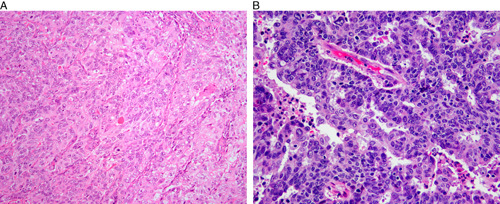
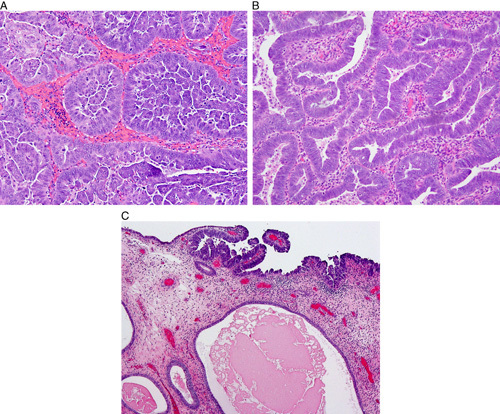
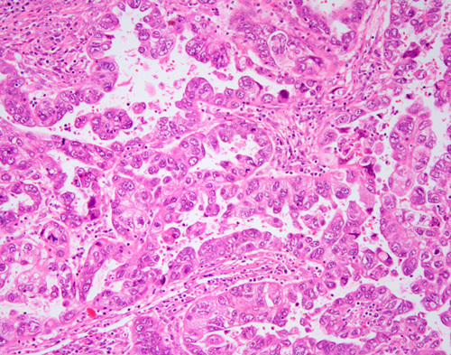
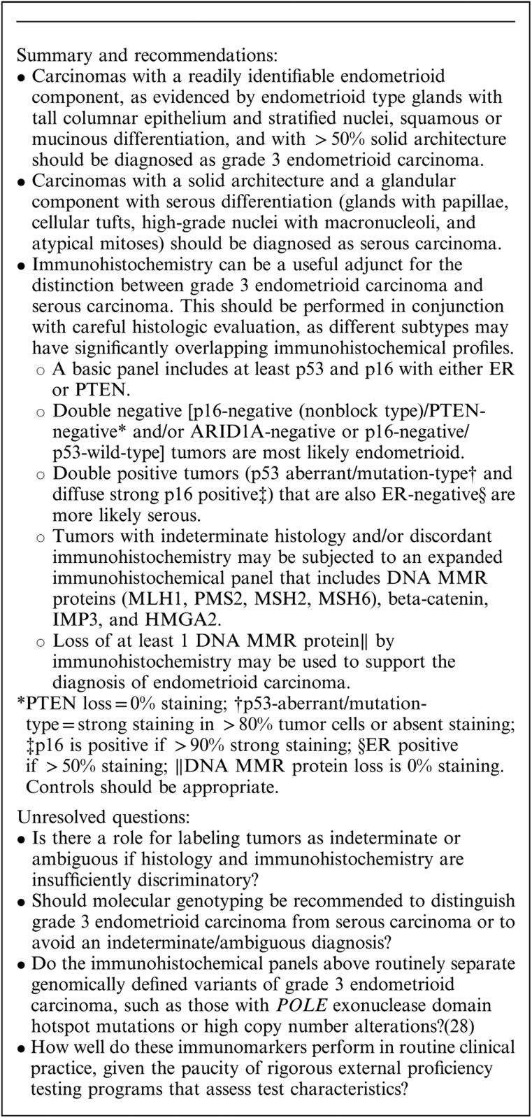
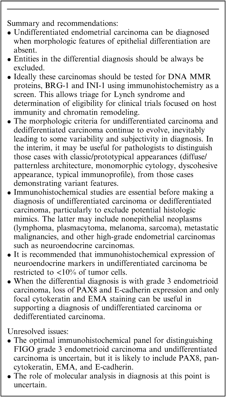
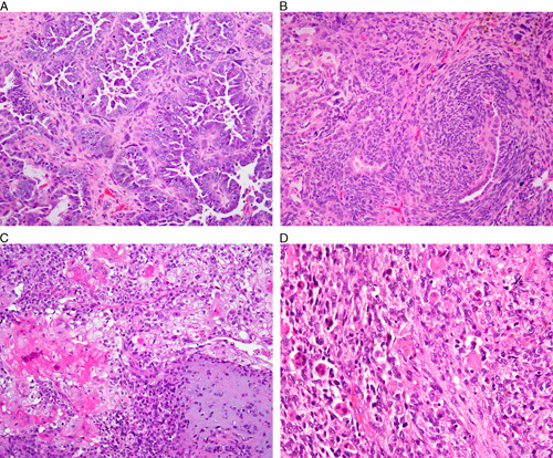
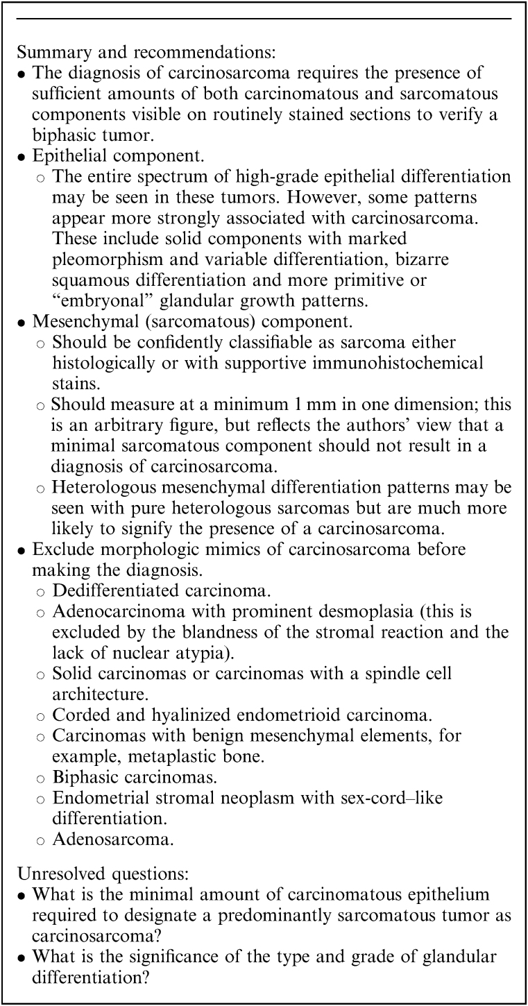
This review of challenging diagnostic issues concerning high-grade endometrial carcinomas is derived from the authors’ review of the literature followed by discussions at the Endometrial Cancer Workshop sponsored by the International Society of Gynecological Pathologists in 2016. Recommendations presented are evidence-based, insofar as this is possible, given that the levels of evidence are weak or moderate due to small sample sizes and nonuniform diagnostic criteria used in many studies. High-grade endometrioid carcinomas include FIGO grade 3 endometrioid carcinomas, serous carcinomas, clear cell carcinomas, undifferentiated carcinomas, and carcinosarcomas. FIGO grade 3 endometrioid carcinoma is diagnosed when an endometrioid carcinoma exhibits >50% solid architecture (excluding squamous areas), or when an architecturally FIGO grade 2 endometrioid carcinoma exhibits marked cytologic atypia, provided that a glandular variant of serous carcinoma has been excluded. The most useful immunohistochemical studies to make the distinction between these 2 histotypes are p53, p16, DNA mismatch repair proteins, PTEN, and ARID1A. Endometrial clear cell carcinomas must display prototypical architectural and cytologic features for diagnosis. Immunohistochemical stains, including, Napsin A and p504s can be used as ancillary diagnostic tools; p53 expression is aberrant in a minority of clear cell carcinomas. Of note, clear cells are found in all types of high-grade endometrial carcinomas, leading to a tendency to overdiagnose clear cell carcinoma. Undifferentiated carcinoma (which when associated with a component of low-grade endometrioid carcinoma is termed “dedifferentiated carcinoma”) is composed of sheets of monotonous, typically dyscohesive cells, which can have a rhabdoid appearance; they often exhibit limited expression of cytokeratins and epithelial membrane antigen, are usually negative for PAX8 and hormone receptors, lack membranous e-cadherin and commonly demonstrate loss of expression of DNA mismatch repair proteins and SWI-SNF chromatin remodeling proteins. Carcinosarcomas must show unequivocal morphologic evidence of malignant epithelial and mesenchymal differentiation.
Overview
The claims and statistics of this paper haven’t been checked yet.
Run the check — free, no account needed. It reads the open-access text and checks the sources it cites.
Integrity signals
Registry
Cites
Statistics
Figures
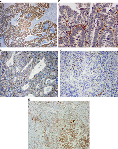
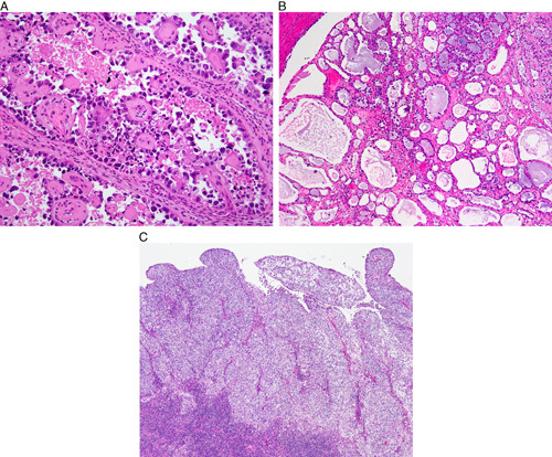
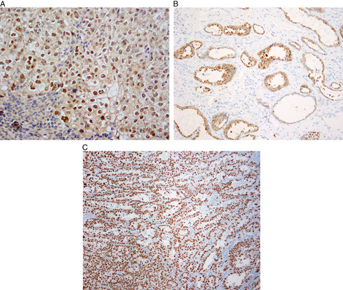
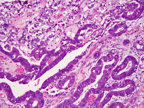
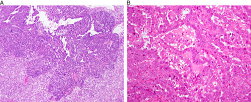
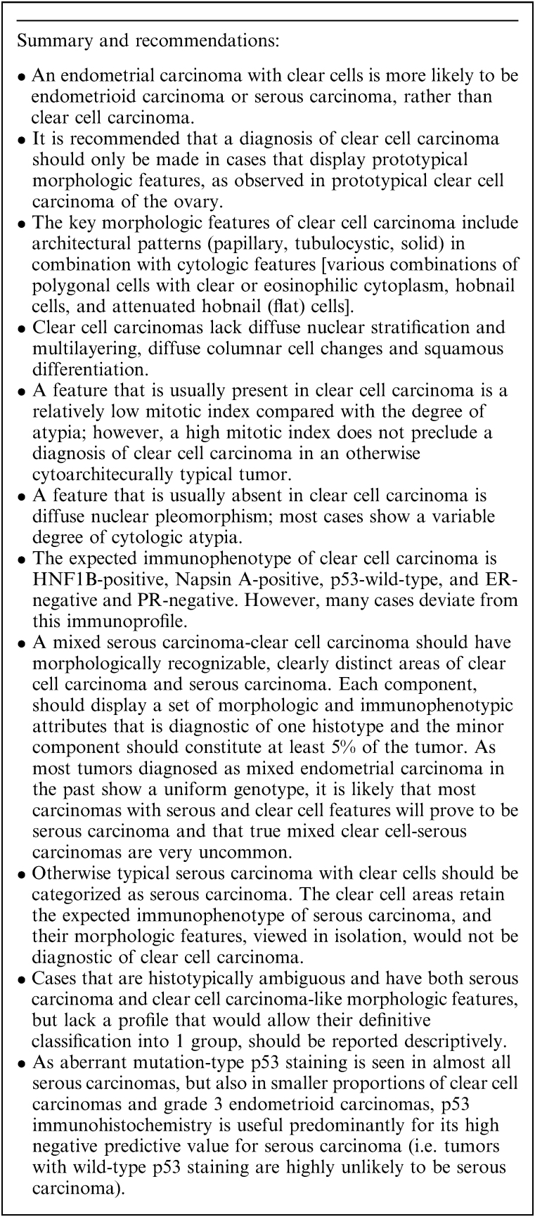
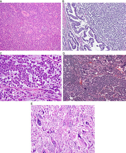
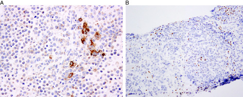
Verification
Claims the paper makes
The claims and statistics of this paper haven’t been checked yet.
Run the check — free, no account needed. It reads the open-access text and checks the sources it cites.
Claims other papers make about this one
Each is checked against this paper’s own text: does it say what the citing paper says it does?
In the citing paper
“Moreover, immunohistochemistry may demonstrate positive stains for HNF-1B, Napsin-A, AMACR, CK-7, and p53 [21].”
From Clear cell carcinoma arising from abdominal wall endometriosis—A report on two cases and literature review (2022) · cites this paper as [21]
Self-check
Percentages, sums and reported differences re-computed from the paper’s own numbers.
The self-check needs the paper’s full text.
Citations
Cites
A clinically applicable molecular-based classification for endometrial cancers
Talhouk, A., McConechy, M. K., Leung, S. C. Y., Li-Chang, H. H., Kwon, J. S., Melnyk, N., Yang, W., Senz, J., Boyd, N., Karnezis, A. N., et al. (2015) · British Journal of Cancer · doi:10.1038/bjc.2015.190 · Cited by 989
Self-citationTashiro, H., Blazes, M. S., Wu, R., Cho, K. R., Bose, S., Wang, S. I., Li, J., Parsons, R. E., & Ellenson, L. H. (1997) · PubMed · Cited by 701
Hamilton, C. A., Cheung, M. K., Osann, K., Chen, L., Teng, N. N. H., Longacre, T. A., Powell, M. A., Hendrickson, M. R., Kapp, D. S., & Chan, J. K. (2006) · British Journal of Cancer · doi:10.1038/sj.bjc.6603012 · Cited by 647
Integrated Molecular Characterization of Uterine Carcinosarcoma
Cherniack, A. D., Shen, H., Walter, V., Stewart, C., Murray, B. A., Bowlby, R., Hu, X., Ling, S., Soslow, R. A., Broaddus, R. R., et al. (2017) · Cancer Cell · doi:10.1016/j.ccell.2017.02.010 · Cited by 440
Self-citationCarcinosarcoma (Malignant Mixed Mesodermal Tumor) of the Uterus
Silverberg, S. G., Major, F. J., Blessing, J. A., Fetter, B. F., Askin, F. B., Liao, S., & Miller, A. W. (1990) · International Journal of Gynecological Pathology · doi:10.1097/00004347-199001000-00001 · Cited by 406
Poor Interobserver Reproducibility in the Diagnosis of High-grade Endometrial Carcinoma
Gilks, C. B., Oliva, E., & Soslow, R. A. (2013) · The American Journal of Surgical Pathology · doi:10.1097/pas.0b013e31827f576a · Cited by 404
Self-citationMalignant biphasic uterine tumours: carcinosarcomas or metaplastic carcinomas?
McCluggage, W. G. (2002) · Journal of Clinical Pathology · doi:10.1136/jcp.55.5.321 · Cited by 381
Self-citationZorn, K. K., Bonome, T., Gangi, L., Chandramouli, G. V. R., Awtrey, C. S., Gardner, G. J., Barrett, J. C., Boyd, J. A., & Birrer, M. J. (2005) · Clinical Cancer Research · doi:10.1158/1078-0432.ccr-05-0508 · Cited by 378
p53 gene mutations are common in uterine serous carcinoma and occur early in their pathogenesis.
Tashiro, H., Isacson, C., Levine, R. L., Kurman, R. J., Cho, K. R., & Hedrick, L. (1997) · Europe PMC (PubMed Central) · Cited by 345
Uterine Serous Carcinoma A Morphologically Diverse Neoplasm With Unifying Clinicopathologic Features
Sherman, M. E., Bitterman, P., Rosenshein, N. B., Delgado, G., & Kurman, R. J. (1992) · The American Journal of Surgical Pathology · doi:10.1097/00000478-199206000-00008 · Cited by 326
Research works printed but not linked: 3
- Holmes, 2014, Napsin A: a sensitive immunohistochemical marker for uterine clear cell carcinoma, Mod Pathol, 27, 536
- O'Keefe, 2015, Napsin A has utility in the diagnosis of clear cell carcinoma in the ovary but may be less valuable in the endometrium, Mod Pathol, 28, 530
- DeLair, 2012, Endometrial clear cell carcinoma with and without aberrant p53 expression: a study of 16 cases, Mod Pathol, 25, 265A
Cited by
Sorted by citations
TCGA molecular groups of endometrial cancer: Pooled data about prognosis
Raffone, A., Travaglino, A., Mascolo, M., Carbone, L., Guida, M., Insabato, L., & Zullo, F. (2019) · Gynecologic Oncology · doi:10.1016/j.ygyno.2019.08.019 · Cited by 198
Santoro, A., Angelico, G., Travaglino, A., Inzani, F. S., Arciuolo, D., Valente, M., D’Alessandris, N., Scaglione, G., Fiorentino, V., Raffone, A., et al. (2021) · Cancers · doi:10.3390/cancers13112623 · Cited by 182
Hong, R., Liu, W., DeLair, D., Razavian, N., & Fenyö, D. (2021) · Cell Reports Medicine · doi:10.1016/j.xcrm.2021.100400 · Cited by 154
Endometrial carcinoma: molecular subtypes, precursors and the role of pathology in early diagnosis
Huvila, J. K., Pors, J., Thompson, E. F., & Gilks, C. B. (2020) · The Journal of Pathology · doi:10.1002/path.5608 · Cited by 149
Self-citationTCGA Molecular Prognostic Groups of Endometrial Carcinoma: Current Knowledge and Future Perspectives
Arciuolo, D., Travaglino, A., Raffone, A., Raimondo, D., Santoro, A., Russo, D., Varricchio, S., Casadio, P., Inzani, F. S., Seracchioli, R., et al. (2022) · International Journal of Molecular Sciences · doi:10.3390/ijms231911684 · Cited by 121
TCGA Classification of Endometrial Cancer: the Place of Carcinosarcoma
Travaglino, A., Raffone, A., Gencarelli, A., Mollo, A., Guida, M., Insabato, L., Santoro, A., Zannoni, G. F., & Zullo, F. (2020) · Pathology & Oncology Research · doi:10.1007/s12253-020-00829-9 · Cited by 104
Genomic landscape of endometrial carcinomas of no specific molecular profile
Boroujeni, A. M., Nguyen, B., Vanderbilt, C., Ladanyi, M., Abu‐Rustum, N. R., Aghajanian, C. A., Ellenson, L. H., Weigelt, B., & Soslow, R. A. (2022) · Modern Pathology · doi:10.1038/s41379-022-01066-y · Cited by 98
Self-citationTCGA Molecular Subgroups in Endometrial Undifferentiated/Dedifferentiated Carcinoma
Travaglino, A., Raffone, A., Mascolo, M., Guida, M., Insabato, L., Zannoni, G. F., & Zullo, F. (2019) · Pathology & Oncology Research · doi:10.1007/s12253-019-00784-0 · Cited by 96
Organoids of the female reproductive tract
Chumduri, C., & Turco, M. Y. (2021) · Journal of Molecular Medicine · doi:10.1007/s00109-020-02028-0 · Cited by 110
Histopathologic features and molecular genetic landscape of HER2-amplified endometrial carcinomas
Ross, D. S., Devereaux, K. A., Jin, C., Lin, D. Y., Zhang, Y., Marra, A., Makker, V., Weigelt, B., Ellenson, L. H., & Chui, M. H. (2021) · Modern Pathology · doi:10.1038/s41379-021-00997-2 · Cited by 84
Current Approaches to the Management of Patients with Endometrial Cancer
Kalampokas, E., Giannis, G., Kalampokas, T., Papathanasiou, A.-A., Mitsopoulou, D., Tsironi, E. E., Triantafyllidou, O., Gurumurthy, M., Parkin, D. E., Cairns, M. R., et al. (2022) · Cancers · doi:10.3390/cancers14184500 · Cited by 74
Impact of endometrial carcinoma histotype on the prognostic value of the TCGA molecular subgroups
Travaglino, A., Raffone, A., Stradella, C., Esposito, R., Moretta, P., Gallo, C., Orlandi, G., Insabato, L., & Zullo, F. (2020) · Archives of Gynecology and Obstetrics · doi:10.1007/s00404-020-05542-1 · Cited by 73
Casey, L., & Singh, N. (2020) · International Journal of Gynecological Pathology · doi:10.1097/pgp.0000000000000710 · Cited by 68
Brett, M. A., Atenafu, E. G., Singh, N., Ghatage, P., Clarke, B. A., Nelson, G. S., Bernardini, M. Q., & Köbel, M. (2020) · International Journal of Gynecological Pathology · doi:10.1097/pgp.0000000000000674 · Cited by 65
Clear cell endometrial carcinoma and the TCGA classification
Travaglino, A., Raffone, A., Mascolo, M., Guida, M., Insabato, L., Zannoni, G. F., & Zullo, F. (2019) · Histopathology · doi:10.1111/his.13976 · Cited by 65
Fbxw7 is a driver of uterine carcinosarcoma by promoting epithelial-mesenchymal transition
Cuevas, I., Sahoo, S. S., Kumar, V., Zhang, H., Westcott, J. M., Aguilar, M., Cortez, J. D., Sullivan, S. A., Xing, C., Hayes, D. N., et al. (2019) · Proceedings of the National Academy of Sciences · doi:10.1073/pnas.1911310116 · Cited by 64
TCGA molecular subgroups and FIGO grade in endometrial endometrioid carcinoma
Travaglino, A., Raffone, A., Mollo, A., Borrelli, G., Alfano, P., Zannoni, G. F., Insabato, L., & Zullo, F. (2020) · Archives of Gynecology and Obstetrics · doi:10.1007/s00404-020-05531-4 · Cited by 59
Prognostic value of the TCGA molecular classification in uterine carcinosarcoma
Travaglino, A., Raffone, A., Raimondo, D., Arciuolo, D., Angelico, G., Valente, M., Scaglione, G., D’Alessandris, N., Casadio, P., Inzani, F. S., et al. (2021) · International Journal of Gynecology & Obstetrics · doi:10.1002/ijgo.13937 · Cited by 62
Conlon, N., Paula, A. D. C., Ashley, C. W., Segura, S. E., Brot, L. D., da Silva, E. M., Soslow, R. A., Weigelt, B., & DeLair, D. F. (2020) · The American Journal of Surgical Pathology · doi:10.1097/pas.0000000000001461 · Cited by 57
Self-citationClear cell carcinoma of the endometrium
Bogani, G., Ray-Coquard, I. L., Concin, N., Ngoi, N. Y. L., Morice, P., Enomoto, T., Takehara, K., Denys, H., Lorusso, D., Coleman, R., et al. (2022) · Gynecologic Oncology · doi:10.1016/j.ygyno.2022.01.012 · Cited by 57
Endometrial carcinoma: changes to classification (WHO 2020)
Masood, M., & Singh, N. (2021) · Diagnostic histopathology · doi:10.1016/j.mpdhp.2021.09.003 · Cited by 44
Plotkin, A., Kuzeljevic, B., Villa, V. D., Thompson, E. F., Gilks, C. B., Clarke, B. A., Köbel, M., & McAlpine, J. N. (2019) · International Journal of Gynecological Pathology · doi:10.1097/pgp.0000000000000654 · Cited by 43
Self-citationTravaglino, A., Raffone, A., Santoro, A., Raimondo, D., Angelico, G., Valente, M., Arciuolo, D., Scaglione, G., D’Alessandris, N., Casadio, P., et al. (2021) · Gynecologic Oncology · doi:10.1016/j.ygyno.2021.07.007 · Cited by 42
Translating biological insights into improved management of endometrial cancer
How, J., Jazaeri, A. A., Westin, S. N., Lawson, B. C., Klopp, A. H., Soliman, P. T., & Lu, K. H. (2024) · Nature Reviews Clinical Oncology · doi:10.1038/s41571-024-00934-7 · Cited by 44
Rabban, J. T., Garg, K., Ladwig, N. R., Zaloudek, C. J., & Devine, W. P. (2021) · The American Journal of Surgical Pathology · doi:10.1097/pas.0000000000001713 · Cited by 41
Molecular Characterization of Neuroendocrine Carcinomas of the Endometrium
Howitt, B. E., Dong, F., Vivero, M., Shah, V., Lindeman, N. I., Schoolmeester, J. K., Baltay, M., MacConaill, L. E., Sholl, L. M., Nucci, M. R., et al. (2020) · The American Journal of Surgical Pathology · doi:10.1097/pas.0000000000001560 · Cited by 40
Self-citationEvrard, C., & Alexandre, J. (2021) · Cancers · doi:10.3390/cancers13102434 · Cited by 40
New Trends in the Detection of Gynecological Precancerous Lesions and Early-Stage Cancers
Holčáková, J., Bartošík, M., Anton, M., Minář, L., Hausnerová, J., Bednaříková, M., Weinberger, V., & Hrstka, R. (2021) · Cancers · doi:10.3390/cancers13246339 · Cited by 41
Applying Artificial Intelligence to Gynecologic Oncology: A Review
Mysona, D., Kapp, D. S., Rohatgi, A. P., Lee, D., Mann, A. K., Tran, P. M. H., Tran, L. K. H., She, J., & Chan, J. K. (2021) · Obstetrical & Gynecological Survey · doi:10.1097/ogx.0000000000000902 · Cited by 41
Wong, R. W.-C., Palicelli, A., Hoang, L. N., & Singh, N. (2020) · Diagnostic histopathology · doi:10.1016/j.mpdhp.2020.03.002 · Cited by 37
Segura, S. E., Nobre, S. P., Hussein, Y. R., Abu‐Rustum, N. R., Weigelt, B., Soslow, R. A., & DeLair, D. F. (2020) · The American Journal of Surgical Pathology · doi:10.1097/pas.0000000000001561 · Cited by 33
Self-citationLi, X., Fan, Y., Wang, J., Zhou, R., Tian, L., Wang, Y., & Wang, Y. (2021) · Frontiers in Oncology · doi:10.3389/fonc.2021.744689 · Cited by 31
Androgen Signaling in Uterine Diseases: New Insights and New Targets
Lv, M., Yu, J., Huang, Y., Ma, J., Xiang, J., Wang, Y., Li, L., Zhang, Z., & Liao, H. (2022) · Biomolecules · doi:10.3390/biom12111624 · Cited by 31
Buza, N. (2021) · Seminars in Diagnostic Pathology · doi:10.1053/j.semdp.2021.10.006 · Cited by 30
BRG1, INI1, and ARID1B Deficiency in Endometrial Carcinoma
Kihara, A., Amano, Y., Matsubara, D., Fukushima, N., Fujiwara, H., & Niki, T. (2020) · The American Journal of Surgical Pathology · doi:10.1097/pas.0000000000001581 · Cited by 28
Raffone, A., Travaglino, A., Raimondo, D., Maletta, M., Vivo, V. D., Visiello, U., Casadio, P., Seracchioli, R., Zullo, F., Insabato, L., et al. (2021) · International Journal of Gynecology & Obstetrics · doi:10.1002/ijgo.14033 · Cited by 29
Lin, D. I., Fine, A. D., Danziger, N. A., Huang, R. S. P., Mata, D. A., Decker, B. J., Killian, J. K., Ramkissoon, S. H., Lechpammer, M., Janovitz, T., et al. (2022) · Gynecologic Oncology · doi:10.1016/j.ygyno.2021.12.030 · Cited by 27
HER2 Expression in Endometrial Cancers Diagnosed as Clear Cell Carcinoma
Cagaanan, A., Stelter, B., Vu, N., Rhode, E., Stewart, T., Hui, P., Buza, N., Al‐Niaimi, A., Flynn, C. M., Weisman, P. S., et al. (2021) · International Journal of Gynecological Pathology · doi:10.1097/pgp.0000000000000783 · Cited by 26
Selection of endometrial carcinomas for p53 immunohistochemistry based on nuclear features
Kang, E. Y., Wiebe, N. J. P., Aubrey, C., Lee, C., Anglesio, M. S., Tilley, D. G., Ghatage, P., Nelson, G. S., Lee, S., & Köbel, M. (2021) · The Journal of Pathology Clinical Research · doi:10.1002/cjp2.243 · Cited by 26
Tung, H., Wu, R., Lin, C., & Lai, C. (2022) · International Journal of Molecular Sciences · doi:10.3390/ijms23073794 · Cited by 26
Arend, R. C., Dholakia, J., Castro, C., Matulonis, U. A., Hamilton, E. P., Jackson, C. G., Lybarger, K., Goodman, H. M., Duska, L., Mahdi, H., et al. (2023) · Gynecologic Oncology · doi:10.1016/j.ygyno.2023.03.013 · Cited by 30
Unique Molecular Features in High-Risk Histology Endometrial Cancers
Pandita, P., Wang, X., Jones, D. E., Collins, K. E., & Hawkins, S. M. (2019) · Cancers · doi:10.3390/cancers11111665 · Cited by 23
Dedifferentiated and undifferentiated neoplasms: A conceptual approach
Baraban, E. G., & Cooper, K. (2020) · Seminars in Diagnostic Pathology · doi:10.1053/j.semdp.2020.09.002 · Cited by 25
Matías‐Guiu, X., Selinger, C. I., Anderson, L. A., Buza, N., Ellenson, L. H., Fadare, O., Ganesan, R., Ip, P. P. C., Palacios, J., Parra‐Herran, C., et al. (2022) · International Journal of Gynecological Pathology · doi:10.1097/pgp.0000000000000901 · Cited by 24
Self-citationSaglam, O., Tang, Z., Tang, G., Medeiros, L. J., & Törüner, G. (2020) · PLoS ONE · doi:10.1371/journal.pone.0238477 · Cited by 24
Bahall, V., De Barry, L., & Rampersad, A. (2022) · World Journal of Surgical Oncology · doi:10.1186/s12957-022-02553-x · Cited by 22
Ghosh, A., Jaaback, K., Boulton, A., Wong‐Brown, M. W., Raymond, S., Dutta, P., Bowden, N. A., & Ghosh, A. (2024) · Cells · doi:10.3390/cells13080717 · Cited by 23
Clear Cell Carcinoma of the Endometrium: Evaluation of Prognostic Parameters in 27 Cases
Zhang, Z., Gao, P., Bao, Z., Zeng, L., Yao, J., Chai, D., & Li, T. X. (2021) · Frontiers in Oncology · doi:10.3389/fonc.2021.732782 · Cited by 22
Pten and Dicer1 loss in the mouse uterus causes poorly differentiated endometrial adenocarcinoma
Wang, X., Wendel, J. R. H., Emerson, R. E., Broaddus, R. R., Creighton, C. J., Rusch, D. B., Buechlein, A. M., DeMayo, F. J., Lydon, J. P., & Hawkins, S. M. (2020) · Oncogene · doi:10.1038/s41388-020-01434-5 · Cited by 21
Kobayashi, Y., Kitazono, I., Akahane, T., Yanazume, S., Kamio, M., Togami, S., Nohara, S., Sakamoto, I., Yokoyama, S., Tabata, K., et al. (2021) · Pathology & Oncology Research · doi:10.3389/pore.2021.1610013 · Cited by 21
A 4-gene signature predicts prognosis of uterine serous carcinoma
Chen, H., Li, L., Qin, P., Xiong, H., Chen, R., Zhang, M., & Jiang, Q. (2021) · BMC Cancer · doi:10.1186/s12885-021-07834-4 · Cited by 20
Giordano, G., Ferioli, E., Guareschi, D., & Tafuni, A. (2023) · Cancers · doi:10.3390/cancers15215155 · Cited by 23
Novel Therapies in Gynecologic Cancer
Grau-Béjar, J. F., Oaknin, A., Williamson, C. W., Mayadev, J., Peters, P., Secord, A. A., Wield, A. M., & Coffman, L. G. (2022) · American Society of Clinical Oncology Educational Book · doi:10.1200/edbk_351294 · Cited by 19
Jones, T. E., Pradhan, D., Dabbs, D. J., Bhargava, R., Oniśko, A., & Jones, M. W. (2019) · International Journal of Gynecological Pathology · doi:10.1097/pgp.0000000000000662 · Cited by 19
High-grade endometrial carcinomas: Morphologic spectrum and molecular classification
Zhang, C., & Zheng, W. (2021) · Seminars in Diagnostic Pathology · doi:10.1053/j.semdp.2021.11.002 · Cited by 19
Zannoni, G. F., Angelico, G., Spadola, S., Bragantini, E., Troncone, G., Fraggetta, F., & Santoro, A. (2025) · Gynecologic Oncology · doi:10.1016/j.ygyno.2025.01.012 · Cited by 18
Chimeric Antigen Receptor Design and Efficacy in Ovarian Cancer Treatment
Terlikowska, K. M., Dobrzycka, B., & Terlikowski, S. J. (2021) · International Journal of Molecular Sciences · doi:10.3390/ijms22073495 · Cited by 19
Lucas, E., & Carrick, K. S. (2022) · Seminars in Diagnostic Pathology · doi:10.1053/j.semdp.2022.02.002 · Cited by 17
Travaglino, A., Arciuolo, D., Santoro, A., Raffone, A., Anchora, L. P., Piermattei, A., Martinelli, M., Mollo, A., Onori, M. E., Minucci, A., et al. (2022) · Archiv für Pathologische Anatomie und Physiologie und für Klinische Medicin · doi:10.1007/s00428-022-03472-8 · Cited by 17
Practical Updates and Diagnostic Challenges in Endometrial Carcinoma
Turashvili, G., & Hanley, K. Z. (2023) · Archives of Pathology & Laboratory Medicine · doi:10.5858/arpa.2022-0280-ra · Cited by 17
Przewoźny, S., Rogaliński, J., de Mezer, M., Markowska, A., Markowska, J., & Żurawski, J. (2024) · Diagnostics · doi:10.3390/diagnostics14030322 · Cited by 17
Hacking, S. M., Chavarria, H., Jin, C., Perry, A. C., & Nasim, M. M. (2020) · Pathology - Research and Practice · doi:10.1016/j.prp.2020.152847 · Cited by 17
Sentinel Lymph Node Mapping in High-Grade Endometrial Cancer
Salman, L., Cusimano, M. C., Marchocki, Z., & Ferguson, S. E. (2022) · Current Oncology · doi:10.3390/curroncol29020096 · Cited by 17
Kalinkova, L., Kajo, K., Karhánek, M., Wachsmannová, L., Suran, P., Zmetakova, I., & Fridrichová, I. (2020) · International Journal of Molecular Sciences · doi:10.3390/ijms21176071 · Cited by 17
Travaglino, A., Arciuolo, D., Santoro, A., Raffone, A., Raimondo, D., Casadio, P., Seracchioli, R., Fulgione, C., Guida, M., Mollo, A., et al. (2023) · Pathology - Research and Practice · doi:10.1016/j.prp.2023.154515 · Cited by 16
Ida, N., Nakamura, K., Saijo, M., Nasu, A., Yoshino, T., Masuyama, H., & Yanai, H. (2021) · Pathology - Research and Practice · doi:10.1016/j.prp.2021.153383 · Cited by 15
Chaudet, K. M., Kem, M., Lerwill, M. F., Young, R. H., Mino-Kenudson, M. A., Agaimy, A., McCluggage, W. G., & Oliva, E. (2020) · Histopathology · doi:10.1111/his.14110 · Cited by 15
Self-citationMori, T., Kato, H., Kawaguchi, M., Hatano, Y., Ishihara, T., Noda, Y., Hyodo, F., Matsuo, M., Furui, T., & Morishige, K. (2022) · European Radiology · doi:10.1007/s00330-021-08512-6 · Cited by 15
Sauriol, S. A., Simeone, K., Portelance, L., Meunier, L., Leclerc-Desaulniers, K., de Ladurantaye, M., Chergui, M., Kendall‐Dupont, J., Rahimi, K., Carmona, E., et al. (2020) · Cancers · doi:10.3390/cancers12082222 · Cited by 15
The role of adjuvant treatment for early-stage uterine clear cell carcinomas
Orellana, T. J., Garrett, A. A., Soong, T. R., Rives, T. A., Courtney-Brooks, M. B., Taylor, S. E., Lesnock, J. L., Berger, J., Boisen, M. M., Coffman, L. G., et al. (2023) · Gynecologic Oncology · doi:10.1016/j.ygyno.2022.12.024 · Cited by 13
Budget impact analysis of molecular subtype profiling in endometrial cancer
Plotkin, A., Olkhov‐Mitsel, E., Nofech‐Mozes, S., Djordjevic, B., Mirković, J., Fitzpatrick, M. N., Krizova, A., & Hong, N. J. L. (2023) · Gynecologic Oncology · doi:10.1016/j.ygyno.2023.09.006 · Cited by 13
Hammer, P. M., Wang, A., Vermij, L., Zdravkovic, S., Heilbroner, L. M., Ryan, E. E., Geisick, R. L. P., Charu, V., Longacre, T. A., Suarez, C. J., et al. (2024) · The American Journal of Surgical Pathology · doi:10.1097/pas.0000000000002250 · Cited by 14
Akçay, A., Gültekin, M. A., Altıntaş, F., Peker, A. A., Balsak, S., Atasoy, B., Toluk, Ö., & Toprak, H. (2024) · Abdominal Radiology · doi:10.1007/s00261-024-04398-2 · Cited by 13
Wang, M., & Hui, P. (2021) · Archives of Pathology & Laboratory Medicine · doi:10.5858/arpa.2021-0098-ra · Cited by 14
Lin, C., Wu, R., Huang, C., Lai, C., Chao, A., Li, H., Tsai, C.-L., Kuek, E. J.-W., Hsu, C., & Chao, A. (2021) · Cancers · doi:10.3390/cancers13235962 · Cited by 12
Pallatt, S., Nambidi, S., Adhikary, S., Banerjee, A., Pathak, S., & Duttaroy, A. K. (2025) · Oncology Reviews · doi:10.3389/or.2025.1549416 · Cited by 13
Stockhammer, P., Okumus, Ö., Hegedüs, L., Rittler, D., Plönes, T., Herold, T., Kalbourtzis, S., Bánkfalvi, Á., Sucker, A., Kimmig, R., et al. (2021) · Pathology & Oncology Research · doi:10.3389/pore.2021.636088 · Cited by 12
Li, J. J. X., Ng, J. K. M., Aphivatanasiri, C., Chan, R., Poon, I. K., Tsang, J. Y., & Tse, G. M.-K. (2022) · Cytopathology · doi:10.1111/cyt.13110 · Cited by 12
The Role of Sentinel Lymph Node Mapping in High-grade Endometrial Cancer
Yu, R., Matthews, B. J., & Beavis, A. (2022) · Current Treatment Options in Oncology · doi:10.1007/s11864-022-00999-5 · Cited by 12
Wang, C., Kuang, W., Zeng, J., Ren, Y., Liu, Q., Sun, H., Feng, M., & Liang, D. (2023) · PeerJ · doi:10.7717/peerj.15920 · Cited by 12
Travaglino, A., Raffone, A., Arciuolo, D., Santoro, A., Inzani, F. S., Maio, A. D., Visiello, U., Fulgione, C., Guida, M., Mollo, A., et al. (2022) · Pathology - Research and Practice · doi:10.1016/j.prp.2022.154019 · Cited by 13
Hashem, S., Zare, S. Y., & Fadare, O. (2023) · International Journal of Gynecological Pathology · doi:10.1097/pgp.0000000000000943 · Cited by 9
Self-citationMolefi, T., Mabonga, L., Hull, R., Mwazha, A., Sebitloane, M., & Dlamini, Z. (2025) · Cells · doi:10.3390/cells14050382 · Cited by 12
Jin, C., Hacking, S. M., Chavarria, H., Bhuiya, T. A., & Khutti, S. (2021) · International Journal of Gynecological Pathology · doi:10.1097/pgp.0000000000000756 · Cited by 11
la Rubia, E. C.-D., Martínez-García, E., Dittmar, G. A. G., Nazarov, P. V., Bebia, V., Cabrera, S., Gil‐Moreno, A., & Colás, E. (2021) · Cancers · doi:10.3390/cancers13205052 · Cited by 11
Zong, L., Mo, S., Sun, Z., Lu, Z., Chen, J., Yu, S., & Xiang, Y. (2023) · Journal of Clinical Medicine · doi:10.3390/jcm12020530 · Cited by 11
Diagnostic Performance of Preoperative Imaging in Endometrial Cancer
Hashimoto, C., Shigeta, S., Shimada, M., Shibuya, Y., Ishibashi, M., Kageyama, S., Sato, T., Tokunaga, H., Takase, K., & Yaegashi, N. (2023) · Current Oncology · doi:10.3390/curroncol30090597 · Cited by 11
Immunohistochemical Markers and TILs Evaluation for Endometrial Carcinoma
Bounous, V. E., Ferrero, A., Campisi, P., Fuso, L., Sanjinez, J. O. S. P., Manassero, S., Rosa, G. D., & Biglia, N. (2022) · Journal of Clinical Medicine · doi:10.3390/jcm11195678 · Cited by 10
The clinico pathological features and survival in serous endometrial cancers
Datta, A., Thomas, V., Sebastian, A., George, R., Thomas, A. A., Ram, T. S., Daniel, S., Reka, K., & Peedicayil, A. (2023) · Gynecologic Oncology Reports · doi:10.1016/j.gore.2023.101194 · Cited by 10
Sentinel lymph node biopsy in endometrial cancer: When, how and in which patients
Maggiore, U. L. R., Bascio, L. S., Alboni, C., Chiarello, G., Savelli, L., Bogani, G., Martinelli, F., Chiappa, V., Ditto, A., & Raspagliesi, F. (2024) · European Journal of Surgical Oncology · doi:10.1016/j.ejso.2024.107956 · Cited by 10
Santoro, A., Angelico, G., Travaglino, A., Inzani, F. S., Arciuolo, D., d’Amati, A., D’Alessandris, N., Scaglione, G., Valente, M., Urtueta, B. P., et al. (2024) · International Journal of Molecular Sciences · doi:10.3390/ijms251910320 · Cited by 10
Nigon, E., Lefeuvre‐Plesse, C., Martínez, A., Chauleur, C., Lortholary, A. H., Favier, L., Bats, A., Guillé, A., Adélaı̈de, J., Finetti, P., et al. (2023) · Journal of Translational Medicine · doi:10.1186/s12967-023-04264-7 · Cited by 10
Sarmadi, S., Izadi‐Mood, N., Mansourzadeh, N., & Motevalli, D. (2019) · Iranian journal of pathology · doi:10.30699/ijp.2019.90831.1867 · Cited by 10
High-resolution copy number analysis of clear cell endometrial carcinoma
O’Hara, A. J., Gallo, M. L., Rudd, M. L., & Bell, D. W. (2019) · Cancer Genetics · doi:10.1016/j.cancergen.2019.10.005 · Cited by 10
Undifferentiated and dedifferentiated neoplasms of the female genital tract
Bennett, J. A., & Oliva, E. (2020) · Seminars in Diagnostic Pathology · doi:10.1053/j.semdp.2020.11.002 · Cited by 10
Self-citationPOLE-Mutated Uterine Carcinosarcomas: A Clinicopathologic and Molecular Study of 11 Cases
Hammer, P. M., Boroujeni, A. M., Kolin, D. L., Kingsley, L. G., Folkins, A. K., Geisick, R. L. P., Ho, C. C., Suarez, C. J., & Howitt, B. E. (2024) · Modern Pathology · doi:10.1016/j.modpat.2024.100676 · Cited by 9
Pappa, C., Thanh, V. L., Smyth, S. L., Zouridis, A., Kashif, A., Sadeghi, N., Sattar, A., Damato, S., Abdalla, M., Laganà, A. S., et al. (2023) · Journal of Clinical Medicine · doi:10.3390/jcm12196373 · Cited by 10
Norimatsu, Y., Irino, S., Maeda, Y., Yanoh, K., Kurokawa, T., Hirai, Y., Kobayashi, T. K., & Fulciniti, F. (2020) · Cytopathology · doi:10.1111/cyt.12902 · Cited by 9
Vasilevska, D., Rudaitis, V., Adamiak, A., Semczuk‐Sikora, A., Lewkowicz, D., Vasilevska, D., & Semczuk, A. (2022) · Journal of Cancer · doi:10.7150/jca.70550 · Cited by 9
Expression of microRNA in tumor cells of endmetrioid carcinoma of endometrium
Buchynska, L. G., Borykun, T. V., Iurchenko, N. P., Nespryadko, S. V., & Nesina, I. P. (2020) · Experimental Oncology · doi:10.32471/exp-oncology.2312-8852.vol-42-no-4.15522 · Cited by 9
Metadata
- Authors
- Murali, R., Davidson, B., Fadare, O., Carlson, J. A., Crum, C. P., Gilks, C. B., Irving, J. A., Malpica, A., Matias-Guiu, X., McCluggage, W. G., Mittal, K., Oliva, E., Parkash, V., Rutgers, J. K. L., Staats, P. N., Stewart, C. J. R., Tornos, C., & Soslow, R. A.
- Affiliations
- Oslo University Hospital (NO)
- Corresponding author
- Soslow, Robert A.
- ORCID
- Venue
- International Journal of Gynecological Pathology
- Published
- 2019-01
- Volume
- 38
- Issue
- Supplement 1
- Pages
- S40-S63
- ISSN
- 0277-1691 (ISSN-L)
- PMC
- PMC6296248
- OpenAlex
- W2904313334
- Cited by
- 278
- References
- 152
- Citations by year
- 2026: 20 · 2025: 38 · 2024: 33 · 2023: 53 · 2022: 42 · 2021: 42 · 2020: 33 · 2019: 17
- FWCI
- 3.71
- Type
- journal article
- Language
- en
- Open access
- hybrid
- doi.org
- Licence
- Locations
- International Journal of Gynecological Pathology (publishedVersion, cc-by)
- Europe PMC (PubMed Central) (publishedVersion, other-oa)
- PubMed Central (publishedVersion, cc-by)
- PubMed (publishedVersion)
- Repositori ObertUDL (University of Lleida) (publishedVersion, other-oa)
- RECERCAT (Consorci de Serveis Universitaris de Catalunya) (submittedVersion, cc-by)
- UWA Profiles and Research Repository (UWA) (submittedVersion, cc-by)
- Dipòsit Digital de la Universitat de Barcelona (Universitat de Barcelona) (submittedVersion, public-domain)
- LA Referencia (Red Federada de Repositorios Institucionales de Publicaciones Científicas) (submittedVersion, cc-by)
- NORA - Norwegian Open Research Archives (submittedVersion, cc-by)
- Funders
- National Institutes of Health (CA008748)
- National Cancer Institute (CA008748, P30CA008748)
- Topics
- Health Sciences › Medicine › Obstetrics and Gynecology › Endometrial and Cervical Cancer Treatments
- Health Sciences › Medicine › Oncology › Neuroendocrine Tumor Research Advances
- Health Sciences › Medicine › Epidemiology › Cervical Cancer and HPV Research
- Keywords
- high-grade endometrial cancer, endometrioid carcinoma, serous carcinoma, clear cell carcinoma, undifferentiated carcinoma, dedifferentiated carcinoma, carcinosarcoma, immunohistochemistry, p53, p16, DNA mismatch repair, PTEN, ARID1A, Napsin A, AMACR, diagnostic criteria, overdiagnosis, SWI/SNF chromatin remodeling, E-cadherin
- MeSH terms
- Female, Humans, Immunohistochemistry, Societies, Medical, Biomarkers, Tumor, Endometrial Neoplasms, Practice Guidelines as Topic, Carcinoma, Endometrioid, Neoplasm Grading
About these checks
Registry, retraction, Cites and statistics results are computed, not judged, with one exception: a printed reference that no index lists is read once by a model before it counts as a citation error. Claims are extracted by a model and kept only when their quote appears verbatim in the text; each cited source is then checked, with a model’s help, for whether it says what the claim relies on. Those verdicts can be wrong — read the claim’s quote and the cited source before relying on one.
Statistics and arithmetic are recomputed, not judged.
Confidence bands are shown only for fields calibrated on our annotated test set. None is calibrated yet, so no band is shown.
What counts as a claim ›How we measure confidence ›How we check this ›
Are you an author, or do you see a problem with this page? Request a review
