

The DOI matches its Crossref record; 148 of the 152 references it cites were found in a registry or an index.
High-grade Endometrial Carcinomas: Morphologic and Immunohistochemical Features, Diagnostic Challenges and Recommendations
Open accessAbstract
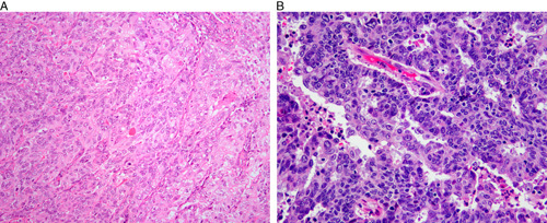
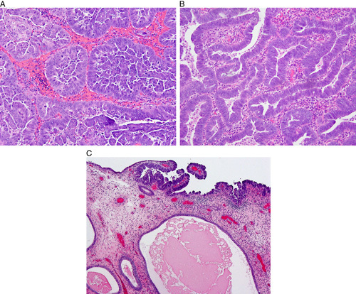
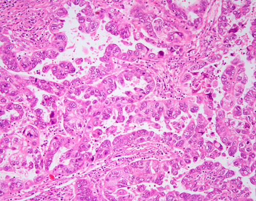
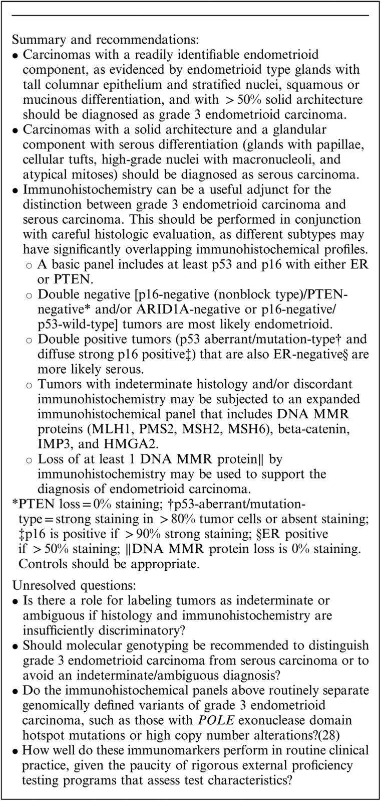
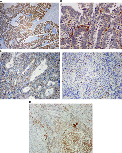
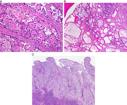
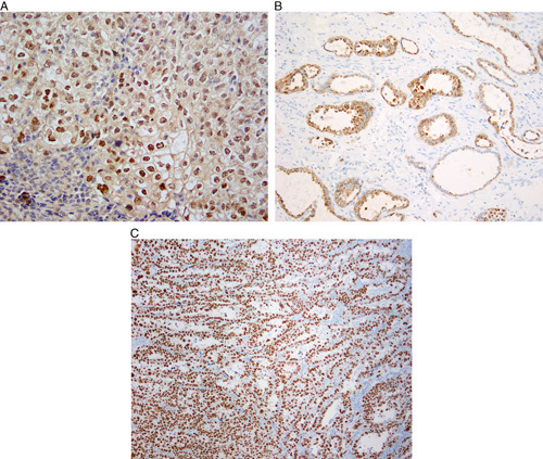
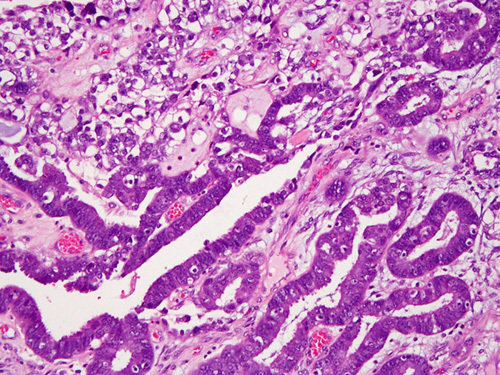
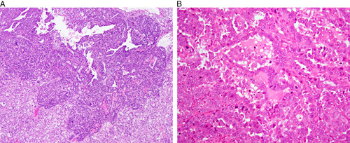
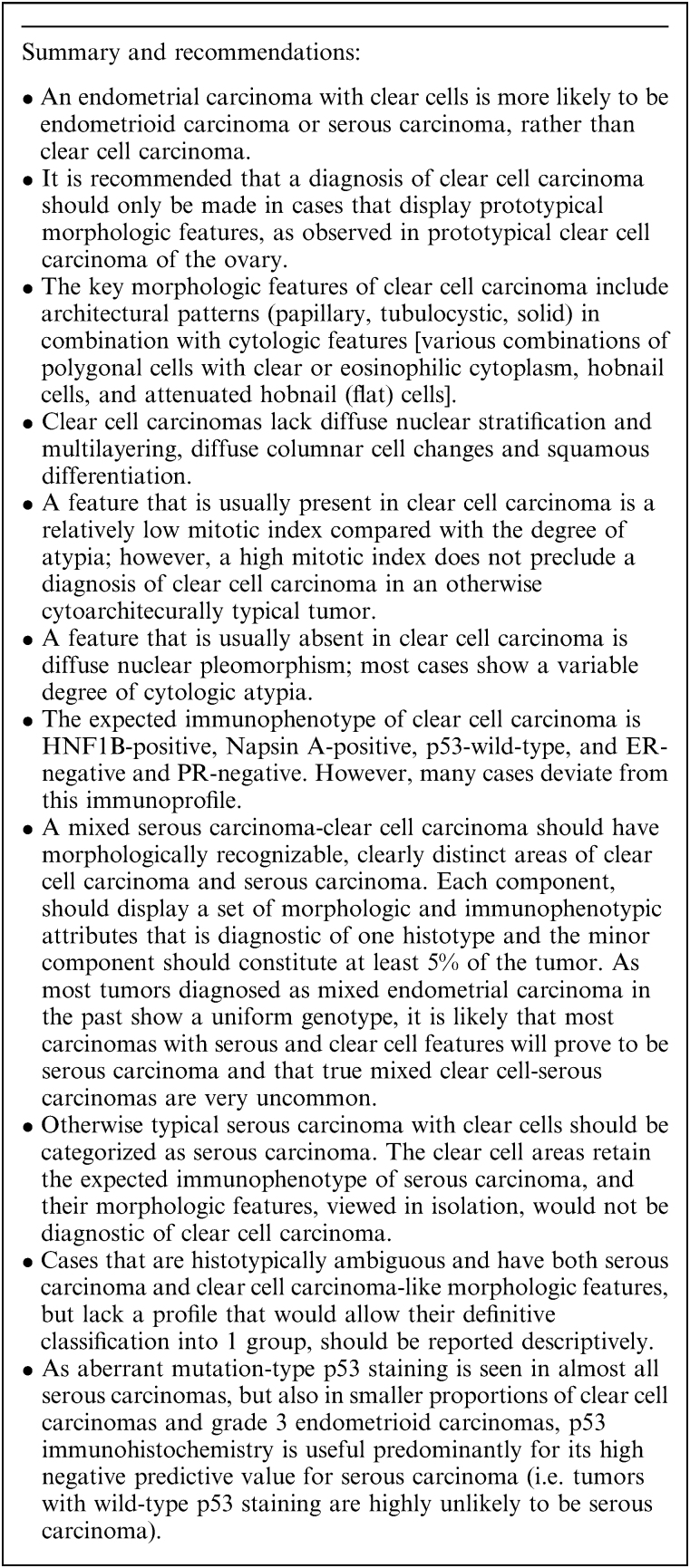
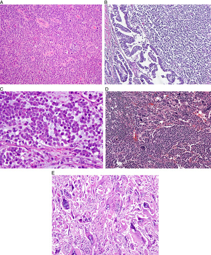
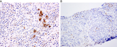
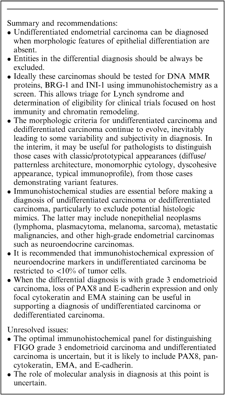
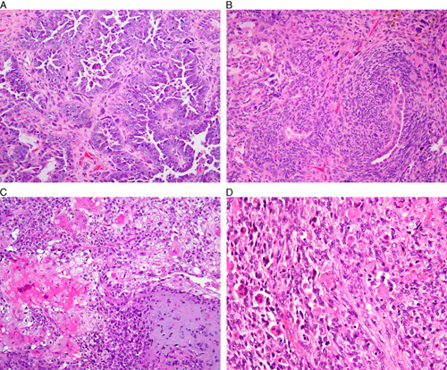
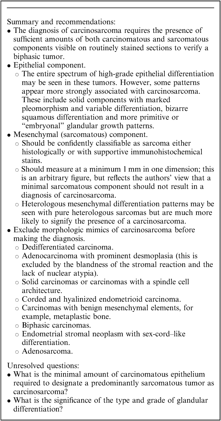
This review of challenging diagnostic issues concerning high-grade endometrial carcinomas is derived from the authors’ review of the literature followed by discussions at the Endometrial Cancer Workshop sponsored by the International Society of Gynecological Pathologists in 2016. Recommendations presented are evidence-based, insofar as this is possible, given that the levels of evidence are weak or moderate due to small sample sizes and nonuniform diagnostic criteria used in many studies. High-grade endometrioid carcinomas include FIGO grade 3 endometrioid carcinomas, serous carcinomas, clear cell carcinomas, undifferentiated carcinomas, and carcinosarcomas. FIGO grade 3 endometrioid carcinoma is diagnosed when an endometrioid carcinoma exhibits >50% solid architecture (excluding squamous areas), or when an architecturally FIGO grade 2 endometrioid carcinoma exhibits marked cytologic atypia, provided that a glandular variant of serous carcinoma has been excluded. The most useful immunohistochemical studies to make the distinction between these 2 histotypes are p53, p16, DNA mismatch repair proteins, PTEN, and ARID1A. Endometrial clear cell carcinomas must display prototypical architectural and cytologic features for diagnosis. Immunohistochemical stains, including, Napsin A and p504s can be used as ancillary diagnostic tools; p53 expression is aberrant in a minority of clear cell carcinomas. Of note, clear cells are found in all types of high-grade endometrial carcinomas, leading to a tendency to overdiagnose clear cell carcinoma. Undifferentiated carcinoma (which when associated with a component of low-grade endometrioid carcinoma is termed “dedifferentiated carcinoma”) is composed of sheets of monotonous, typically dyscohesive cells, which can have a rhabdoid appearance; they often exhibit limited expression of cytokeratins and epithelial membrane antigen, are usually negative for PAX8 and hormone receptors, lack membranous e-cadherin and commonly demonstrate loss of expression of DNA mismatch repair proteins and SWI-SNF chromatin remodeling proteins. Carcinosarcomas must show unequivocal morphologic evidence of malignant epithelial and mesenchymal differentiation.
Overview
The claims and statistics of this paper haven’t been checked yet.
Run the check — free, no account needed. It reads the open-access text and checks the sources it cites.
Integrity signals
Registry
Cites
Statistics
Figures
Verification
Claims the paper makes
The claims and statistics of this paper haven’t been checked yet.
Run the check — free, no account needed. It reads the open-access text and checks the sources it cites.
Claims other papers make about this one
Each is checked against this paper’s own text: does it say what the citing paper says it does?
In the citing paper
“Moreover, immunohistochemistry may demonstrate positive stains for HNF-1B, Napsin-A, AMACR, CK-7, and p53 [21].”
From Clear cell carcinoma arising from abdominal wall endometriosis—A report on two cases and literature review (2022) · cites this paper as [21]
Self-check
Percentages, sums and reported differences re-computed from the paper’s own numbers.
The self-check needs the paper’s full text.
Citations
Cites
A clinically applicable molecular-based classification for endometrial cancers
Talhouk, A., McConechy, M. K., Leung, S. C. Y., Li-Chang, H. H., Kwon, J. S., Melnyk, N., Yang, W., Senz, J., Boyd, N., Karnezis, A. N., et al. (2015) · British Journal of Cancer · doi:10.1038/bjc.2015.190 · Cited by 989
Self-citationTashiro, H., Blazes, M. S., Wu, R., Cho, K. R., Bose, S., Wang, S. I., Li, J., Parsons, R. E., & Ellenson, L. H. (1997) · PubMed · Cited by 701
Hamilton, C. A., Cheung, M. K., Osann, K., Chen, L., Teng, N. N. H., Longacre, T. A., Powell, M. A., Hendrickson, M. R., Kapp, D. S., & Chan, J. K. (2006) · British Journal of Cancer · doi:10.1038/sj.bjc.6603012 · Cited by 647
Integrated Molecular Characterization of Uterine Carcinosarcoma
Cherniack, A. D., Shen, H., Walter, V., Stewart, C., Murray, B. A., Bowlby, R., Hu, X., Ling, S., Soslow, R. A., Broaddus, R. R., et al. (2017) · Cancer Cell · doi:10.1016/j.ccell.2017.02.010 · Cited by 440
Self-citationCarcinosarcoma (Malignant Mixed Mesodermal Tumor) of the Uterus
Silverberg, S. G., Major, F. J., Blessing, J. A., Fetter, B. F., Askin, F. B., Liao, S., & Miller, A. W. (1990) · International Journal of Gynecological Pathology · doi:10.1097/00004347-199001000-00001 · Cited by 406
Poor Interobserver Reproducibility in the Diagnosis of High-grade Endometrial Carcinoma
Gilks, C. B., Oliva, E., & Soslow, R. A. (2013) · The American Journal of Surgical Pathology · doi:10.1097/pas.0b013e31827f576a · Cited by 404
Self-citationMalignant biphasic uterine tumours: carcinosarcomas or metaplastic carcinomas?
McCluggage, W. G. (2002) · Journal of Clinical Pathology · doi:10.1136/jcp.55.5.321 · Cited by 381
Self-citationZorn, K. K., Bonome, T., Gangi, L., Chandramouli, G. V. R., Awtrey, C. S., Gardner, G. J., Barrett, J. C., Boyd, J. A., & Birrer, M. J. (2005) · Clinical Cancer Research · doi:10.1158/1078-0432.ccr-05-0508 · Cited by 378
p53 gene mutations are common in uterine serous carcinoma and occur early in their pathogenesis.
Tashiro, H., Isacson, C., Levine, R. L., Kurman, R. J., Cho, K. R., & Hedrick, L. (1997) · Europe PMC (PubMed Central) · Cited by 345
Uterine Serous Carcinoma A Morphologically Diverse Neoplasm With Unifying Clinicopathologic Features
Sherman, M. E., Bitterman, P., Rosenshein, N. B., Delgado, G., & Kurman, R. J. (1992) · The American Journal of Surgical Pathology · doi:10.1097/00000478-199206000-00008 · Cited by 326
Research works printed but not linked: 3
- Holmes, 2014, Napsin A: a sensitive immunohistochemical marker for uterine clear cell carcinoma, Mod Pathol, 27, 536
- O'Keefe, 2015, Napsin A has utility in the diagnosis of clear cell carcinoma in the ovary but may be less valuable in the endometrium, Mod Pathol, 28, 530
- DeLair, 2012, Endometrial clear cell carcinoma with and without aberrant p53 expression: a study of 16 cases, Mod Pathol, 25, 265A
Cited by
Sorted by citations
Items 101–200 of 295
Jia, H., Wu, S., Ma, G., Yang, P., Li, X., Zeng, M., Ji, X., & Xing, X. (2024) · Pathology - Research and Practice · doi:10.1016/j.prp.2024.155595 · Cited by 8
Role of signaling pathways in endometrial cancer
Balhara, N., Yadav, R., & Chauhan, M. B. (2025) · Molecular Biology Reports · doi:10.1007/s11033-025-10523-1 · Cited by 9
Molecular Classification of Endometrial Carcinomas: Review and Recent Updates
Kumari, A., Kumar, H., Harvey, S. E., Xing, D., & Li, Z. (2025) · Cancers · doi:10.3390/cancers18010051 · Cited by 8
Alexandre, J., Belda, M. A. L. F., Prulhière, K., Treilleux, I., Leary, A. F., Pomel, C., Chargari, C., Ducassou, A., & Joly, F. (2020) · Bulletin du Cancer · doi:10.1016/j.bulcan.2020.06.006 · Cited by 8
Monsur, M., Yamaguchi, M., Tashiro, H., Yoshinobu, K., Saito, F., Erdenebaatar, C., Li, C., Iwagoi, Y., Ohba, T., Iyama, K., et al. (2021) · Medical Molecular Morphology · doi:10.1007/s00795-020-00273-3 · Cited by 9
Clinical pathological characteristics and survival of high‐grade endometrioid carcinoma
Liang, S., & Zhang, Y. (2021) · Journal of obstetrics and gynaecology research · doi:10.1111/jog.14839 · Cited by 8
Meixner, E., Lang, K., König, L., Sandrini, E., Lischalk, J. W., Debus, J., & Hörner‐Rieber, J. (2021) · Cancers · doi:10.3390/cancers13246264 · Cited by 8
Dedifferentiated endometrioid adenocarcinoma of the uterus: A case series and review of literature
Goh, C. S. Y., Farah, B. L., Ho, W. Y., Wong, S. L., Goh, C. S. Y., Chew, S. H., Nadarajah, R., Lim, Y. K., & Hồ, T. H. P. (2020) · Gynecologic Oncology Reports · doi:10.1016/j.gore.2020.100538 · Cited by 8
Uno, K., Yoshikawa, N., Kitami, K., Mori, S., Shibata, T., Iyoshi, S., Fujimoto, H., Mogi, K., Yoshihara, M., Tamauchi, S., et al. (2023) · Japanese Journal of Clinical Oncology · doi:10.1093/jjco/hyad086 · Cited by 7
Vrede, S., Hulsman, A. M. C., Reijnen, C., de Vijver, K. K. V., Colás, E., Mancebo, G., Moiola, C. P., Gil‐Moreno, A., Huvila, J. K., Koskas, M., et al. (2022) · Gynecologic Oncology · doi:10.1016/j.ygyno.2022.08.016 · Cited by 7
PGT-M, a Useful Tool to Manage the Lynch Syndrome Transmission
Listorti, I., Manzo, R., Arrivi, C., Mencacci, C., Biricik, A., Greco, E., & Greco, P. (2023) · International Journal of Molecular Sciences · doi:10.3390/ijms242216114 · Cited by 7
Lyndіn, M., Kravtsova, O., Sikora, K., Lyndina, Y., Sikora, Y., Awuah, W. A., Hasan, M. M., Abdul‐Rahman, T., Sikora, V., Hyriavenko, N., et al. (2023) · Annals of Medicine and Surgery · doi:10.1097/ms9.0000000000000939 · Cited by 7
Boennelycke, M., Peters, E. E. M., León‐Castillo, A., Smit, V. T. H. B. M., Bosse, T., Christensen, I. J., Ørtoft, G., Høgdall, C. K., & Høgdall, E. V. S. (2021) · Archiv für Pathologische Anatomie und Physiologie und für Klinische Medicin · doi:10.1007/s00428-021-03133-2 · Cited by 7
Mourtzoukou, D., Thomakos, N., Lazaris, A. C., Vlachodimitropoulos, D. G., Goutas, N. D., Sotiropoulou, M. G., Rodolakis, A., & Nonni, A. (2023) · Apmis · doi:10.1111/apm.13313 · Cited by 6
Deciphering the Therapeutic Applications of Nanomedicine in Ovarian Cancer Therapy: An Overview
Mathur, P., Bhatt, S., Kumar, S., Kamboj, S., Kamboj, R., Rana, A., Kumar, H., & Verma, R. (2023) · Current Drug Delivery · doi:10.2174/0115672018253815230922070558 · Cited by 6
A Primer on the Role of TP53 Mutation and Targeted Therapy in Endometrial Cancer
Zhang, B., Zhang, H., & Qin, Y. (2025) · Frontiers in Bioscience-Landmark · doi:10.31083/fbl25447 · Cited by 6
Ţurcan, N., Baroş, A., Zugravu, C., Mergeanu, M., Sajin, M., Andreescu, C. V., Frîncu, F., Carp-Velişcu, A., Edu, A., Mehedințu, C., et al. (2021) · Romanian Journal of Medical Practice · doi:10.37897/rjmp.2021.s6.14 · Cited by 6
Kitazono, I., Akahane, T., Yokoyama, S., Kobayashi, Y., Togami, S., Yanazume, S., Tasaki, T., Noguchi, H., Tabata, K., Kobayashi, H., et al. (2023) · Pathology - Research and Practice · doi:10.1016/j.prp.2023.154563 · Cited by 6
Namazov, A., Helpman, L., Eitan, R., Vaknin, Z., Lavie, O., Amit, A., Levy, T., Shachar, I. B., Atlas, I., Bruchim, I., et al. (2023) · European Journal of Obstetrics & Gynecology and Reproductive Biology · doi:10.1016/j.ejogrb.2023.12.021 · Cited by 6
Interobserver diagnostic reproducibility in advanced-stage endometrial carcinoma
Jung, H. J., Lee, S. Y., Hong, J. H., & Chun, Y. K. (2020) · Journal of Pathology and Translational Medicine · doi:10.4132/jptm.2020.10.04 · Cited by 6
Matson, D. R., Accola, M. A., Henderson, L., Shao, X., Frater-Rubsam, L., Horner, V. L., Rehrauer, W. M., Weisman, P. S., & Xu, J. Y. (2021) · International Journal of Gynecological Pathology · doi:10.1097/pgp.0000000000000817 · Cited by 6
Clear cell endometrial carcinoma precursors: presentation of two cases and diagnostic issues
Santoro, A., Travaglino, A., Inzani, F. S., Arciuolo, D., Angelico, G., D’Alessandris, N., Scaglione, G., Valente, M., Martini, M., Raffone, A., et al. (2021) · Diagnostic Pathology · doi:10.1186/s13000-021-01154-8 · Cited by 6
Salibay, C. J., & Fadare, O. (2020) · International Journal of Surgical Pathology · doi:10.1177/1066896920913114 · Cited by 6
Self-citationMolecular phenotype of high-grade endometrioid carcinoma of the endometrium
Buchynska, L. G., Glushchenko, N. М., Nesina, I. P., Brieieva, O., & Iurchenko, N. P. (2020) · Experimental Oncology · doi:10.32471/exp-oncology.2312-8852.vol-42-no-4.15450 · Cited by 5
Prieto‐Potín, I., Idrovo, F., Suárez‐Gauthier, A., Díaz-Blázquez, M., de Córdova, L. A. B., Chamizo, C., Zazo, S., Carvajal, N., López‐Sánchez, A., Pérez‐Buira, S., et al. (2022) · Diagnostics · doi:10.3390/diagnostics12081903 · Cited by 5
Reijnen, C., Vrede, S., Eijkelenboom, A., Draak, R., Sweegers, S., Snijders, M. P. L. M., van Gestel, P., Pijnenborg, J. M. A., Bulten, J., & Küsters‐Vandevelde, H. V. N. (2023) · Cancer Medicine · doi:10.1002/cam4.5937 · Cited by 5
Comparative analysis of EZH2, p16 and p53 expression in uterine carcinosarcomas
Makk, E., Bohonyi, N., Oszter, A., Éles, K., Tornóczky, T., Tóth, A., Kálmán, E., & Kovács, K. (2023) · Pathology & Oncology Research · doi:10.3389/pore.2023.1611547 · Cited by 5
Histomolecular features of high-grade endometrial cancers
Morotti, M., majd, H. S., Casarin, J., Alazzam, M., & Damato, S. (2021) · Minerva Medica · doi:10.23736/s0026-4806.20.07082-2 · Cited by 5
Santoro, A., Travaglino, A., Arciuolo, D., Fulgione, C., Pasquini, S., Minucci, A., Raffone, A., Inzani, F. S., & Zannoni, G. F. (2022) · Pathology - Research and Practice · doi:10.1016/j.prp.2022.154209 · Cited by 5
Bennett, J. A., Fleming, G. F., Kurnit, K. C., Mills, K. A., & van Weelden, W. J. (2022) · CA A Cancer Journal for Clinicians · doi:10.3322/caac.21715 · Cited by 5
Moreno, S. V., Khatun, M., Pasanen, A. M., Bützow, R. C., Salumets, A., Loukovaara, M. J., & Modhukur, V. (2025) · Computational and Structural Biotechnology Journal · doi:10.1016/j.csbj.2025.12.018 · Cited by 4
Russell, D. H., Epstein, J. I., Kryvenko, O. N., Schlumbrecht, M. P., Jordà, M., & Pinto, A. (2023) · Archives of Pathology & Laboratory Medicine · doi:10.5858/arpa.2022-0469-oa · Cited by 4
Menshikova, E., Deeb, K. K., Genega, E. M., Hanley, K. Z., & Turashvili, G. (2025) · Laboratory Investigation · doi:10.1016/j.labinv.2025.104240 · Cited by 4
Chung, C., Yueh, B., Subhash, S., Eskiocak, O., Nizam, A., Viola, M. G., Belleau, P., Kredentser, A., Kapedani, A., Krasnitz, A., et al. (2026) · Cancer Research Communications · doi:10.1158/2767-9764.crc-25-0193 · Cited by 4
Saglam, O., Cao, B., Wang, X., Törüner, G., & Conejo-Garcia, J. R. (2022) · PLoS ONE · doi:10.1371/journal.pone.0264014 · Cited by 4
The role of TCGA molecular classification in clear cell endometrial carcinoma
Tang, X.-Y., & Hu, Y. (2023) · Frontiers in Oncology · doi:10.3389/fonc.2023.1147394 · Cited by 4
Zhang, M., Li, R., Zhang, J., Wang, Y., Wang, Y., & Guo, Y. (2023) · BMC Cancer · doi:10.1186/s12885-023-11682-9 · Cited by 4
Dahl, M. J., Lofgren, K. A., Haugen, C., Harmon, G. E., Hughes, S. P., & Dahl, K. D. C. (2024) · Frontiers in Oncology · doi:10.3389/fonc.2024.1368592 · Cited by 4
Quddus, M. R., Ribeiro, R. R. E., DiSilvestro, J. B., Sung, C. J., & Singh, K. (2024) · Archiv für Pathologische Anatomie und Physiologie und für Klinische Medicin · doi:10.1007/s00428-024-03828-2 · Cited by 4
Wang, Y., Zheng, Y.-T., Zhang, L., Cao, X., Lin, Z., Liu, H., & Hu, Q. (2024) · Frontiers in Oncology · doi:10.3389/fonc.2024.1440246 · Cited by 4
Prognostic implication of CD47 and CTLA-4 expressions in endometrial carcinoma
Ahmed, M. M., Hussein, S., AbdAlsalam, M., Sameh, H., Waley, A. B., Ebian, H. F., Sakr, M. M. H., Attia, R. N., & Sameh, R. (2024) · Human Immunology · doi:10.1016/j.humimm.2024.111210 · Cited by 4
Undifferentiated and dedifferentiated carcinoma of the endometrium
Connolly, C., & Riain, C. Ó. (2023) · International Journal of Gynecological Cancer · doi:10.1136/ijgc-2022-004106 · Cited by 4
Role of radiotherapy in the management of rare gynaecological cancers
Morcet-Delattre, R., Espenel, S., Tas, P., Chargari, C., & Escande, A. (2023) · Cancer/Radiothérapie · doi:10.1016/j.canrad.2023.08.006 · Cited by 4
Cytologic features of undifferentiated and dedifferentiated carcinomas of the endometrium
Akbari, A., Wang, L., Soslow, R. A., & Murali, R. (2020) · Cancer Cytopathology · doi:10.1002/cncy.22351 · Cited by 4
Self-citationWillman, G. H., Podder, V., Westin, S. N., Corr, B. R., Coleman, R. L., Pothuri, B., Moore, K. N., & Slomovitz, B. M. (2025) · International Journal of Gynecological Cancer · doi:10.1016/j.ijgc.2025.102859 · Cited by 3
Lengyel, K., Musick, A., Fisch, A. S., Lin, L. H., Turashvili, G., Young, R. H., Oliva, E., & Devins, K. M. (2026) · The American Journal of Surgical Pathology · doi:10.1097/pas.0000000000002537 · Cited by 3
Self-citationEndometrial Mesonephric-Like Carcinoma With Prominent Clear Cell Carcinoma-Like Features
Valencia, M. F., Protopsaltis, N. J., Song, W., & Fadare, O. (2025) · International Journal of Surgical Pathology · doi:10.1177/10668969251361180 · Cited by 3
Self-citationRabban, J. T. (2020) · Diagnostic histopathology · doi:10.1016/j.mpdhp.2020.03.001 · Cited by 3
Hormone Therapy and Replacement in Cancer and Aging-related Diseases
Rangel, L. B. A., Kirubamani, H., Silva, I. V., & Junior, P. C. M. L. (2019) · IntechOpen eBooks · doi:10.5772/intechopen.77810 · Cited by 3
Güngördük, K., Muallem, J., Aşıcıoğlu, O., Gülseren, V., Güleç, Ü. K., Meydanlı, M. M., Sehouli, J., Özdemir, A., Şahin, H., Khatib, G., et al. (2021) · Archives of Gynecology and Obstetrics · doi:10.1007/s00404-021-06187-4 · Cited by 3
The statuses of HER2 expression and mismatch repair in endometrial clear cell carcinoma
Bayramoğlu, Z., Erozbek, S. Y., Özdemir, İ. A., Ülker, V., & Müezzínoğlu, B. (2022) · Pathology - Research and Practice · doi:10.1016/j.prp.2022.154258 · Cited by 3
Zhi, W., Zhan, Y., He, C., & Jin, Y. (2023) · Annals of Diagnostic Pathology · doi:10.1016/j.anndiagpath.2023.152173 · Cited by 3
Molecular subtypes and genomic landscape of undifferentiated and dedifferentiated endometrial cancer
Dagher, C., Selenica, P., Boroujeni, A. M., Ellenson, L. H., Mueller, J. J., Abu‐Rustum, N. R., Chui, M. H., & Weigelt, B. (2025) · International Journal of Gynecological Cancer · doi:10.1016/j.ijgc.2025.101815 · Cited by 3
The Role of PTEN Tumor Suppressor Genes in Endometrial Cancer
Paz, C. S. D. L., Viana, L. V. C., & Bueno, M. M. (2025) · International Healthcare Review (online) · doi:10.56226/114 · Cited by 3
Uncommon and Difficult High-Grade Endometrial Carcinomas
Mirković, J. (2022) · Surgical pathology clinics · doi:10.1016/j.path.2022.02.007 · Cited by 3
Primary retroperitoneal nodal endometrioid carcinoma associated with Lynch syndrome: A case report
Fischerová, D., Scovazzi, U., de Sousa, N. Q., Hovhannisyan, T., Burgetová, A., Dundr, P., Němejcová, K., Bennett, R. J., Vočka, M., Frühauf, F., et al. (2023) · Frontiers in Oncology · doi:10.3389/fonc.2023.1092044 · Cited by 3
Li, C., Yu, J., & Fu, Z. (2021) · PubMed Central · Cited by 3
Савченко, П. В., Shponka, I. S., Hrytsenko, P. O., & Savchenko, O. A. (2024) · Pathologia · doi:10.14739/2310-1237.2024.1.299170 · Cited by 2
NTRK fusion cervical sarcoma with rhabdoid cells and misleading molecular testing
Thellman, C. J., Halling, K. C., & Saglam, O. (2024) · Histopathology · doi:10.1111/his.15306 · Cited by 2
Pradhan, R., Biswas, R. R., Mondal, S., & Mukherjee, U. (2024) · The Journal of Obstetrics and Gynecology of India · doi:10.1007/s13224-024-02046-9 · Cited by 2
Kuang, W., Zeng, J., Tong, L., Liu, Q., Sun, H., Feng, M., Liang, D., Wang, W., & Wang, C. (2025) · Frontiers in Immunology · doi:10.3389/fimmu.2025.1567824 · Cited by 2
Almperi, E.-A., Margioula‐Siarkou, C., Almperis, A., Margioula‐Siarkou, G., Flindris, S., Daponte, A. I., Papamitsou, T., Dinas, K. D., & Petousis, S. (2025) · Cureus · doi:10.7759/cureus.99364 · Cited by 2
Das un- und dedifferenzierte Endometriumkarzinom
Höhn, A. K., Brambs, C., Opitz, S., Erber, R., Hartmann, A., & Horn, L. (2019) · Der Pathologe · doi:10.1007/s00292-019-00670-1 · Cited by 2
Buchynska, L. G., Glushchenko, N. М., Nespryadko, S. V., Marchenko, I. O., & Iurchenko, N. P. (2023) · Oncology · doi:10.15407/oncology.2023.03.194 · Cited by 2
Das, S., Ahlawat, S., Jain, P., Panda, A. K., Sarangi, J., Joshi, R., Gogi, R., & Jain, A. (2024) · Indian Journal of Surgical Oncology · doi:10.1007/s13193-024-02047-0 · Cited by 2
Güngördük, K., Plett, H., Gülseren, V., Meydanli, M. M., Boyraz, G., Özdemir, İ. A., Şahin, H., Şenol, T., Yıldırım, N., Turan, T., et al. (2020) · Oncology Research and Treatment · doi:10.1159/000511288 · Cited by 2
Rajanbabu, A., Nair, I. R., Ambikakumari, S., Kunneri, B., & Pavithran, K. (2022) · Journal of South Asian Federation of Obstetrics and Gynaecology · doi:10.5005/jp-journals-10006-2122 · Cited by 2
MLH1 promoter hypermethylation in uterine carcinosarcoma rarely coexists with TP53 mutation
Kunc, M., Gabrych, A., Rękawiecki, B., Gorczyński, A., Franke, S., Haybaeck, J., Biernat, W., & Czapiewski, P. (2019) · Współczesna Onkologia · doi:10.5114/wo.2019.89635 · Cited by 2
Jeanne, C., Treilleux, I., Frère‐Belda, M. L., Alexandre, J., Joly, F., & Rouleau, É. (2023) · Bulletin du Cancer · doi:10.1016/s0007-4551(23)00330-2 · Cited by 1
Destegül, E., Peköz, B. Ç., Seyfettinoğlu, S., Baş, S., Adıgüzel, F. I., & Narin, M. A. (2023) · Journal of Health Sciences and Medicine · doi:10.32322/jhsm.1330040 · Cited by 1
Pham, E. N. B., van den Berg, C. B., van Es, R., van Doorn, H. C., Groenendijk, F. H., & van Beekhuizen, H. J. (2025) · Journal of Gynecologic Oncology · doi:10.3802/jgo.2025.36.e52 · Cited by 1
Keyhanian, K., Busca, A., Howitt, B. E., Hammer, P. M., Kingsley, L. G., & Lo, B. (2025) · The American Journal of Surgical Pathology · doi:10.1097/pas.0000000000002412 · Cited by 1
Endometrial Carcinoma:Low-Grade Endometrioid Carcinoma
Lucas, E., Mills, A. M., & Carrick, K. S. (2025) · Springer eBooks · doi:10.1007/978-981-97-4822-8_58 · Cited by 1
Lee, K. J., Chokshi, S., Joshi, T., Cummings, M., Ramírez‐Alcántara, V., Turbat‐Herrera, E. A., Rocconi, R. P., Scalici, J. M., Pierce, J. Y., & Jones, N. (2025) · Gynecologic Oncology · doi:10.1016/j.ygyno.2025.07.016 · Cited by 1
Han, F., Humaria, S., & Dardik, M. (2025) · Cureus · doi:10.7759/cureus.90168 · Cited by 1
Shi, S., Dai, C., Liu, D., & Liu, X. (2025) · Frontiers in Oncology · doi:10.3389/fonc.2025.1567024 · Cited by 1
Blanc-Durand, F., Genestie, C., Nikolaev, S. I., Leman, R., Church, D. N., Beinse, G., Trédan, O., Bengrine-Lefèvre, L., Brachet, P.-E., Lebreton, C., et al. (2026) · Clinical Cancer Research · doi:10.1158/1078-0432.ccr-25-2758 · Cited by 1
Takahashi, Y., Takei, Y., Takadera, M., Koyanagi, T., Taneichi, A., Saga, Y., & Fujiwara, H. (2026) · Japanese Journal of Clinical Oncology · doi:10.1093/jjco/hyag036 · Cited by 1
Yağcı, S., Erdemoğlu, E., Erdogan, M., Mustafa Asım, A. V. C. I., Tunc, A., Ozkoç, I., & Şengül, S. S. (2026) · Cancers · doi:10.3390/cancers18060905 · Cited by 1
Russu, M. C. (2019) · IntechOpen eBooks · doi:10.5772/intechopen.89046 · Cited by 1
Sadiq, Q., & Khan, F. A. (2021) · Gynecologic Oncology Reports · doi:10.1016/j.gore.2021.100905 · Cited by 1
Arciuolo, D., Travaglino, A., Santoro, A., Raffone, A., Anchora, L. P., Fanfani, F., Inzani, F. S., & Zannoni, G. F. (2022) · Pathology - Research and Practice · doi:10.1016/j.prp.2022.154122 · Cited by 1
Travaglino, A., Arciuolo, D., Raffone, A., Santoro, A., Piermattei, A., Navarra, E., Minucci, A., Mascolo, M., Scaglione, G., D’Alessandris, N., et al. (2022) · Archiv für Pathologische Anatomie und Physiologie und für Klinische Medicin · doi:10.1007/s00428-022-03411-7 · Cited by 1
Lin, S., & Tang, Y. (2023) · Therapeutic Radiology and Oncology · doi:10.21037/tro-22-35 · Cited by 1
Weiß, F., Kaltofen, T., Kanitz, V., Schröder, L., Kost, B., König, A., Delius, M., Mahner, S., & Alejandre, I. A. (2023) · Journal of Medical Case Reports · doi:10.1186/s13256-023-03994-y · Cited by 1
Jassar, A., Hemali, N., & Bhatnagar, A. (2023) · Indian Journal of Pathology and Microbiology · doi:10.4103/ijpm.ijpm_748_22 · Cited by 1
Kitazono, I., Akahane, T., Yokoyama, S., KUBOTA, E., Kirita, Y., Noguchi, H., Murakami, M., Yanazume, S., Kobayashi, H., & Tanimoto, A. (2023) · In Vivo · doi:10.21873/invivo.13442 · Cited by 1
Napsin A Immunoreactivity in “Hobnail” Epithelium in Benign Endometrial and Endocervical Polyps
Al-Hussaini, M. A., Talia, K. L., & McCluggage, W. G. (2024) · International Journal of Gynecological Pathology · doi:10.1097/pgp.0000000000001037 · Cited by 1
Self-citationDai, Y., Wu, H., Cao, J., Li, Y., Cheng, W., & Luo, C. (2024) · Cancer Management and Research · doi:10.2147/cmar.s466022 · Cited by 1
Travaglino, A., Santoro, A., Arciuolo, D., Scaglione, G., Raffone, A., Piermattei, A., Onori, M. E., Minucci, A., Anchora, L. P., Fanfani, F., et al. (2024) · Pathologica · doi:10.32074/1591-951x-974 · Cited by 1
Kumar, H., Deshwal, A., Datwani, S., & Li, Z. (2025) · Journal of Clinical and Translational Pathology · doi:10.14218/jctp.2025.00021 · Cited by 1
Qilin, W., Liu, Y., Li, J., Shengying, Y., & Yong, L. (2025) · Frontiers in Medicine · doi:10.3389/fmed.2025.1619601 · Cited by 1
Thelagathoti, R. K., Chandel, D. S., Jiang, C. Q., Tom, W. A., Krzyzanowski, G. D., Olou, A. A., & Fernando, M. R. (2025) · Cancers · doi:10.3390/cancers17213509 · Cited by 1
Sullivan, S. A., Hawkins, G. M., Zhao, X., Jo, H., Hayes, D. N., Deng, X., Bandyopadhyay, D., Bae-Jump, V. L., & Rossi, E. C. (2021) · Gynecologic Oncology Reports · doi:10.1016/j.gore.2021.100720 · Cited by 1
Margari, N., Lorito, A. D., & Panayiotides, I. G. (2022) · Springer eBooks · doi:10.1007/978-981-16-5011-6_14 · Cited by 1
Feng, C., Cui, L., Jin, Z., Sun, L., Wang, X., Chi, X., Sun, Q., & Lian, S. (2022) · BMC Genomic Data · doi:10.1186/s12863-022-01028-y · Cited by 1
Uno, K., Yoshikawa, N., Kitami, K., Mori, S., Shibata, T., Iyoshi, S., Fujimoto, H., Mogi, K., Yoshihara, M., Tamauchi, S., et al. (2023) · Research Square · doi:10.21203/rs.3.rs-2775069/v1 · Cited by 1
Ondič, O. (2025) · Archiv für Pathologische Anatomie und Physiologie und für Klinische Medicin · doi:10.1007/s00428-025-04058-w · Cited by 1
Epithelial and Mixed Epithelial/Mesenchymal Malignancies of the Uterine Corpus
Fadare, O., & Roma, A. A. (2019) · Atlas of anatomic pathology · doi:10.1007/978-3-030-17931-1_4
Self-citation
Metadata
- Authors
- Murali, R., Davidson, B., Fadare, O., Carlson, J. A., Crum, C. P., Gilks, C. B., Irving, J. A., Malpica, A., Matias-Guiu, X., McCluggage, W. G., Mittal, K., Oliva, E., Parkash, V., Rutgers, J. K. L., Staats, P. N., Stewart, C. J. R., Tornos, C., & Soslow, R. A.
- Affiliations
- Oslo University Hospital (NO)
- Corresponding author
- Soslow, Robert A.
- ORCID
- Venue
- International Journal of Gynecological Pathology
- Published
- 2019-01
- Volume
- 38
- Issue
- Supplement 1
- Pages
- S40-S63
- ISSN
- 0277-1691 (ISSN-L)
- PMC
- PMC6296248
- OpenAlex
- W2904313334
- Cited by
- 278
- References
- 152
- Citations by year
- 2026: 20 · 2025: 38 · 2024: 33 · 2023: 53 · 2022: 42 · 2021: 42 · 2020: 33 · 2019: 17
- FWCI
- 3.71
- Type
- journal article
- Language
- en
- Open access
- hybrid
- doi.org
- Licence
- Locations
- International Journal of Gynecological Pathology (publishedVersion, cc-by)
- Europe PMC (PubMed Central) (publishedVersion, other-oa)
- PubMed Central (publishedVersion, cc-by)
- PubMed (publishedVersion)
- Repositori ObertUDL (University of Lleida) (publishedVersion, other-oa)
- RECERCAT (Consorci de Serveis Universitaris de Catalunya) (submittedVersion, cc-by)
- UWA Profiles and Research Repository (UWA) (submittedVersion, cc-by)
- Dipòsit Digital de la Universitat de Barcelona (Universitat de Barcelona) (submittedVersion, public-domain)
- LA Referencia (Red Federada de Repositorios Institucionales de Publicaciones Científicas) (submittedVersion, cc-by)
- NORA - Norwegian Open Research Archives (submittedVersion, cc-by)
- Funders
- National Institutes of Health (CA008748)
- National Cancer Institute (CA008748, P30CA008748)
- Topics
- Health Sciences › Medicine › Obstetrics and Gynecology › Endometrial and Cervical Cancer Treatments
- Health Sciences › Medicine › Oncology › Neuroendocrine Tumor Research Advances
- Health Sciences › Medicine › Epidemiology › Cervical Cancer and HPV Research
- Keywords
- high-grade endometrial cancer, endometrioid carcinoma, serous carcinoma, clear cell carcinoma, undifferentiated carcinoma, dedifferentiated carcinoma, carcinosarcoma, immunohistochemistry, p53, p16, DNA mismatch repair, PTEN, ARID1A, Napsin A, AMACR, diagnostic criteria, overdiagnosis, SWI/SNF chromatin remodeling, E-cadherin
- MeSH terms
- Female, Humans, Immunohistochemistry, Societies, Medical, Biomarkers, Tumor, Endometrial Neoplasms, Practice Guidelines as Topic, Carcinoma, Endometrioid, Neoplasm Grading
About these checks
Registry, retraction, Cites and statistics results are computed, not judged, with one exception: a printed reference that no index lists is read once by a model before it counts as a citation error. Claims are extracted by a model and kept only when their quote appears verbatim in the text; each cited source is then checked, with a model’s help, for whether it says what the claim relies on. Those verdicts can be wrong — read the claim’s quote and the cited source before relying on one.
Statistics and arithmetic are recomputed, not judged.
Confidence bands are shown only for fields calibrated on our annotated test set. None is calibrated yet, so no band is shown.
What counts as a claim ›How we measure confidence ›How we check this ›
Are you an author, or do you see a problem with this page? Request a review
