PreprintCited by 8Monoclonal and Polyclonal Antibodies ResearchCancer Immunotherapy and Biomarkersvaccines and immunoinformatics approaches

The DOI matches its Crossref record; 1 of the 1 references it cites were found in a registry or an index.

Simultaneous selection of nanobodies for accessible epitopes on immune cells in the tumor microenvironment

Open access

Preprint — published as 10.1038/s41467-023-43038-z · Paper page

Abstract

In the rapidly advancing field of synthetic biology, there is a critical need for technology to discover targeting moieties for therapeutic biologics. We developed INSPIRE-seq, an approach that utilizes a nanobody library and next-generation sequencing to identify nanobodies selected for complex environments. INSPIRE-seq enables the parallel enrichment of immune cell-binding nanobodies that penetrate the tumor microenvironment. Clone enrichment and specificity varies across immune cell subtypes in the tumor, lymph node, and spleen. INSPIRE-seq identified a dendritic cell binding clone that binds PHB2. Single-cell RNA sequencing revealed a connection with cDC1s, and immunofluorescence confirmed nanobody-PHB2 colocalization along cell membranes. Structural modeling and docking studies assisted binding predictions and will guide nanobody selection. In this work, we demonstrate that INSPIRE-seq offers an unbiased approach to examine complex microenvironments and assist in the development of nanobodies, which could serve as active drugs, modified to become drugs, or used as targeting moieties.

Overview

The claims and statistics of this paper haven’t been checked yet.

Run the check — free, no account needed. It reads the open-access text and checks the sources it cites.

A few free checks a day · papers already verified open instantly · results are public

Integrity signals

Registry

Crossref · 10.1101/2021.05.26.445719

Cites

1 of 1 references verified0 citation errors · 0 not checkable

Statistics

Figures

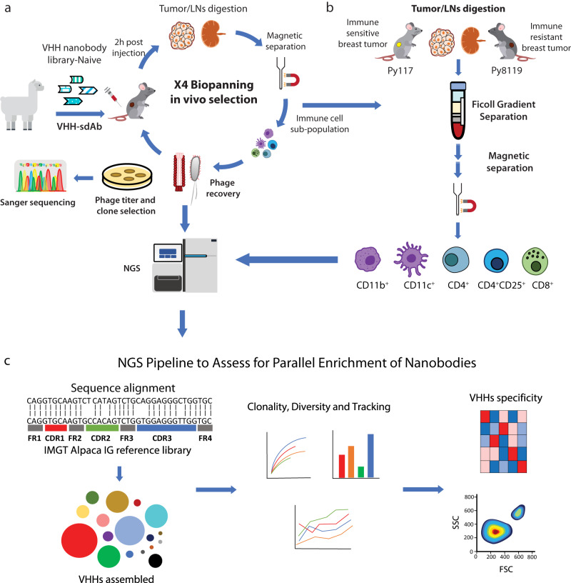
Fig. 1 In vivo biopanning for selection of VHH nanobodies that target the immune TME and assessment. a In vivo biopanning schema of VHH-nanobody phage display harvesting tumor and draining LN analyzed by NGS and viral titer then amplified CD45+ cells for a total of four rounds. b Each immune cell sub-population was sorted from immune-sensitive (Py117) and immune resistant (Py8119) tumors and LNs via ficoll gradient followed by magnetic beads separation for each round, then samples were sequenced to evaluate selection and enrichment. c NGS analysis pipeline for each round of biopanning. Reads were aligned to Alpaca IG reference, all regions of VHHs CDRs were assembled, followed by clonality assessment, diversity assessment, and clone tracking for enrichment. Llama and mouse illustrations were adapted from https://www.svgrepo.com/svg/162/llama, https://creazilla.com/nodes/7772730-mouse-clipart under creative commons license (CC0).
Fig. 1 In vivo biopanning for selection of VHH nanobodies that target the immune TME and assessment. a In vivo biopanning schema of VHH-nanobody phage display harvesting tumor and draining LN analyzed by NGS and viral titer then amplified CD45+ cells for a total of four rounds. b Each immune cell sub-population was sorted from immune-sensitive (Py117) and immune resistant (Py8119) tumors and LNs via ficoll gradient followed by magnetic beads separation for each round, then samples were sequenced to evaluate selection and enrichment. c NGS analysis pipeline for each round of biopanning. Reads were aligned to Alpaca IG reference, all regions of VHHs CDRs were assembled, followed by clonality assessment, diversity assessment, and clone tracking for enrichment. Llama and mouse illustrations were adapted from https://www.svgrepo.com/svg/162/llama, https://creazilla.com/nodes/7772730-mouse-clipart under creative commons license (CC0).CC BY · PubMed Central
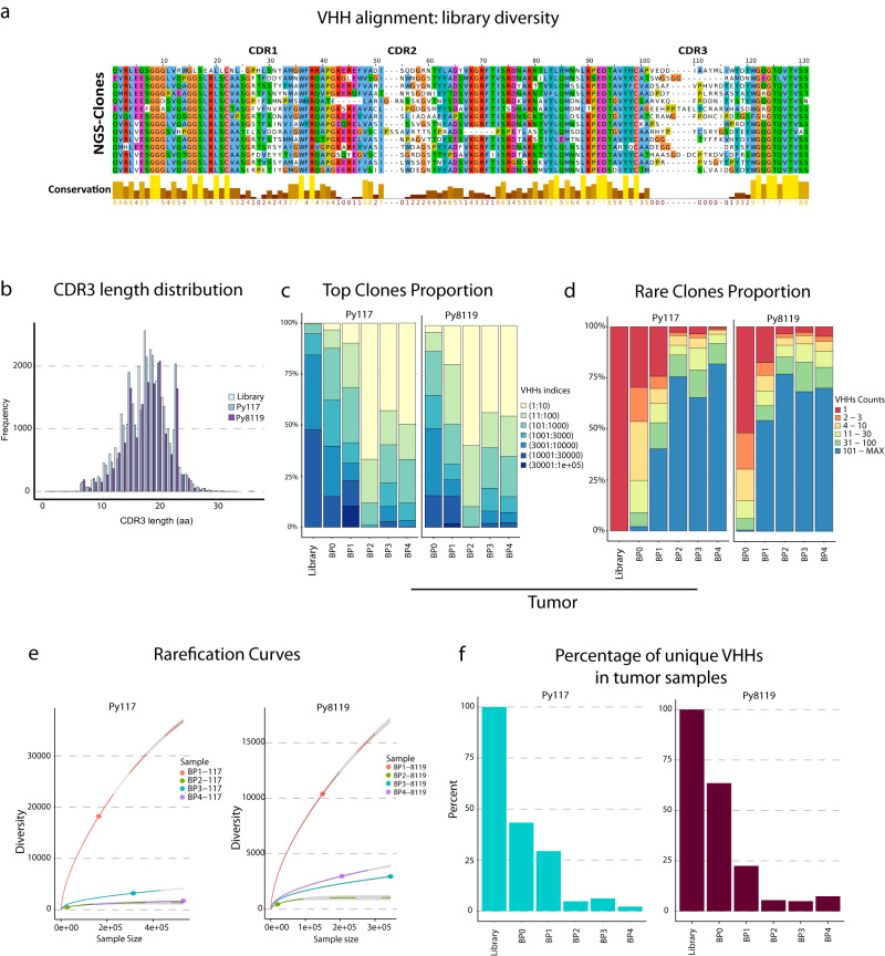
Fig. 2 Diversity and enrichment across in vivo biopanning to validate the approach. a VHH alignment showing the diversity in CDR1, CDR2 and CDR3 regions. b Frequencies of CDR3 amino acid length in the library, Py117 and Py8119 samples demonstrate an equal distribution across the samples with a median length of 18 aa. Top clone proportion (c) and rare clonal proportion (d) showing summary proportion of VHHs with specific indices and counts in Py117 and Py8119 tumor samples from BP0 to BP4. e Rarefication curve assessed the diversity of BP1 to BP4 in Py117 and Py8119 tumor samples through extrapolation and subsampling showing the reduction in diversity after BP1. f Percentage of unique VHH numbers in tumor samples of Py117 and Py8119.
Fig. 2 Diversity and enrichment across in vivo biopanning to validate the approach. a VHH alignment showing the diversity in CDR1, CDR2 and CDR3 regions. b Frequencies of CDR3 amino acid length in the library, Py117 and Py8119 samples demonstrate an equal distribution across the samples with a median length of 18 aa. Top clone proportion (c) and rare clonal proportion (d) showing summary proportion of VHHs with specific indices and counts in Py117 and Py8119 tumor samples from BP0 to BP4. e Rarefication curve assessed the diversity of BP1 to BP4 in Py117 and Py8119 tumor samples through extrapolation and subsampling showing the reduction in diversity after BP1. f Percentage of unique VHH numbers in tumor samples of Py117 and Py8119.CC BY · PubMed Central
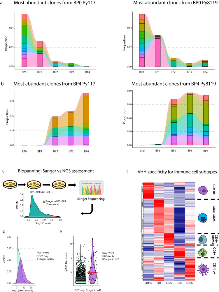
Fig. 3 Selection and evaluation of nanobodies developed by in vivo biopanning. Tracking Top VHHs either in BP0 towards BP4 (a) or in BP4 backward to BP0 (b) demonstrates the biological enrichment of the biopanning process in vivo. c Distribution of VHHs’ density after biopanning, where titration followed by Sanger sequencing theoretically provides the most abundant clones, such as the top 25%. and NGS allows for deeper assessment of biopanning with a boarder range of assessment at a multiplexed scale. d Experimental distribution of abundance by density of all enriched clones in BP3 and BP4 identified by NGS only or clones also identified by Sanger showing deeper identification of enriched clones where Sanger identified the most abundant clones. e Log2 counts of clones identified by NGS in BP3-BP4 from all cell sub-types (n = 2087 clones) in compared to clones selected for Sanger sequencing clones (n = 156 clones), median represented by red line. f Relative enrichment of VHHs in each row across the five cell subtypes from BP3-4 that identify VHHs with cell type specificity.
Fig. 3 Selection and evaluation of nanobodies developed by in vivo biopanning. Tracking Top VHHs either in BP0 towards BP4 (a) or in BP4 backward to BP0 (b) demonstrates the biological enrichment of the biopanning process in vivo. c Distribution of VHHs’ density after biopanning, where titration followed by Sanger sequencing theoretically provides the most abundant clones, such as the top 25%. and NGS allows for deeper assessment of biopanning with a boarder range of assessment at a multiplexed scale. d Experimental distribution of abundance by density of all enriched clones in BP3 and BP4 identified by NGS only or clones also identified by Sanger showing deeper identification of enriched clones where Sanger identified the most abundant clones. e Log2 counts of clones identified by NGS in BP3-BP4 from all cell sub-types (n = 2087 clones) in compared to clones selected for Sanger sequencing clones (n = 156 clones), median represented by red line. f Relative enrichment of VHHs in each row across the five cell subtypes from BP3-4 that identify VHHs with cell type specificity.CC BY · PubMed Central
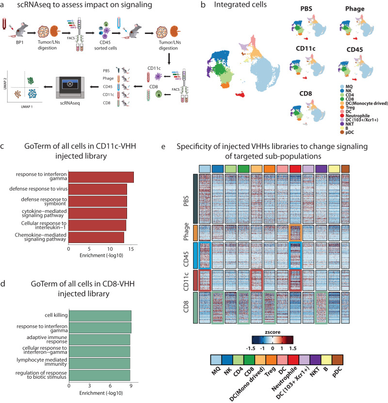
Fig. 4 ScRNAseq to explore functional changes introduced by VHHs in target immune cells. a Schema for scRNAseq experimental outline to determine functional changes in target immune cell populations. b Cell populations across all samples showing harmonized and integrated samples (left), cells clustered by sample type showing comparable distribution of cell populations regardless of the samples (right). c, d GO term enrichment of upregulated genes in CD11c-VHHs and CD8-VHHs library injected mice respectively were performed using enricher package, significance was determined by Fisher exact test (−log10) of adjusted p value by benjamini–hochberg (BH) method. e Heatmap showing differential gene expression analysis (DGE) between samples (rows) and their expression distribution across cell populations (columns). Colored boxes highlighted cell sub-population that showed most DEGs in the respected sample (rows). For example, in CD11c sample most altered cell populations were DC (Monocytes derived) and Neutrophil (red boxes). In the CD8 sample, the most altered cell populations were NK, CD8, Treg, and NKT (green boxes). Source data are provided as a Source Data file. Mouse illustration was adapted https://creazilla.com/nodes/7772730-mouse-clipart under creative commons license (CC0).
Fig. 4 ScRNAseq to explore functional changes introduced by VHHs in target immune cells. a Schema for scRNAseq experimental outline to determine functional changes in target immune cell populations. b Cell populations across all samples showing harmonized and integrated samples (left), cells clustered by sample type showing comparable distribution of cell populations regardless of the samples (right). c, d GO term enrichment of upregulated genes in CD11c-VHHs and CD8-VHHs library injected mice respectively were performed using enricher package, significance was determined by Fisher exact test (−log10) of adjusted p value by benjamini–hochberg (BH) method. e Heatmap showing differential gene expression analysis (DGE) between samples (rows) and their expression distribution across cell populations (columns). Colored boxes highlighted cell sub-population that showed most DEGs in the respected sample (rows). For example, in CD11c sample most altered cell populations were DC (Monocytes derived) and Neutrophil (red boxes). In the CD8 sample, the most altered cell populations were NK, CD8, Treg, and NKT (green boxes). Source data are provided as a Source Data file. Mouse illustration was adapted https://creazilla.com/nodes/7772730-mouse-clipart under creative commons license (CC0).CC BY · PubMed Central
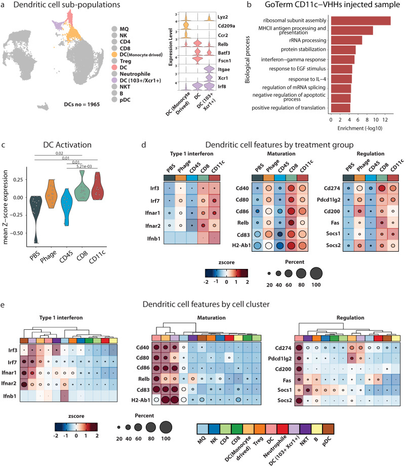
Fig. 5 Transcriptional changes introduced by CD11c VHHs injected library. a DC populations and their example of canonical markers used in the annotation process. b GO term enrichment of upregulated genes in CD11c-VHHs library injected mice in DCs subpopulations were performed using enricher package, significance was determined by Fisher exact test (−log10) of adjusted p value by benjamini-hochberg (BH) method. c Mean expression of DC activation GO term genes identified in VHHs library injected mice (F Welch test with Holm adjusted p value). d, e Heatmap showing Type 1 interferon, maturation, and regulations genes expression in all samples and cell subpopulations respectively. Source data are provided as a Source Data file.
Fig. 5 Transcriptional changes introduced by CD11c VHHs injected library. a DC populations and their example of canonical markers used in the annotation process. b GO term enrichment of upregulated genes in CD11c-VHHs library injected mice in DCs subpopulations were performed using enricher package, significance was determined by Fisher exact test (−log10) of adjusted p value by benjamini-hochberg (BH) method. c Mean expression of DC activation GO term genes identified in VHHs library injected mice (F Welch test with Holm adjusted p value). d, e Heatmap showing Type 1 interferon, maturation, and regulations genes expression in all samples and cell subpopulations respectively. Source data are provided as a Source Data file.CC BY · PubMed Central
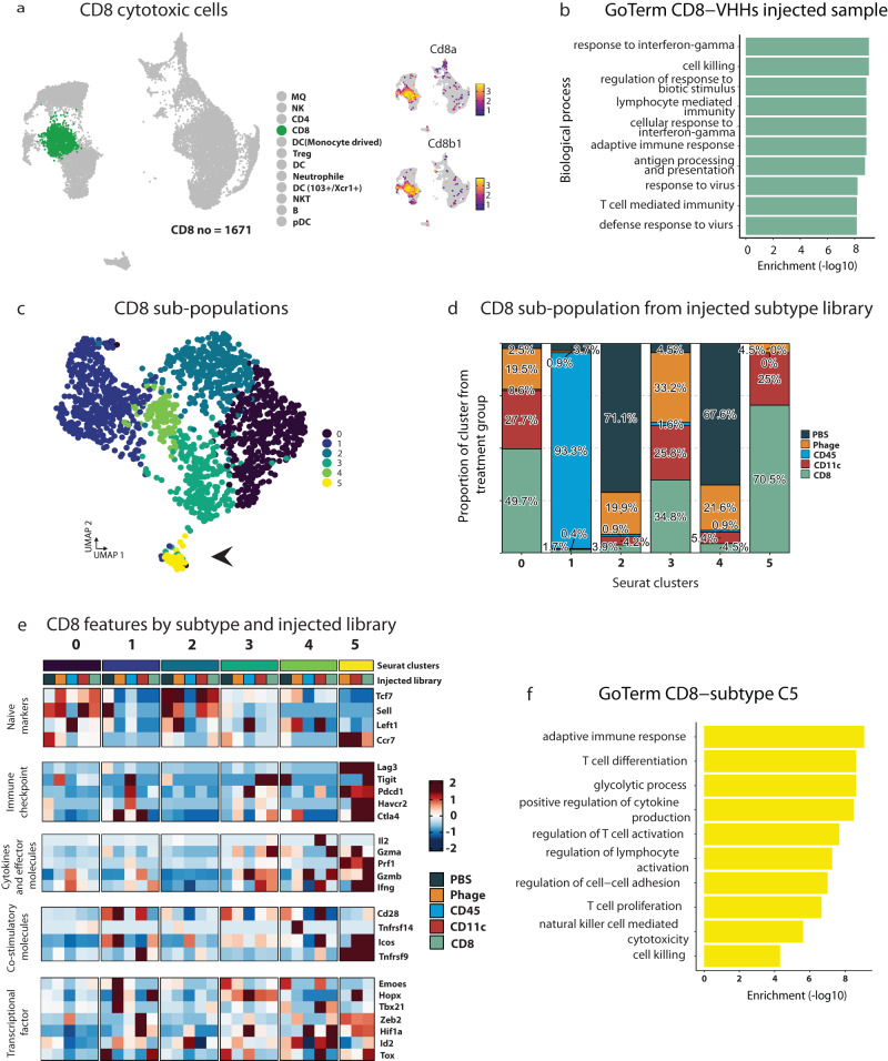
Fig. 6 Transcriptional changes introduced by CD8 VHHs injected library. a CD8 T cells and expression of their canonical markers. b GO term enrichment of upregulated genes in CD8-VHHs library injected mice in CD8 T cells were performed using enricher package, significance was determined by Fisher exact test (−log10) of adjusted p value by benjamini-hochberg (BH) method. c UMAP plot showing the CD8 sub-populations by sub-clustering CD8 T cells. Black arrows refer to sub-population cluster 5. d Histogram showing relative distribution of CD8+ T cell sub-populations based upon sample type. e Heatmap showing naïve markers, immune checkpoint, cytokines and effector molecules, co-stimulatory molecules, and transcriptional factor genes expression in CD8 T cells sub-populations and injected libraries. f GO term enrichment of upregulated genes in CD8-VHHs library injected mice in CD8 T cells cluster 5, enrichment was performed using enricher package, significance was determined by Fisher exact test (−log10) of adjusted p value by benjamini-hochberg (BH) method. Source data are provided as a Source Data file.
Fig. 6 Transcriptional changes introduced by CD8 VHHs injected library. a CD8 T cells and expression of their canonical markers. b GO term enrichment of upregulated genes in CD8-VHHs library injected mice in CD8 T cells were performed using enricher package, significance was determined by Fisher exact test (−log10) of adjusted p value by benjamini-hochberg (BH) method. c UMAP plot showing the CD8 sub-populations by sub-clustering CD8 T cells. Black arrows refer to sub-population cluster 5. d Histogram showing relative distribution of CD8+ T cell sub-populations based upon sample type. e Heatmap showing naïve markers, immune checkpoint, cytokines and effector molecules, co-stimulatory molecules, and transcriptional factor genes expression in CD8 T cells sub-populations and injected libraries. f GO term enrichment of upregulated genes in CD8-VHHs library injected mice in CD8 T cells cluster 5, enrichment was performed using enricher package, significance was determined by Fisher exact test (−log10) of adjusted p value by benjamini-hochberg (BH) method. Source data are provided as a Source Data file.CC BY · PubMed Central
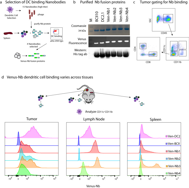
Fig. 7 DC-VHHs selection and differential binding of VHHs across immune cells and tissues. a Pipeline to select VHHs that preferably bind to dendritic cells and then generate Venus-VHH fusions. b Purified fusion protein by ÄKTA pure detected by Coomassie blue staining, Venus fluorescence, and Immunoblot using α−His antibody (representative of 3 independent experiments). c Flowcytometry gating strategy to select cells sub-populations CD11c+, CD11b+, CD8+, and CD4+. d Detection of the selected Venus fusion DC-VHHs, BCII10-VHH, and DC2.1-VHH in the tumor, spleen, and LN of Py117 tumor-paired mice (representative experiment) from three independents experiments. Source data are provided as a Source Data file. Mouse illustration was adapted https://creazilla.com/nodes/19275-cartoon-grey-mouse-clipart under creative commons license (CC0).
Fig. 7 DC-VHHs selection and differential binding of VHHs across immune cells and tissues. a Pipeline to select VHHs that preferably bind to dendritic cells and then generate Venus-VHH fusions. b Purified fusion protein by ÄKTA pure detected by Coomassie blue staining, Venus fluorescence, and Immunoblot using α−His antibody (representative of 3 independent experiments). c Flowcytometry gating strategy to select cells sub-populations CD11c+, CD11b+, CD8+, and CD4+. d Detection of the selected Venus fusion DC-VHHs, BCII10-VHH, and DC2.1-VHH in the tumor, spleen, and LN of Py117 tumor-paired mice (representative experiment) from three independents experiments. Source data are provided as a Source Data file. Mouse illustration was adapted https://creazilla.com/nodes/19275-cartoon-grey-mouse-clipart under creative commons license (CC0).CC BY · PubMed Central
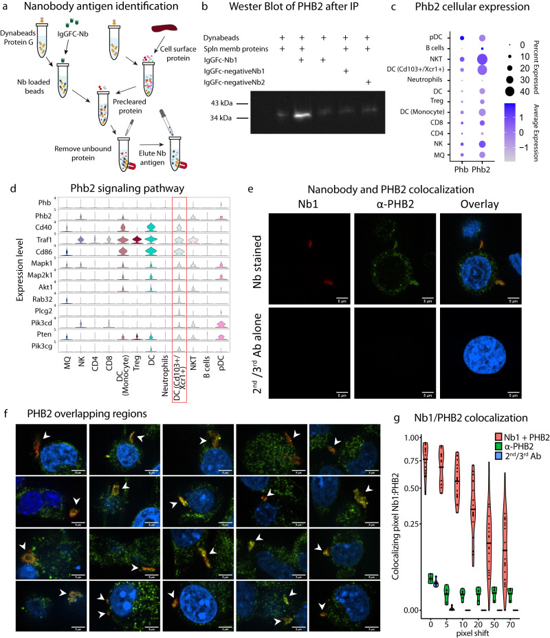
Fig. 8 Identification and verification of Nb1 binding to Phb2. a Nanobody pull-down and antigen identification pipeline using immunoprecipitation. b Western blot of α-PHB2 protein after pulled-down by Nb-FC fusion protein (IgGFC-Nb1) (representative of 3 independent experiments) c Dot plot for Phb and Phb2 expression across immune cell populations. d Violin plot showing the Phb2 signaling pathway across immune cells populations, revealing the greatest expression in the DC CD103+/Xcr1+ population (red box). e Representative confocal images from two independent experiments of Nb1 and PHB2 colocalization in MC38 cells with the upper row Nb1/α-PHB2 stained and lower row secondary and tertiary antibody alone control. The first column is the nanobody channel red, then α-PHB2 green, and the overlay with Höchst 33342 blue (scale 5 μm). f Representative confocal images from two independent experiments of Nb1 colocalization with membranous regions expressing PHB2 from 20 different cells (white arrows). g Mander’s colocalization coefficient M1 of Nb1 pixel overlapping PHB2 pixel (mean represented by black line) for Nb1 and α-PHB2 (n = 20 images), α-PHB2 antibody alone (n = 6) and 2nd/3rd antibodies alone (n = 6) from two independent experiments with six incrementing pixel shift configurations. Source data are provided as a Source Data file.
Fig. 8 Identification and verification of Nb1 binding to Phb2. a Nanobody pull-down and antigen identification pipeline using immunoprecipitation. b Western blot of α-PHB2 protein after pulled-down by Nb-FC fusion protein (IgGFC-Nb1) (representative of 3 independent experiments) c Dot plot for Phb and Phb2 expression across immune cell populations. d Violin plot showing the Phb2 signaling pathway across immune cells populations, revealing the greatest expression in the DC CD103+/Xcr1+ population (red box). e Representative confocal images from two independent experiments of Nb1 and PHB2 colocalization in MC38 cells with the upper row Nb1/α-PHB2 stained and lower row secondary and tertiary antibody alone control. The first column is the nanobody channel red, then α-PHB2 green, and the overlay with Höchst 33342 blue (scale 5 μm). f Representative confocal images from two independent experiments of Nb1 colocalization with membranous regions expressing PHB2 from 20 different cells (white arrows). g Mander’s colocalization coefficient M1 of Nb1 pixel overlapping PHB2 pixel (mean represented by black line) for Nb1 and α-PHB2 (n = 20 images), α-PHB2 antibody alone (n = 6) and 2nd/3rd antibodies alone (n = 6) from two independent experiments with six incrementing pixel shift configurations. Source data are provided as a Source Data file.CC BY · PubMed Central
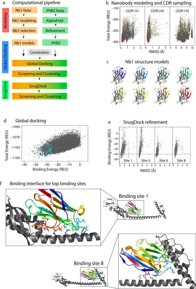
Fig. 9 Computational modeling for docking predictions of Nb1 to PHB2. a Computational protocol for modeling and docking using Rosetta and AlphaFold. b Modeling of Nb1: Rosetta total energy vs. RMSD for each individual CDR. The best representative model for each cluster is shown in orange circles, and the top eight are shown in red. c Cartoon representation of the eight selected Nb1 candidates for computational docking. d Rosetta total energy vs. Rosetta binding energy after global docking. The best representative model for each cluster is shown in cyan circles, and the top 11 are shown in blue circles. e SnugDock binding energy vs RMSD results for binding sites 1, 5, 6 and 8 (full results in Supplementary Fig. 7e). f Cartoon representation of binding sites 1 (left) and 8 (right). PHB2 is shown in gray, while Nb1 is shown with rainbow color. The residues forming critical interactions are shown as sticks, and hydrogen bonds are shown as yellow dash lines.
Fig. 9 Computational modeling for docking predictions of Nb1 to PHB2. a Computational protocol for modeling and docking using Rosetta and AlphaFold. b Modeling of Nb1: Rosetta total energy vs. RMSD for each individual CDR. The best representative model for each cluster is shown in orange circles, and the top eight are shown in red. c Cartoon representation of the eight selected Nb1 candidates for computational docking. d Rosetta total energy vs. Rosetta binding energy after global docking. The best representative model for each cluster is shown in cyan circles, and the top 11 are shown in blue circles. e SnugDock binding energy vs RMSD results for binding sites 1, 5, 6 and 8 (full results in Supplementary Fig. 7e). f Cartoon representation of binding sites 1 (left) and 8 (right). PHB2 is shown in gray, while Nb1 is shown with rainbow color. The residues forming critical interactions are shown as sticks, and hydrogen bonds are shown as yellow dash lines.CC BY · PubMed Central

Verification

Claims the paper makes

The claims and statistics of this paper haven’t been checked yet.

Run the check — free, no account needed. It reads the open-access text and checks the sources it cites.

A few free checks a day · papers already verified open instantly · results are public

Self-check

Percentages, sums and reported differences re-computed from the paper’s own numbers.

The self-check needs the paper’s full text.

Citations

Cites

Show all 1 references

Research works printed but not linked: 50

  1. 10.1016/j.immuni.2019.12.011 · DOI · Paper page
  2. 10.1038/s41422-020-0337-2 · DOI
  3. 2020, The history and advances in cancer immunotherapy: understanding the characteristics of tumor-infiltrating immune cells and their therapeutic implications, Cell Mol Immunol, 17, 807, 10.1038/s41423-020-0488-6 · DOI
  4. Sodir, N.M. et al. MYC Instructs and Maintains Pancreatic Adenocarcinoma Phenotype. Cancer Discov (2020). · DOI
  5. 10.1016/j.cell.2017.11.013 · DOI · Paper page
  6. 10.1126/science.1259425 · DOI
  7. 10.1016/j.cell.2016.12.022 · DOI · Paper page
  8. 10.1038/s41422-019-0195-y · DOI · Paper page
  9. 10.1016/j.cell.2018.05.060 · DOI · Paper page
  10. 2018, Nobel work that galvanized an industry, Nat Biotechnol, 36, 1023, 10.1038/nbt.4301 · DOI
  11. 10.1038/380364a0 · DOI
  12. 10.1016/S1525-0016(03)00181-3 · DOI
  13. 10.1073/pnas.0403317101 · DOI
  14. 10.1074/jbc.M110.138297 · DOI
  15. 10.1038/nbt.1764 · DOI
  16. Iezzi, M.E. , Policastro, L. , Werbajh, S. , Podhajcer, O. & Canziani, G.A . Single-Domain Antibodies and the Promise of Modular Targeting in Cancer Imaging and Treatment. Front Immunol 9, 273 (2018). · DOI
  17. Gonzalez-Sapienza, G. , Rossotti, M.A. & Tabares-da Rosa, S . Single-Domain Antibodies As Versatile Affinity Reagents for Analytical and Diagnostic Applications. Front Immunol 8, 977 (2017). · DOI
  18. 10.1146/annurev-biochem-063011-092449 · DOI · Paper page
  19. 10.1038/363446a0 · DOI · Paper page
  20. 10.1038/ncomms13898 · DOI
  21. Aguilera, T.A. et al. Induced tumor heterogeneity reveals factors informing radiation and immunotherapy combinations. Clin Cancer Res (2020). · DOI
  22. 10.1038/nature14404 · DOI · Paper page
  23. 10.1038/nature14424 · DOI
  24. Santana-Magal, N. et al. Melanoma-secreted lysosomes trigger monocyte-derived dendritic cell apoptosis and limit cancer immunotherapy. Cancer research (2020). · DOI
  25. Maier, B. et al. A conserved dendritic-cell regulatory program limits antitumour immunity. Nature (2020). · DOI
  26. 10.2967/jnumed.109.070078 · DOI
  27. 10.4049/jimmunol.1201646 · DOI
  28. 2020, Macromolecular modeling and design in Rosetta: recent methods and frameworks, Nature methods, 17, 665, 10.1038/s41592-020-0848-2 · DOI · Paper page
  29. 2021, Modeling Immunity with Rosetta: Methods for Antibody and Antigen Design, Biochemistry, 60, 825, 10.1021/acs.biochem.0c00912 · DOI
  30. 2021, Ensuring scientific reproducibility in bio-macromolecular modeling via extensive, automated benchmarks, Nat Commun, 12, 6947, 10.1038/s41467-021-27222-7 · DOI
  31. Adolf-Bryfogle, J. et al. RosettaAntibodyDesign (RAbD): A general framework for computational antibody design. PLoS Comput Biol 14, e1006112 (2018). · DOI · Paper page
  32. 2017, Modeling and docking of antibody structures with Rosetta, Nat Protoc, 12, 401, 10.1038/nprot.2016.180 · DOI
  33. Finn, J.A. et al. Improving Loop Modeling of the Antibody Complementarity-Determining Region 3 Using Knowledge-Based Restraints. PLoS One 11, e0154811 (2016). · DOI
  34. 10.1126/science.aap7577 · DOI
  35. 2019, Structural Basis of Mitochondrial Scaffolds by Prohibitin Complexes: Insight into a Role of the Coiled-Coil Region, iScience, 19, 1065, 10.1016/j.isci.2019.08.056 · DOI
  36. 10.1016/j.sbi.2006.03.003 · DOI
  37. 10.1093/bioinformatics/bty355 · DOI · Paper page
  38. Sircar, A. & Gray, J.J . SnugDock: paratope structural optimization during antibody-antigen docking compensates for errors in antibody homology models. PLoS Comput Biol 6, e1000644 (2010). · DOI
  39. 2021, An expanded benchmark for antibody-antigen docking and affinity prediction reveals insights into antibody recognition determinants, Structure, 29, 606, 10.1016/j.str.2021.01.005 · DOI
  40. Denisenko, E. et al. Systematic assessment of tissue dissociation and storage biases in single-cell and single-nucleus RNA-seq workflows. Genome Biol 21, 130 (2020). · DOI · Paper page
  41. 2021, The need to reassess single-cell RNA sequencing datasets: the importance of biological sample processing, F1000Res, 10, 767, 10.12688/f1000research.54864.1 · DOI
  42. 10.1126/science.aaz6695 · DOI
  43. Gibby, K. et al. Early vascular deficits are correlated with delayed mammary tumorigenesis in the MMTV-PyMT transgenic mouse following genetic ablation of the NG2 proteoglycan. Breast Cancer Res 14, R67 (2012). · DOI
  44. Shugay, M. et al. VDJtools: Unifying Post-analysis of T Cell Receptor Repertoires. PLoS Comput Biol 11, e1004503 (2015). · DOI
  45. 10.1890/13-0133.1 · DOI
  46. 10.1038/ni1008-1091 · DOI · Paper page
  47. 10.1111/j.1365-2818.2006.01706.x · DOI
  48. 10.1267/ahc.07002 · DOI
  49. 10.1152/ajpcell.00462.2010 · DOI
  50. 2021, Robustification of RosettaAntibody and Rosetta SnugDock, PLoS One, 16, e0234282, 10.1371/journal.pone.0234282 · DOI

Metadata

Authors
Sekar, T. V., Elghonaimy, E. A., Swancutt, K. L., Diegeler, S., Gonzalez, I., Hamilton, C., Leung, P. Q., Meiler, J., Martina, C. E., Whitney, M., & Aguilera, T. A.
Affiliations
  • Pondicherry University (IN)
  • The University of Texas Southwestern Medical Center (US)
  • Vanderbilt University (US)
  • University of California San Diego (US)
Corresponding author
Aguilera, Todd A.
Venue
Nature Communications
Publisher
openRxiv
Published
2021-05-29
Volume
14
Issue
1
Pages
7473-7473
ISSN
2041-1723 (ISSN-L)
OpenAlex
W4388750814
Cited by
8
References
1
Citations by year
2026: 4 · 2025: 3 · 2024: 1
FWCI
1.32
Type
journal article
Language
en
Open access
Licence
cc-by
Locations
  • Nature Communications (publishedVersion, cc-by)
  • bioRxiv (Cold Spring Harbor Laboratory) (acceptedVersion)
  • PubMed Central (publishedVersion, cc-by)
  • PubMed (publishedVersion)
  • RePEc: Research Papers in Economics (submittedVersion)
  • DOAJ (DOAJ: Directory of Open Access Journals) (submittedVersion, cc-by-sa)
Funders
  • Mary Kay Foundation
  • Moody Foundation
  • Cancer Prevention and Research Institute of Texas (RR170051)
  • Vanderbilt University
  • University of Texas Southwestern Medical Center
  • Children’s Hospital of Wisconsin Research Institute
  • National Cancer Institute (T32CA124334)
Topics
  • Life Sciences › Immunology and Microbiology › Immunology › Monoclonal and Polyclonal Antibodies Research
  • Health Sciences › Medicine › Oncology › Cancer Immunotherapy and Biomarkers
  • Life Sciences › Biochemistry, Genetics and Molecular Biology › Molecular Biology › vaccines and immunoinformatics approaches
Keywords
tumor microenvironment, immune cells, PHB2, dendritic cells, cDC1, single-cell RNA sequencing, binding prediction
MeSH terms
Epitopes, Tumor Microenvironment, Single-Domain Antibodies

About these checks

Registry, retraction, Cites and statistics results are computed, not judged, with one exception: a printed reference that no index lists is read once by a model before it counts as a citation error. Claims are extracted by a model and kept only when their quote appears verbatim in the text; each cited source is then checked, with a model’s help, for whether it says what the claim relies on. Those verdicts can be wrong — read the claim’s quote and the cited source before relying on one.

Statistics and arithmetic are recomputed, not judged.

Confidence bands are shown only for fields calibrated on our annotated test set. None is calibrated yet, so no band is shown.

What counts as a claim ›How we measure confidence ›How we check this ›

Are you an author, or do you see a problem with this page? Request a review

How to cite

Cite CoChat for the checks on this page, and the paper for its own findings.

This page
CoChat. (2026). Citation check: Simultaneous selection of nanobodies for accessible epitopes on immune cells in the tumor microenvironment. https://app.cochat.ai/papers/10.1101/2021.05.26.445719
The paper
Sekar, T. V., Elghonaimy, E. A., Swancutt, K. L., Diegeler, S., Gonzalez, I., Hamilton, C., Leung, P. Q., Meiler, J., Martina, C. E., Whitney, M., & Aguilera, T. A. (2021). Simultaneous selection of nanobodies for accessible epitopes on immune cells in the tumor microenvironment. Nature Communications. https://doi.org/10.1101/2021.05.26.445719