PreprintCited by 18Antimicrobial Resistance in StaphylococcusBacterial Genetics and BiotechnologyBiochemical and Structural Characterization

The DOI matches its Crossref record; 1 of the 1 references it cites were found in a registry or an index.

SmdA is a novel cell morphology determinant in Staphylococcus aureus

Open access

Preprint — published as 10.1128/mbio.03404-21

Abstract

Cell division and cell wall synthesis in staphylococci need to be precisely coordinated and controlled to allow the cell to multiply while maintaining their nearly spherical shape. The mechanisms ensuring correct placement of the division plane and synthesis of new cell wall have been studied intensively, however, hitherto unknown factors and proteins are likely to play key roles in this complex interplay. We here identified and investigated a protein with major influence on cell morphology in Staphylococcus aureus . The protein, named SmdA (for staphylococcal morphology determinant A), is a membrane-protein with septum-enriched localization. By CRISPRi knockdown and overexpression combined with different microscopy techniques, we demonstrate that proper levels of SmdA is necessary for cell division, including septum formation and cell splitting. We also identified conserved residues in SmdA that are critical for its functionality. Pulldown- and bacterial two-hybrid interaction experiments showed that SmdA interacts with several known cell division- and cell wall synthesis proteins, including penicillin binding proteins (PBPs) and EzrA. Notably, SmdA also affects susceptibility to cell wall targeting antibiotics, particularly in methicillin-resistant S. aureus (MRSA). Together, our results show that S. aureus is dependent on balanced amounts of membrane-attached SmdA in order to carry out proper cell division. Importance: Staphylococcus aureus is an important human and animal pathogen. Antibiotic resistance is a major problem in treatment of staphylococcal infections, and cell division and cell wall synthesis factors have previously been shown to modulate susceptibility to antibiotics in this species. In the current work we investigated the function of an essential protein named SmdA, which was identified based on its septal localization and knockdown phenotype resulting in defective cellular morphologies. We demonstrate that this protein is critical for normal cell division in S. aureus . Depletion of SmdA sensitize resistant staphylococci to β-lactam antibiotics. This work thus reveals a new staphylococcal cell division factor and a potential future target for narrow spectrum antimicrobials or compounds to resensitize antibiotic resistant staphylococcal strains.

Overview

The claims and statistics of this paper haven’t been checked yet.

Run the check — free, no account needed. It reads the open-access text and checks the sources it cites.

A few free checks a day · papers already verified open instantly · results are public

Integrity signals

Registry

Crossref · 10.1101/2021.11.23.469651

Cites

1 of 1 references verified0 citation errors · 0 not checkable

Statistics

Figures

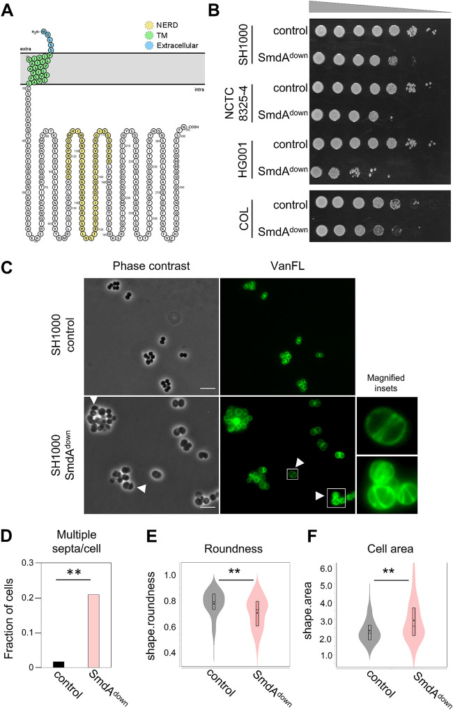
FIG 1 Phenotypes resulting from depletion of SmdA. (A) Predicted topology of SmdA using Protter (77). SmdA is predicted to have one transmembrane (TM) helix with a short extracellular N terminus and a large intracellular domain. The sequence with predicted similarity to the NERD domain is highlighted in yellow. (B) Growth on a solid medium of SmdA knockdown strains (SmdAdown) in S. aureus SH1000 (IM269), NCTC8325-4 (IM311), HG001 (IM312), and COL (IM294). Strains carrying a nontargeting sgRNA were used as controls (IM284, IM307, IM313, and IM295 for the respective strains). From noninduced overnight cultures, 10-fold dilution series were made and spotted onto plates with 300 μM isopropyl-β-d-thiogalactopyranoside (IPTG). (C) SmdAdown (IM269) and control strain (IM284) were analyzed by phase-contrast and fluorescence microscopy of cells stained with the cell wall label VanFL. White arrows point at misshaped cells and cells with perturbed septum formation. Magnified insets of representative cells are shown for the VanFL micrographs. Scale bars, 5 μm. (D) Fraction of cells with multiple septa per cell for the SmdAdown strain IM269 (n = 225) and the nontarget control strain IM284 (n = 242) are plotted. The asterisks indicate a significant difference (Fisher’s exact test, P < 0.001). (E) Cell roundness, as determined using MicrobeJ, was used as a measure of the morphology of the cells. Spherical cells will have values close to 1. Cell roundness measures for the control strain IM284 (n = 198) and the SmdAdown strain IM269 (n = 191) are plotted. (F) Cell area (in μm2) as determined using MicrobeJ of the control strain IM284 (n = 198) and the SmdAdown strain IM269 (n = 191). (E and F) Significant differences between the distributions are indicated by asterisks (**, P < 0.001). P values were derived from a Mann-Whitney test.
FIG 1 Phenotypes resulting from depletion of SmdA. (A) Predicted topology of SmdA using Protter (77). SmdA is predicted to have one transmembrane (TM) helix with a short extracellular N terminus and a large intracellular domain. The sequence with predicted similarity to the NERD domain is highlighted in yellow. (B) Growth on a solid medium of SmdA knockdown strains (SmdAdown) in S. aureus SH1000 (IM269), NCTC8325-4 (IM311), HG001 (IM312), and COL (IM294). Strains carrying a nontargeting sgRNA were used as controls (IM284, IM307, IM313, and IM295 for the respective strains). From noninduced overnight cultures, 10-fold dilution series were made and spotted onto plates with 300 μM isopropyl-β-d-thiogalactopyranoside (IPTG). (C) SmdAdown (IM269) and control strain (IM284) were analyzed by phase-contrast and fluorescence microscopy of cells stained with the cell wall label VanFL. White arrows point at misshaped cells and cells with perturbed septum formation. Magnified insets of representative cells are shown for the VanFL micrographs. Scale bars, 5 μm. (D) Fraction of cells with multiple septa per cell for the SmdAdown strain IM269 (n = 225) and the nontarget control strain IM284 (n = 242) are plotted. The asterisks indicate a significant difference (Fisher’s exact test, P < 0.001). (E) Cell roundness, as determined using MicrobeJ, was used as a measure of the morphology of the cells. Spherical cells will have values close to 1. Cell roundness measures for the control strain IM284 (n = 198) and the SmdAdown strain IM269 (n = 191) are plotted. (F) Cell area (in μm2) as determined using MicrobeJ of the control strain IM284 (n = 198) and the SmdAdown strain IM269 (n = 191). (E and F) Significant differences between the distributions are indicated by asterisks (**, P < 0.001). P values were derived from a Mann-Whitney test.CC BY · PubMed Central
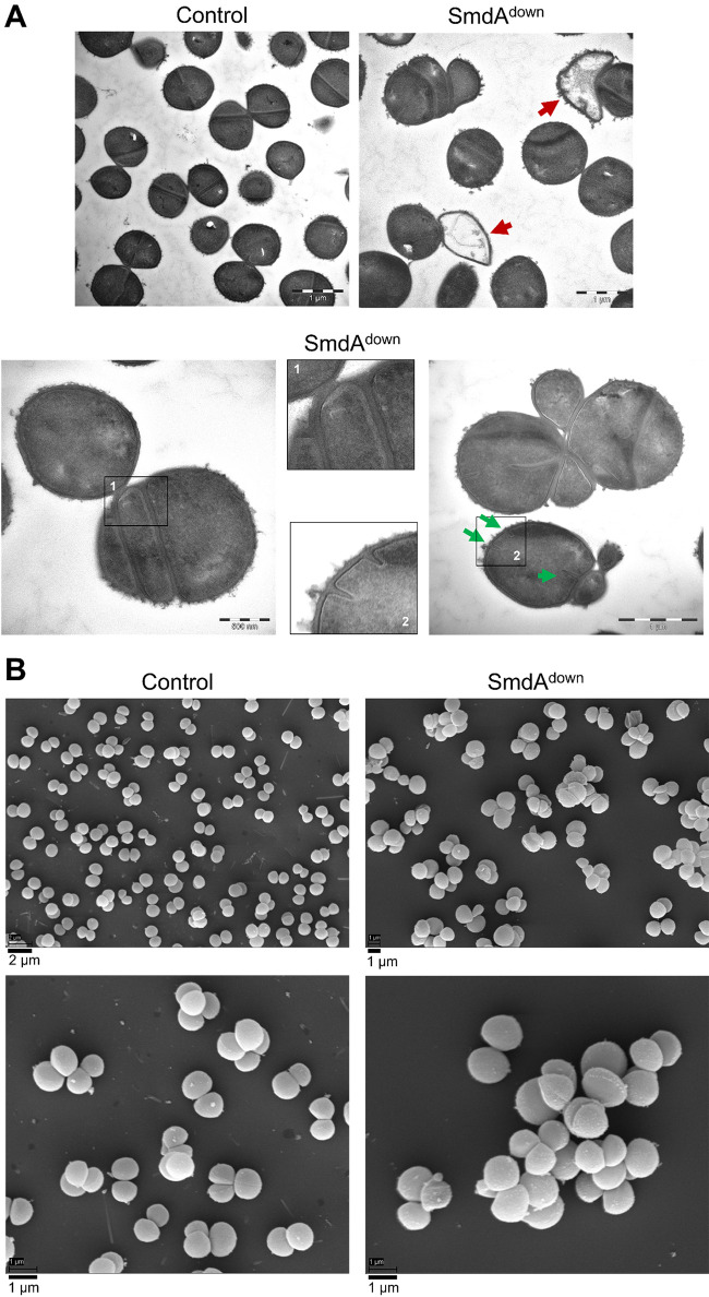
FIG 2 SmdAdown in S. aureus SH1000 visualized by electron microscopy. (A) Transmission electron and (B) scanning electron micrographs of SH1000 CRISPRi control strain (IM284) and SH1000 SmdAdown (IM269). In (A), red arrows in the TEM micrographs point at lysed cells. Representative examples of cells with parallel septa or multiple septa are shown. Green arrows point to the initiation of septum synthesis at multiple sites within the same cell. Different magnifications are shown, indicated by the scale bars.
FIG 2 SmdAdown in S. aureus SH1000 visualized by electron microscopy. (A) Transmission electron and (B) scanning electron micrographs of SH1000 CRISPRi control strain (IM284) and SH1000 SmdAdown (IM269). In (A), red arrows in the TEM micrographs point at lysed cells. Representative examples of cells with parallel septa or multiple septa are shown. Green arrows point to the initiation of septum synthesis at multiple sites within the same cell. Different magnifications are shown, indicated by the scale bars.CC BY · PubMed Central
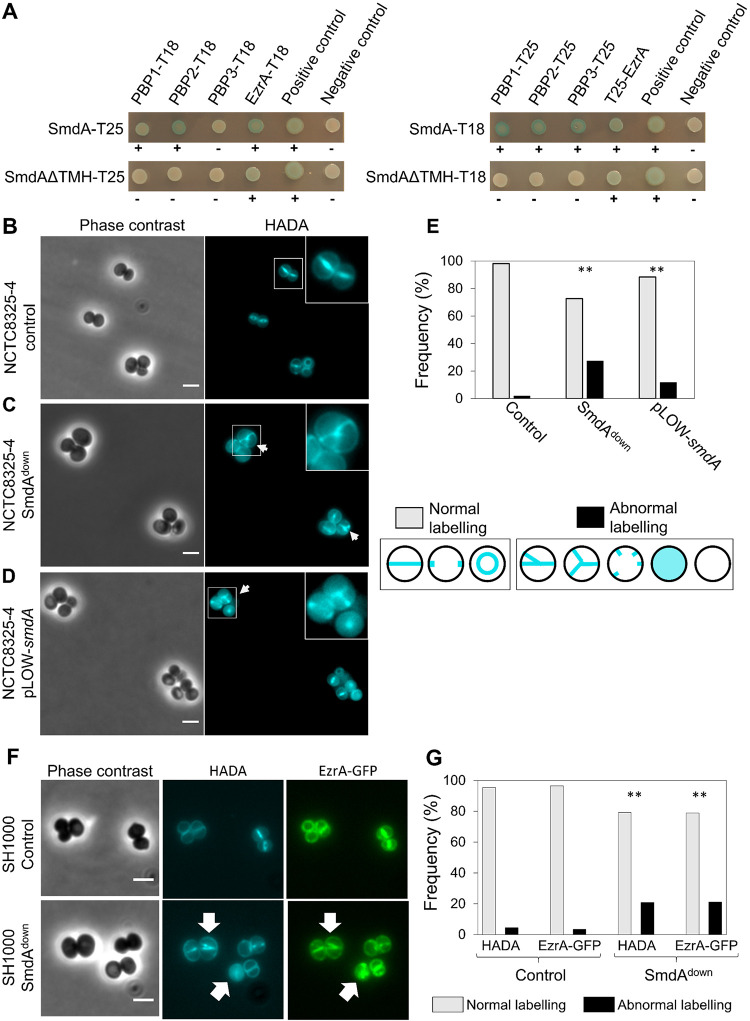
FIG 3 SmdA interacts with important cell division proteins and is necessary for proper localization of EzrA and peptidoglycan synthesis. (A) Protein-protein interactions were tested with bacterial two-hybrid assays, where SmdA and SmdAΔTMH were tested against PBP1, PBP2, PBP3, and EzrA. The proteins were fused to the T18 or T25 domains as indicated. Blue bacterial spots and plus signs indicate positive interactions and white spots and minus symbols indicate no interaction. (B to D) Micrographs of HADA labeled S. aureus NCTC8325-4 with depletion and overexpression of SmdA. Arrows point at cells with misplaced septum synthesis. Scale bars, 2 μm. (B) CRISPRi control strain, IM307 (C) SmdAdown strain, IM311 and (D) SmdA overexpression strain, MK1866. (E) Frequency plot of cells with normal or abnormal HADA labeling pattern. Categorization of normal or abnormal labeling patterns is indicated. The number of cells analyzed was 259, 179, and 189 for (B to D), respectively. The asterisks indicate a significant difference from the control (Fisher’s exact test, P < 0.001). (F) Micrographs showing colocalization of EzrA-GFP and HADA incorporation in S. aureus SH1000 strains with (MK1952) or without (MK1953) knockdown of SmdA. Phase-contrast and fluorescence images of HADA labeling and GFP (EzrA-GFP) are shown. Scale bars, 2 μm. Arrows point to cells with abnormal localization of both HADA and EzrA-GFP. (G) Frequencies of cells (from [F]) with normal or abnormal localization of HADA and EzrA-GFP are plotted. The number of cells analyzed was 285 (for MK1953) and 298 (for MK1952). The asterisks indicate significant differences from the respective controls (Fisher’s exact test, P < 0.001).
FIG 3 SmdA interacts with important cell division proteins and is necessary for proper localization of EzrA and peptidoglycan synthesis. (A) Protein-protein interactions were tested with bacterial two-hybrid assays, where SmdA and SmdAΔTMH were tested against PBP1, PBP2, PBP3, and EzrA. The proteins were fused to the T18 or T25 domains as indicated. Blue bacterial spots and plus signs indicate positive interactions and white spots and minus symbols indicate no interaction. (B to D) Micrographs of HADA labeled S. aureus NCTC8325-4 with depletion and overexpression of SmdA. Arrows point at cells with misplaced septum synthesis. Scale bars, 2 μm. (B) CRISPRi control strain, IM307 (C) SmdAdown strain, IM311 and (D) SmdA overexpression strain, MK1866. (E) Frequency plot of cells with normal or abnormal HADA labeling pattern. Categorization of normal or abnormal labeling patterns is indicated. The number of cells analyzed was 259, 179, and 189 for (B to D), respectively. The asterisks indicate a significant difference from the control (Fisher’s exact test, P < 0.001). (F) Micrographs showing colocalization of EzrA-GFP and HADA incorporation in S. aureus SH1000 strains with (MK1952) or without (MK1953) knockdown of SmdA. Phase-contrast and fluorescence images of HADA labeling and GFP (EzrA-GFP) are shown. Scale bars, 2 μm. Arrows point to cells with abnormal localization of both HADA and EzrA-GFP. (G) Frequencies of cells (from [F]) with normal or abnormal localization of HADA and EzrA-GFP are plotted. The number of cells analyzed was 285 (for MK1953) and 298 (for MK1952). The asterisks indicate significant differences from the respective controls (Fisher’s exact test, P < 0.001).CC BY · PubMed Central
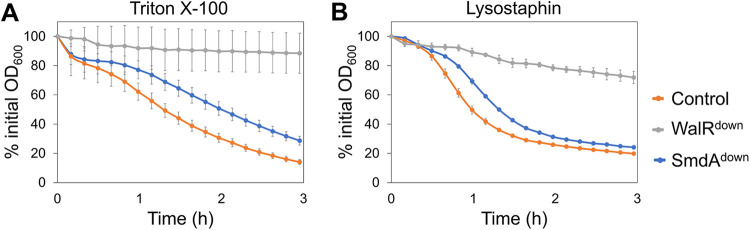
FIG 4 Autolysis of S. aureus SH1000 depletion strains (control = IM284; WalRdown = IM293; SmdAdown = IM269) measured in the presence of (A) 0.5% Triton X-100 and (B) 100 ng/mL lysostaphin. Results presented as % of initial OD600. Error bars represent standard error calculated from four technical replicates.
FIG 4 Autolysis of S. aureus SH1000 depletion strains (control = IM284; WalRdown = IM293; SmdAdown = IM269) measured in the presence of (A) 0.5% Triton X-100 and (B) 100 ng/mL lysostaphin. Results presented as % of initial OD600. Error bars represent standard error calculated from four technical replicates.CC BY · PubMed Central
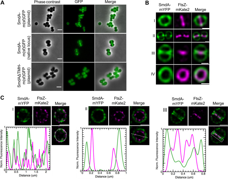
FIG 5 Subcellular localization analysis of SmdA. (A) Micrographs of cells with induced expression of SmdA-m(sf)GFP from a plasmid (top panel, IM104), native chromosomal expression of the SmdA-m(sf)GFP fusion protein (middle panel, IM308), and expression of SmdAΔTMH-m(sf)GFP from a plasmid (bottom panel, IM373). Scale bars, 2 μm. (B) SIM images of fixed S. aureus SH1000 with plasmid-expressed SmdA-mYFP and FtsZ-mKate2 (HC060). (B) Side-view of a cell showing that FtsZ localizes at septum before the arrival of SmdA (I). As cell division progresses, SmdA concentrates at sites where septum formation is initiated (II) and displays a septal localization at the two septal membranes as FtsZ constricts and septum formation proceeds (III). Top-view of a cell showing the FtsZ-ring inside the SmdA-ring (IV). All scale bars, 0.5 μm. (C) STED images of fixed S. aureus SH1000 with plasmid-expressed SmdA-mYFP and FtsZ-mKate2 (HC060), where line scans show fluorescence intensity of selected areas. (C) At an early stage of cell division, the rings of both SmdA and FtsZ had a similar diameter but do not overlap in their distribution, and a heterogeneous distribution with a patchy signal was observed (I). Top-view of a cell as cell division progresses, with the FtsZ-ring laying inside SmdA, approximately 100 to 150 nm apart from each other (II). Side-view of a cell showing how FtsZ was located innermost, and how SmdA was located at the two septal membranes (III). Scale bars, 0.5 μm.
FIG 5 Subcellular localization analysis of SmdA. (A) Micrographs of cells with induced expression of SmdA-m(sf)GFP from a plasmid (top panel, IM104), native chromosomal expression of the SmdA-m(sf)GFP fusion protein (middle panel, IM308), and expression of SmdAΔTMH-m(sf)GFP from a plasmid (bottom panel, IM373). Scale bars, 2 μm. (B) SIM images of fixed S. aureus SH1000 with plasmid-expressed SmdA-mYFP and FtsZ-mKate2 (HC060). (B) Side-view of a cell showing that FtsZ localizes at septum before the arrival of SmdA (I). As cell division progresses, SmdA concentrates at sites where septum formation is initiated (II) and displays a septal localization at the two septal membranes as FtsZ constricts and septum formation proceeds (III). Top-view of a cell showing the FtsZ-ring inside the SmdA-ring (IV). All scale bars, 0.5 μm. (C) STED images of fixed S. aureus SH1000 with plasmid-expressed SmdA-mYFP and FtsZ-mKate2 (HC060), where line scans show fluorescence intensity of selected areas. (C) At an early stage of cell division, the rings of both SmdA and FtsZ had a similar diameter but do not overlap in their distribution, and a heterogeneous distribution with a patchy signal was observed (I). Top-view of a cell as cell division progresses, with the FtsZ-ring laying inside SmdA, approximately 100 to 150 nm apart from each other (II). Side-view of a cell showing how FtsZ was located innermost, and how SmdA was located at the two septal membranes (III). Scale bars, 0.5 μm.CC BY · PubMed Central
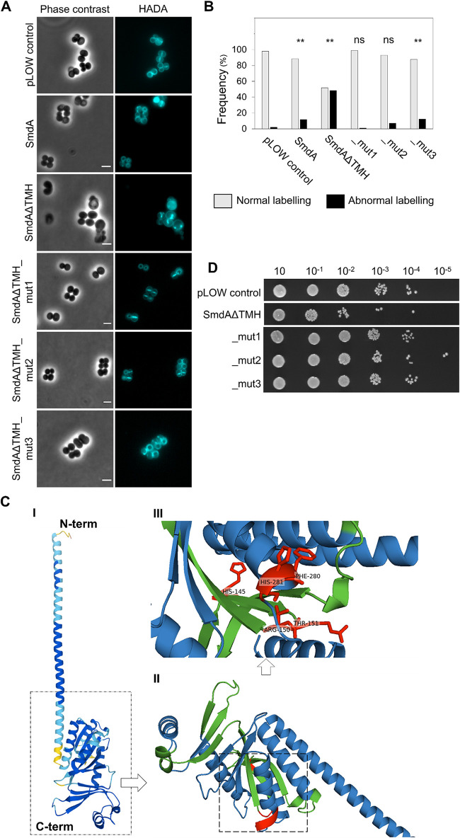
FIG 6 Membrane attachment and the NERD domain are important for SmdA function. (A) HADA stained S. aureus NCTC8325-4 control cells not expressing any SmdA-variant (pLOW control, IM307), cells overexpressing full-length SmdA (MK1866), and cells overexpressing SmdAΔTMH (MK1911), SmdAΔTMH_mut1 (IM377), _mut2 (IM378), and _mut3 (IM379). Scale bars, 2 μm. (B) The frequency of cells from (A) with normal or abnormal HADA staining is plotted. The number of cells plotted were 259 the pLOW control, 180 for SmdA, 82 for SmdAΔTMH, 101 for _mut1, 113 for _mut2, and 122 for _mut3. The asterisks indicate a significant difference from the pLOW control strain (Fisher’s exact test, P < 0.001). ns indicates that there was no significant difference. (C) Structure of SmdA, predicted by AlphaFold (52), where (I) shows the structure colored by a “per-residue confidence score” (pLDDT) with dark blue pLDDT > 90, light blue 90 > pLDDT > 70, and yellow 70 > pLDDT > 50, (II) shows the structured domain magnified with the predicted NERD colored in green and residues that have been mutated in red. A magnified inset of (II) with annotated residues (III). The positions of the mutated residues are indicated in red. (D) Growth assay on a solid medium of S. aureus NCTC8325-4 expressing SmdAΔTMH, SmdAΔTMH_mut1, _mut2, and _mut3.
FIG 6 Membrane attachment and the NERD domain are important for SmdA function. (A) HADA stained S. aureus NCTC8325-4 control cells not expressing any SmdA-variant (pLOW control, IM307), cells overexpressing full-length SmdA (MK1866), and cells overexpressing SmdAΔTMH (MK1911), SmdAΔTMH_mut1 (IM377), _mut2 (IM378), and _mut3 (IM379). Scale bars, 2 μm. (B) The frequency of cells from (A) with normal or abnormal HADA staining is plotted. The number of cells plotted were 259 the pLOW control, 180 for SmdA, 82 for SmdAΔTMH, 101 for _mut1, 113 for _mut2, and 122 for _mut3. The asterisks indicate a significant difference from the pLOW control strain (Fisher’s exact test, P < 0.001). ns indicates that there was no significant difference. (C) Structure of SmdA, predicted by AlphaFold (52), where (I) shows the structure colored by a “per-residue confidence score” (pLDDT) with dark blue pLDDT > 90, light blue 90 > pLDDT > 70, and yellow 70 > pLDDT > 50, (II) shows the structured domain magnified with the predicted NERD colored in green and residues that have been mutated in red. A magnified inset of (II) with annotated residues (III). The positions of the mutated residues are indicated in red. (D) Growth assay on a solid medium of S. aureus NCTC8325-4 expressing SmdAΔTMH, SmdAΔTMH_mut1, _mut2, and _mut3.CC BY · PubMed Central

Verification

Claims the paper makes

The claims and statistics of this paper haven’t been checked yet.

Run the check — free, no account needed. It reads the open-access text and checks the sources it cites.

A few free checks a day · papers already verified open instantly · results are public

Self-check

Percentages, sums and reported differences re-computed from the paper’s own numbers.

The self-check needs the paper’s full text.

Citations

Cites

Show all 1 references

Research works printed but not linked: 77

  1. 10.1016/j.mib.2013.07.006 · DOI
  2. 10.1038/nrmicro3088 · DOI
  3. 10.1128/MMBR.67.1.52-65.2003 · DOI
  4. 2020, Reassessment of the distinctive geometry of Staphylococcus aureus cell division, Nat Comm, 11, 1
  5. 10.1111/j.1365-2958.2011.07651.x · DOI
  6. 2021, CcrZ is a pneumococcal spatiotemporal cell cycle regulator that interacts with FtsZ and controls DNA replication by modulating the activity of DnaA, Nat Microbiol, 6, 1175, 10.1038/s41564-021-00949-1 · DOI
  7. 10.1111/j.1574-6976.2008.00104.x · DOI
  8. 10.1111/j.1574-6976.2007.00089.x · DOI
  9. 10.1128/AAC.47.3.837-846.2003 · DOI
  10. 10.1126/science.1254522 · DOI
  11. 10.1074/jbc.RA117.001068 · DOI
  12. 2019, SEDS-bPBP pairs direct lateral and septal peptidoglycan synthesis in Staphylococcus aureus, Nat Microbiol, 4, 1368, 10.1038/s41564-019-0437-2 · DOI
  13. 10.1038/nature19331 · DOI
  14. 10.1111/j.1432-1033.1981.tb05620.x · DOI
  15. 10.1073/pnas.1004304107 · DOI
  16. 10.1111/j.1365-2958.2004.04420.x · DOI
  17. 10.1128/JB.158.2.513-516.1984 · DOI
  18. 10.1128/AAC.27.5.851 · DOI
  19. 10.1073/pnas.92.1.285 · DOI
  20. 2020, New insights in the coordinated amidase and glucosaminidase activity of the major autolysin (Atl) in Staphylococcus aureus, Commun Biol, 3, 1
  21. 2019, The Sle1 cell wall amidase is essential for β-lactam resistance in community-acquired methicillin-resistant Staphylococcus aureus USA300, Antimicrob Agents Chemother, 64, e01931
  22. 10.1128/JB.00645-07 · DOI
  23. 10.1126/science.aaa1511 · DOI
  24. 2015, Cell shape dynamics during the staphylococcal cell cycle, Nat Comm, 6, 1
  25. 10.1146/annurev-micro-092412-155620 · DOI · Paper page
  26. 2020, The length of lipoteichoic acid polymers controls Staphylococcus aureus cell size and envelope integrity, J Bacteriol, 202, e00149
  27. 10.1128/mBio.01006-20 · DOI
  28. 10.1021/pr300394r · DOI
  29. 10.1371/journal.ppat.1008044 · DOI
  30. 10.1186/1471-2164-10-291 · DOI
  31. 10.1186/1471-2164-16-1 · DOI
  32. 2014, Genes contributing to Staphylococcus aureus fitness in abscess-and infection-related ecologies, mBio, 5, e01729
  33. 10.1016/j.tibs.2004.01.002 · DOI
  34. 10.1128/AEM.70.11.6887-6891.2004 · DOI
  35. 10.1099/mic.0.045146-0 · DOI
  36. 10.1007/BF01967563 · DOI · Paper page
  37. 10.1021/cb100269f · DOI
  38. 10.1128/AAC.05938-11 · DOI
  39. 10.1021/jacs.7b11704 · DOI
  40. 10.1038/ja.2007.62 · DOI
  41. 10.1002/cbic.200900557 · DOI
  42. 10.1073/pnas.1404099111 · DOI
  43. 10.1128/JB.01221-08 · DOI
  44. 10.1016/j.ijantimicag.2021.106283 · DOI
  45. 10.1093/jac/dks184 · DOI
  46. 10.1074/jbc.M114.567669 · DOI
  47. 10.1038/s41598-018-23276-8 · DOI
  48. 10.1146/annurev-biochem-062917-012921 · DOI · Paper page
  49. 10.1111/j.1574-6968.2006.00281.x · DOI
  50. 10.1371/journal.pone.0027542 · DOI
  51. 2018, Molecular coordination of Staphylococcus aureus cell division, eLife, 7, e32057, 10.7554/eLife.32057 · DOI
  52. 10.1016/j.mib.2013.06.012 · DOI
  53. 10.1126/scitranslmed.3003592 · DOI
  54. 10.1016/j.chembiol.2011.08.015 · DOI
  55. 10.1073/pnas.1209126109 · DOI
  56. 10.1186/1471-2180-11-72 · DOI
  57. 2019, Thermosensitive PBP2a requires extracellular folding factors PrsA and HtrA1 for Staphylococcus aureus MRSA β-lactam resistance, Commun Biol, 2, 417, 10.1038/s42003-019-0667-0 · DOI
  58. 2016, FtsZ-dependent elongation of a coccoid bacterium, mBio, 7, e00908
  59. 10.1111/j.1365-2958.2011.07591.x · DOI
  60. 10.1093/nar/gks382 · DOI
  61. 10.1126/scisignal.aav3810 · DOI
  62. 10.1128/IAI.00088-10 · DOI
  63. 10.1128/m Bio.00308-15 · DOI
  64. 10.1111/j.1365-2672.2006.03127.x · DOI
  65. 10.1073/pnas.1620608114 · DOI
  66. 10.1111/mmi.12517 · DOI
  67. 10.1128/AEM.00144-13 · DOI
  68. 10.1111/mmi.13999 · DOI
  69. 10.1128/AEM.70.10.6076-6085.2004 · DOI
  70. 10.1038/nmeth.2019 · DOI · Paper page
  71. 2016, MicrobeJ, a tool for high throughput bacterial cell detection and quantitative analysis, Nat Microbiol, 1, 16077, 10.1038/nmicrobiol.2016.77 · DOI
  72. 10.1128/AAC.00431-07 · DOI
  73. 10.1016/j.mimet.2012.05.008 · DOI
  74. 10.1002/pmic.201300553 · DOI
  75. 10.1073/pnas.95.10.5752 · DOI
  76. 10.1093/bioinformatics/btt607 · DOI
  77. 10.1038/msb.2011.75 · DOI

Metadata

Authors
Myrbråten, I. S., Stamsås, G. A., Chan, H., Angeles, D. M., Knutsen, T. M., Salehian, Z., Shapaval, V., Straume, D., & Kjos, M.
Affiliations
  • Norwegian University of Life Sciences (NO)
  • University of Technology Sydney (AU)
Corresponding author
Kjos, Morten
Venue
mBio
Publisher
openRxiv
Published
2021-11-23
Volume
13
Issue
2
Pages
e0340421-e0340421
ISSN
2150-7511 (ISSN-L)
OpenAlex
W4226060396
Cited by
18
References
1
Citations by year
2026: 2 · 2025: 5 · 2024: 4 · 2023: 4 · 2022: 3
FWCI
1.96
Type
journal article
Language
en
Open access
Licence
cc-by
Locations
  • mBio (publishedVersion, cc-by)
  • bioRxiv (Cold Spring Harbor Laboratory) (acceptedVersion, cc-by-nc-nd)
  • PubMed Central (publishedVersion, cc-by)
  • PubMed (publishedVersion)
  • DOAJ (DOAJ: Directory of Open Access Journals) (submittedVersion, cc-by-sa)
Funders
  • Joint Programming Initiative on Antimicrobial Resistance (296906)
  • Okinawa Institute of Science and Technology Graduate University
  • Norges Forskningsråd (295 910, 296906, 250976)
  • Norges Miljø- og Biovitenskapelige Universitet
Topics
  • Health Sciences › Medicine › Infectious Diseases › Antimicrobial Resistance in Staphylococcus
  • Life Sciences › Biochemistry, Genetics and Molecular Biology › Genetics › Bacterial Genetics and Biotechnology
  • Life Sciences › Biochemistry, Genetics and Molecular Biology › Molecular Biology › Biochemical and Structural Characterization
Keywords
Staphylococcus aureus, cell division, cell morphology, antibiotic resistance, beta-lactam antibiotics, antibiotic susceptibility, cell wall biosynthesis, antimicrobial drug targets
MeSH terms
Anti-Bacterial Agents, Humans, Staphylococcal Infections, Staphylococcus, Staphylococcus aureus, Penicillin-Binding Proteins, Methicillin-Resistant Staphylococcus aureus

About these checks

Registry, retraction, Cites and statistics results are computed, not judged, with one exception: a printed reference that no index lists is read once by a model before it counts as a citation error. Claims are extracted by a model and kept only when their quote appears verbatim in the text; each cited source is then checked, with a model’s help, for whether it says what the claim relies on. Those verdicts can be wrong — read the claim’s quote and the cited source before relying on one.

Statistics and arithmetic are recomputed, not judged.

Confidence bands are shown only for fields calibrated on our annotated test set. None is calibrated yet, so no band is shown.

What counts as a claim ›How we measure confidence ›How we check this ›

Are you an author, or do you see a problem with this page? Request a review

How to cite

Cite CoChat for the checks on this page, and the paper for its own findings.

This page
CoChat. (2026). Citation check: SmdA is a novel cell morphology determinant in Staphylococcus aureus. https://app.cochat.ai/papers/10.1101/2021.11.23.469651
The paper
Myrbråten, I. S., Stamsås, G. A., Chan, H., Angeles, D. M., Knutsen, T. M., Salehian, Z., Shapaval, V., Straume, D., & Kjos, M. (2021). SmdA is a novel cell morphology determinant in Staphylococcus aureus. mBio. https://doi.org/10.1101/2021.11.23.469651