

The DOI matches its Crossref record; 1 of the 1 references it cites were found in a registry or an index.
patcHwork: A user-friendly pH sensitivity analysis web server for protein sequences and structures
Open accessPreprint — published as 10.1093/nar/gkac252 · Paper page
Abstract
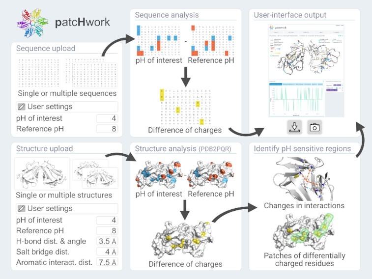
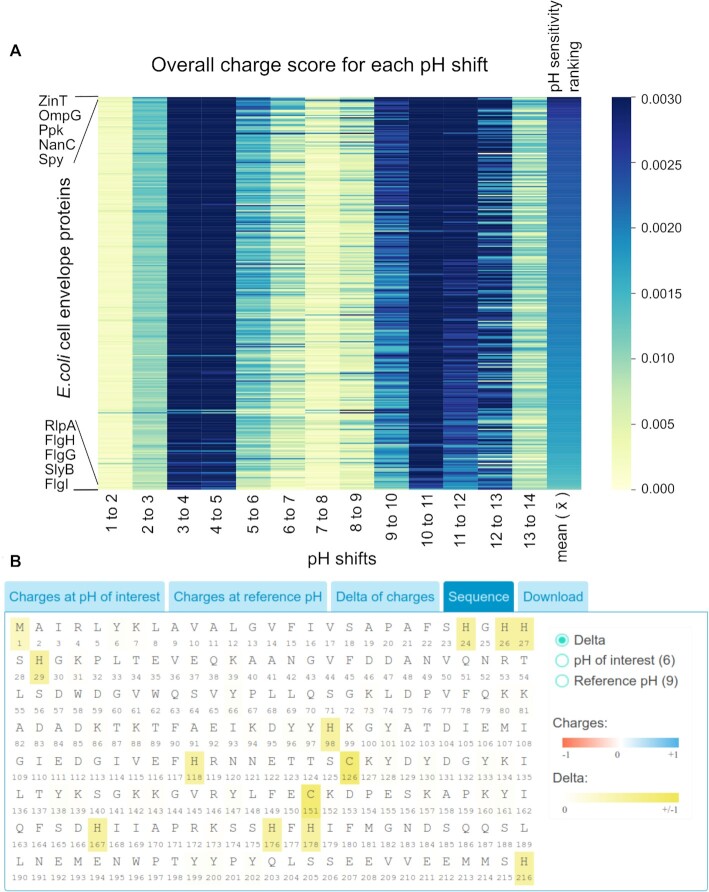
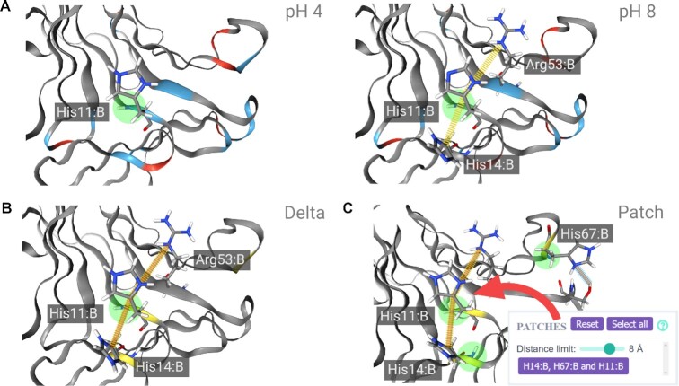
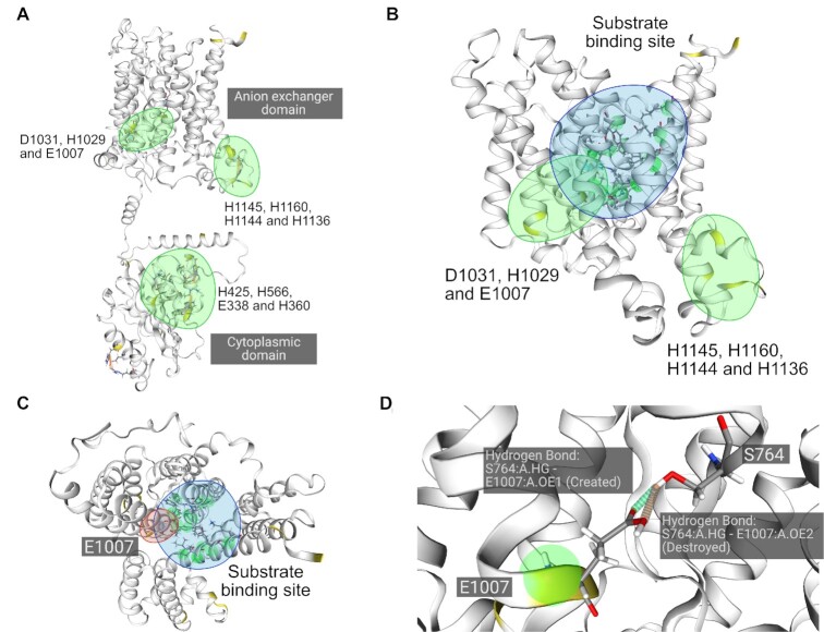
pH regulates protein function and interactions by altering the charge of individual residues causing the loss or gain of intra-molecular non-covalent bonds, which may additionally lead to structural rearrangements. While tools to analyze residue-specific charge distribution of protein sequences and structures at a given pH exist, currently no tool is available to investigate non-covalent bond changes at two different pH values. In an effort to make protein pH sensitivity analysis more accessible to researchers without computational structural biology background, we developed patcHwork, a web server that combines the identification of amino acids undergoing a charge shift with the determination of affected non-covalent bonds at two user-defined pH values. At the sequence-only level, patcHwork applies the Henderson-Hasselbalch equation to determine pH-sensitive residues. When the 3D protein structure is available, patcHwork can be employed to gain a deeper mechanistic understanding of the effect of pH on a protein of interest. This is achieved using the PDB2PQR and PROPKA tools and non-covalent bond determination algorithms. A user-friendly interface allows visualizing pH-sensitive residues as well as affected salt bridges, hydrogen bonds and aromatic (pi-pi and cation-pi) interactions. Importantly, patcHwork can be used to identify patches, a new concept we propose of pH-sensitive residues in close proximity on the protein structure, which may have a major impact on function. We demonstrate the attractiveness of patcHwork studying experimentally investigated pH-sensitive proteins. (Access: https://patchwork.biologie.uni-freiburg.de/ ) Graphical abstract:
Overview
The claims and statistics of this paper haven’t been checked yet.
Run the check — free, no account needed. It reads the open-access text and checks the sources it cites.
Integrity signals
Registry
Cites
Statistics
Figures
Verification
Claims the paper makes
The claims and statistics of this paper haven’t been checked yet.
Run the check — free, no account needed. It reads the open-access text and checks the sources it cites.
Self-check
Percentages, sums and reported differences re-computed from the paper’s own numbers.
The self-check needs the paper’s full text.
Citations
Cites
Highly accurate protein structure prediction with AlphaFold
Jumper, J., Evans, R., Pritzel, A., Green, T., Figurnov, M., Ronneberger, O., Tunyasuvunakool, K., Bates, R., Žídek, A., Potapenko, A., et al. (2021) · Nature · doi:10.1038/s41586-021-03819-2 · Cited by 43,764
Research works printed but not linked: 40
- 10.1074/jbc.R800080200 · DOI · Paper page
- 10.1021/jp962972s · DOI · Paper page
- 2021, The active repertoire of Escherichia coli peptidoglycan amidases varies with physiochemical environment, Mol. Microbiol, 116, 311, 10.1111/mmi.14711 · DOI
- 2018, Effect of pH on the hinge region of influenza viral protein: a combined constant pH and well-tempered molecular dynamics study, J. Phys. Condens. Matter, 30, 195101, 10.1088/1361-648X/aab98c · DOI
- 2021, Acidic pH irreversibly activates the signaling enzyme SARM1, FEBS J, 288, 6783, 10.1111/febs.16104 · DOI
- 10.1146/annurev-biophys-050511-102349 · DOI · Paper page
- 2008, Salt Bridges and Conformational Flexibility: Effect on Protein Stability, Biotechnol. Biotechnol. Equip, 22, 606, 10.1080/13102818.2008.10817520 · DOI
- 2019, Web-based display of protein surface and pH-dependent properties for assessing the developability of biotherapeutics, Sci. Rep, 9, 1969, 10.1038/s41598-018-36950-8 · DOI
- 2019, VOLPES: an interactive web-based tool for visualizing and comparing physicochemical properties of biological sequences, Nucleic Acids Res, 47, W632, 10.1093/nar/gkz407 · DOI
- 10.1002/jcc.21720 · DOI · Paper page
- 1986, Derivation and use of a formula to calculate the net charge of acid-base compounds. Its application to amino acids, proteins and nucleotides, Biochem. Educ, 14, 131, 10.1016/0307-4412(86)90176-7 · DOI
- 10.1073/pnas.1010750108 · DOI · Paper page
- 2011, Progress in the prediction of pKa values in proteins, Proteins Struct. Funct. Bioinforma, 79, 3260, 10.1002/prot.23189 · DOI · Paper page
- 10.1093/nar/gkm276 · DOI · Paper page
- 10.1021/ct100578z · DOI · Paper page
- 10.1186/1472-6807-11-6 · DOI · Paper page
- 10.1016/0263-7855(96)00018-5 · DOI · Paper page
- 10.1093/nar/gkw315 · DOI · Paper page
- 10.1016/j.jmb.2016.12.004 · DOI · Paper page
- 2021, ProteinTools: a toolkit to analyze protein structures, Nucleic Acids Res, 49, W559, 10.1093/nar/gkab375 · DOI
- 2015, GROMACS: High performance molecular simulations through multi-level parallelism from laptops to supercomputers, SoftwareX, 1–2, 19
- 10.1016/S0022-2836(83)80079-5 · DOI · Paper page
- 10.1093/protein/15.2.91 · DOI
- 10.1073/pnas.96.17.9459 · DOI · Paper page
- 10.1371/journal.pone.0019448 · DOI
- 2015, Structural Basis of pH Dependence of Neoculin, a Sweet Taste-Modifying Protein, PLOS ONE, 10, e0126921, 10.1371/journal.pone.0126921 · DOI
- 10.1152/ajpcell.00109.2008 · DOI
- 10.1074/jbc.270.48.28751 · DOI
- 2013, Substitution of transmembrane domain Cys residues alters pHo-sensitive anion transport by AE2/SLC4A2 anion exchanger, Pflüg. Arch. - Eur. J. Physiol, 465, 839, 10.1007/s00424-012-1196-6 · DOI
- 1992, Anion-proton cotransport through the human red blood cell band 3 protein, Role of glutamate 681. J. Biol. Chem, 267, 13964
- 10.1085/jgp.109.3.345 · DOI
- 10.1271/bbb.68.1403 · DOI
- 2008, Acid-induced sweetness of neoculin is ascribed to its pH-dependent agonistic-antagonistic interaction with human sweet taste receptor, FASEB J. Off. Publ. Fed. Am. Soc. Exp. Biol, 22, 2323
- 10.1016/j.jmb.2006.03.030 · DOI
- 2004, The SLC4 family of HCO3-transporters, Pflüg. Arch, 447, 495, 10.1007/s00424-003-1180-2 · DOI · Paper page
- 10.1085/jgp.92.2.161 · DOI
- 2022, AlphaFold Protein Structure Database: massively expanding the structural coverage of protein-sequence space with high-accuracy models, Nucleic Acids Res, 50, D439, 10.1093/nar/gkab1061 · DOI · Paper page
- 2021, Identification of multiple substrate binding sites in SLC4 transporters in the outward-facing conformation: Insights into the transport mechanism, J. Biol. Chem, 296, 100724, 10.1016/j.jbc.2021.100724 · DOI
- 2016, Band 3, the human red cell chloride/bicarbonate anion exchanger (AE1, SLC4A1), in a structural context, Biochim. Biophys. Acta BBA - Biomembr, 1858, 1507, 10.1016/j.bbamem.2016.03.030 · DOI · Paper page
- 10.1038/srep22523 · DOI
Metadata
- Authors
- Schmitz, M., Schultze, A., Vanags, R., Voigt, K., Di Ventura, B., & Öztürk, M. A.
- Affiliations
- University of Freiburg (DE)
- Corresponding author
- Öztürk, Mehmet Ali
- Venue
- Nucleic Acids Research
- Publisher
- openRxiv
- Published
- 2022-02-04
- Volume
- 50
- Issue
- W1
- Pages
- W560-W567
- ISSN
- 0305-1048 (ISSN-L)
- PMC
- PMC9252814
- OpenAlex
- W4225463176
- Cited by
- 8
- References
- 1
- Citations by year
- 2026: 2 · 2025: 4 · 2024: 2
- FWCI
- 0.49
- Type
- journal article
- Language
- en
- Open access
- gold
- doi.org
- Licence
- cc-by
- Locations
- Nucleic Acids Research (publishedVersion, cc-by)
- bioRxiv (Cold Spring Harbor Laboratory) (acceptedVersion, cc-by-nc-nd)
- PubMed Central (publishedVersion, cc-by)
- PubMed (publishedVersion)
- Funders
- European Commission (HORIZON2020)
- Bundesministerium für Bildung und Forschung (031L0079)
- H2020 European Research Council (101002044)
- Topics
- Life Sciences › Biochemistry, Genetics and Molecular Biology › Molecular Biology › Protein Structure and Dynamics
- Life Sciences › Biochemistry, Genetics and Molecular Biology › Structural Biology › Enzyme Structure and Function
- Life Sciences › Biochemistry, Genetics and Molecular Biology › Molecular Biology › Machine Learning in Bioinformatics
- Keywords
- pH sensitivity, patchwork, web server, protein charge distribution, noncovalent bonding, salt bridges, hydrogen bonding, π-π stacking, cation-π interactions, Henderson-Hasselbalch equation, protein structural rearrangement
- MeSH terms
- Amino Acid Sequence, Amino Acids, Computers, Hydrogen-Ion Concentration, Proteins, Software, Internet
About these checks
Registry, retraction, Cites and statistics results are computed, not judged, with one exception: a printed reference that no index lists is read once by a model before it counts as a citation error. Claims are extracted by a model and kept only when their quote appears verbatim in the text; each cited source is then checked, with a model’s help, for whether it says what the claim relies on. Those verdicts can be wrong — read the claim’s quote and the cited source before relying on one.
Statistics and arithmetic are recomputed, not judged.
Confidence bands are shown only for fields calibrated on our annotated test set. None is calibrated yet, so no band is shown.
What counts as a claim ›How we measure confidence ›How we check this ›
Are you an author, or do you see a problem with this page? Request a review
How to cite
Cite CoChat for the checks on this page, and the paper for its own findings.
- This page
- CoChat. (2026). Citation check: patcHwork: A user-friendly pH sensitivity analysis web server for protein sequences and structures. https://app.cochat.ai/papers/10.1101/2022.02.02.478804
- The paper
- Schmitz, M., Schultze, A., Vanags, R., Voigt, K., Di Ventura, B., & Öztürk, M. A. (2022). patcHwork: A user-friendly pH sensitivity analysis web server for protein sequences and structures. Nucleic Acids Research. https://doi.org/10.1101/2022.02.02.478804
Data sources
Bibliographic data: Semantic Scholar, OpenAlex, Crossref.
Contains information from the Semantic Scholar Academic Graph Datasets, made available under the ODC Attribution License (ODC-BY 1.0).
Semantic Scholar’s data is described in: Kinney, R., et al. (2023). The Semantic Scholar Open Data Platform. arXiv:2301.10140.
