

The DOI matches its Crossref record; 2 of the 2 references it cites were found in a registry or an index.
Chaperones facilitate heterologous expression of naturally evolved putative de novo proteins
Open accessPreprint — published as 10.1002/pro.4371 · Paper page
Abstract
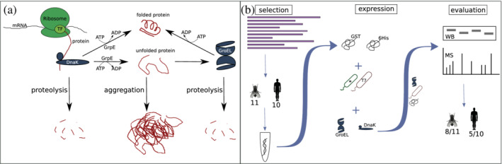
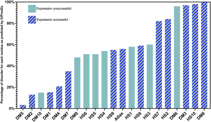
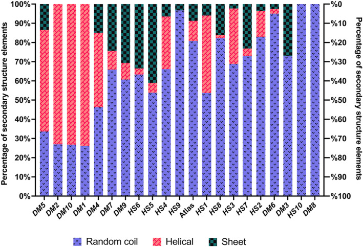
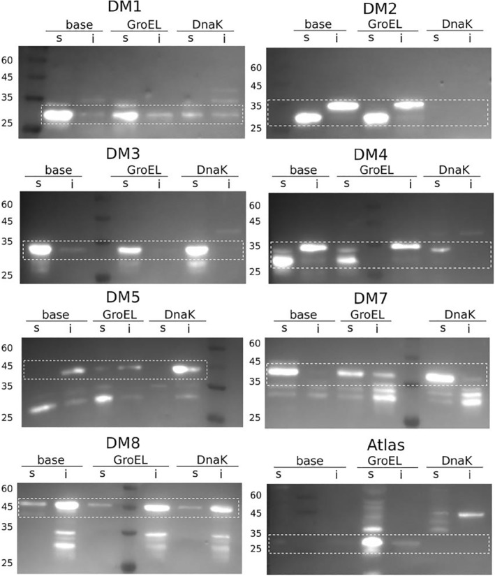
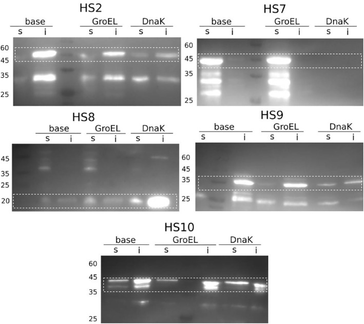
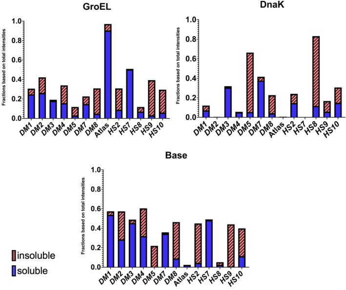
Over the past decade, evidence has accumulated that new protein coding genes can emerge de novo from previously non-coding DNA. Most studies have focused on large scale computational predictions of de novo protein coding genes across a wide range of organisms. In contrast, experimental data concerning the folding and function of de novo proteins is scarce. This might be due to difficulties in handling de novo proteins in vitro , as most are predicted to be short and disordered. Here we propose a guideline for the effective expression of eukaryotic de novo proteins in Escherichia coli . We used 11 sequences from Drosophila melanogaster and 10 from Homo sapiens , that are predicted de novo proteins from former studies, for heterologous expression. The candidate de novo proteins have varying secondary structure and disorder content. Using multiple combinations of purification tags, E. coli expression strains and chaperone systems, we were able to increase the number of solubly expressed putative de novo proteins from 30 % to 62 %. Our findings indicate that the best combination for expressing putative de novo proteins in E.coli is a GST-tag with T7 Express cells and co-expressed chaperones. We found that, overall, proteins with higher predicted disorder were easier to express.
Overview
The claims and statistics of this paper haven’t been checked yet.
Run the check — free, no account needed. It reads the open-access text and checks the sources it cites.
Integrity signals
Registry
Cites
Statistics
Figures
Verification
Claims the paper makes
The claims and statistics of this paper haven’t been checked yet.
Run the check — free, no account needed. It reads the open-access text and checks the sources it cites.
Self-check
Percentages, sums and reported differences re-computed from the paper’s own numbers.
The self-check needs the paper’s full text.
Citations
Cites
Highly accurate protein structure prediction with AlphaFold
Jumper, J., Evans, R., Pritzel, A., Green, T., Figurnov, M., Ronneberger, O., Tunyasuvunakool, K., Bates, R., Žídek, A., Potapenko, A., et al. (2021) · Nature · doi:10.1038/s41586-021-03819-2 · Cited by 43,764
AlphaFold and Implications for Intrinsically Disordered Proteins
Ruff, K. M., & Pappu, R. V. (2021) · Journal of Molecular Biology · doi:10.1016/j.jmb.2021.167208 · Cited by 635
Research works printed but not linked: 66
- 10.1038/nrg3053 · DOI · Paper page
- 10.1038/nrg.2016.78 · DOI
- 10.12688/f1000research.10079.1 · DOI
- 10.1371/journal.pgen.1008160 · DOI
- 10.1016/j.tig.2019.08.007 · DOI
- 10.1016/j.sbi.2020.11.010 · DOI
- 10.1101/2022.01.14.476368 · DOI
- 10.1002/9780470619902.ch1 · DOI
- 10.1002/tera.1420090224 · DOI
- 10.1016/j.sbi.2013.02.012 · DOI
- 10.7554/eLife.53500 · DOI
- 10.1534/genetics.105.050336 · DOI
- 10.1534/genetics.107.084491 · DOI
- 10.1186/1471-2164-14-117 · DOI
- 10.1098/rstb.2014.0332 · DOI
- 10.1016/j.tig.2015.02.007 · DOI
- 10.1038/s41559-018-0639-7 · DOI
- 10.1093/molbev/msx315 · DOI
- 10.1534/g3.119.400326 · DOI
- 10.1038/s41559-019-0822-5 · DOI · Paper page
- 10.1007/s00239-020-09939-z · DOI
- 10.1093/gbe/evaa194 · DOI
- 10.1101/2022.01.04.474757 · DOI
- 10.1093/molbev/msw284 · DOI
- 10.1038/s41467-021-21667-6 · DOI
- 10.1016/j.str.2017.09.006 · DOI
- 10.1038/nature08009 · DOI
- 10.1126/science.1169375 · DOI
- 10.1016/j.sbi.2009.08.003 · DOI
- 10.1016/j.jmb.2022.167462 · DOI
- 10.1038/nrg2808 · DOI
- 10.1038/s41598-017-15635-8 · DOI
- 10.1186/1475-2859-7-11 · DOI
- 10.1042/BST20150089 · DOI
- 10.1146/annurev-biochem-060815-014124 · DOI
- 10.1073/pnas.1510083112 · DOI
- 10.1038/nsmb.1394 · DOI
- 10.1007/BF02785589 · DOI
- 10.1002/j.1460-2075.1993.tb06097.x · DOI
- 10.1038/nchembio.455 · DOI
- 10.1146/annurev-biochem-060208-092442 · DOI
- 10.1038/nature20137 · DOI
- 10.1038/337044a0 · DOI
- 10.1371/journal.pgen.1009787 · DOI
- 10.1002/cpbi.99 · DOI
- 10.1093/nar/gky384 · DOI
- 10.1101/289033 · DOI
- 10.1038/s41598-019-48786-x · DOI
- 10.1093/molbev/msu286 · DOI
- 10.1101/2022.01.13.476251 · DOI
- 10.2174/138920306775474095 · DOI
- 10.1002/0471140864.ps0519s50 · DOI
- 10.3389/fmicb.2014.00172 · DOI · Paper page
- 10.1007/978-1-60761-913-0_14 · DOI
- 10.1073/pnas.1201380109 · DOI
- 10.1155/2014/176172 · DOI
- 10.1073/pnas.1312296111 · DOI · Paper page
- 10.1007/978-1-0716-0524-0_27 · DOI
- 10.1021/acs.biochem.0c00961 · DOI
- 10.1126/science.abj8754 · DOI · Paper page
- 10.1093/bioadv/vbab043 · DOI
- 10.1093/bioinformatics/btz670 · DOI · Paper page
- 10.1128/AEM.64.5.1694-1699.1998 · DOI
- 10.1128/AEM.66.3.884-889.2000 · DOI
- 10.1016/0378-1119(90)90336-P · DOI
- 10.1101/pdb.prot3944 · DOI
Metadata
- Authors
- Eicholt, L. A., Aubel, M., Berk, K., Bornberg-Bauer, E., & Lange, A.
- Affiliations
- University of Münster (DE)
- Corresponding author
- Lange, Andreas
- Venue
- Protein Science
- Publisher
- openRxiv
- Published
- 2022-03-02
- Volume
- 31
- Issue
- 8
- Pages
- e4371-e4371
- ISSN
- 0961-8368 (ISSN-L)
- PMC
- PMC9278007
- OpenAlex
- W4285395243
- Cited by
- 18
- References
- 2
- Citations by year
- 2026: 2 · 2025: 2 · 2024: 4 · 2023: 8 · 2022: 2
- FWCI
- 1.31
- Type
- journal article
- Language
- en
- Open access
- hybrid
- doi.org
- Licence
- cc-by
- Locations
- Protein Science (publishedVersion, cc-by)
- bioRxiv (Cold Spring Harbor Laboratory) (acceptedVersion, cc-by-nc-nd)
- PubMed Central (publishedVersion, cc-by)
- PubMed (publishedVersion)
- Funders
- Deutsche Forschungsgemeinschaft (GRK 2220, 281125614)
- Volkswagen Foundation (98183)
- Topics
- Life Sciences › Biochemistry, Genetics and Molecular Biology › Molecular Biology › Protein purification and stability
- Life Sciences › Biochemistry, Genetics and Molecular Biology › Molecular Biology › Heat shock proteins research
- Life Sciences › Biochemistry, Genetics and Molecular Biology › Molecular Biology › Protein Structure and Dynamics
- Keywords
- heterologous expression, molecular chaperones, Escherichia coli expression, protein solubility, intrinsically disordered proteins, Drosophila melanogaster, human, protein folding
- MeSH terms
- Animals, Drosophila melanogaster, Escherichia coli, Eukaryotic Cells, Protein Structure, Secondary, Molecular Chaperones
About these checks
Registry, retraction, Cites and statistics results are computed, not judged, with one exception: a printed reference that no index lists is read once by a model before it counts as a citation error. Claims are extracted by a model and kept only when their quote appears verbatim in the text; each cited source is then checked, with a model’s help, for whether it says what the claim relies on. Those verdicts can be wrong — read the claim’s quote and the cited source before relying on one.
Statistics and arithmetic are recomputed, not judged.
Confidence bands are shown only for fields calibrated on our annotated test set. None is calibrated yet, so no band is shown.
What counts as a claim ›How we measure confidence ›How we check this ›
Are you an author, or do you see a problem with this page? Request a review
