Student or educator? Sign up with your school email and get 6 months of Pro free.
PreprintCited by 1Malaria Research and ControlCardiomyopathy and Myosin StudiesErythrocyte Function and Pathophysiology

The DOI matches its Crossref record; 1 of the 1 references it cites were found in a registry or an index.

Characterization of Plasmodium falciparum myosin B in complex with the calmodulin-like domain of its light chain MLC-B

Open access
Pires, Isa, Hung, Yu-Fu, Bergmann, Ulrich, Molloy, Justin E., Kursula, Inari·2022·Journal of Biological Chemistry

Preprint — published as 10.1016/j.jbc.2022.102634 · Paper page

Abstract

Myosin B (MyoB) is a class 14 myosin expressed in all invasive stages of the malaria parasite, Plasmodium falciparum . It is not associated with the glideosome complex that drives motility and invasion of host cells. During red blood cell invasion, it remains at the apical tip of the merozoite but is no longer observed once invasion is completed. MyoB is not essential for parasite survival but, when it is knocked out, merozoites are delayed in the initial stages of red blood cell invasion, giving rise to a growth defect that correlates with reduced invasion success. Here, we have expressed and purified functional MyoB with the help of parasite-specific chaperones Hsp90 and Unc45, characterized its binding to actin and its known light chain MLC-B using biochemical and biophysical methods, and determined its low-resolution structure in solution using small-angle X-ray scattering. In addition to MLC-B, four other putative regulatory light chains were found to bind to the MyoB IQ2 motif in vitro . The purified recombinant MyoB adopted the overall shape of a myosin, exhibited actin-activated ATPase activity, and moved actin filaments in vitro . The ADP release rate was faster than the ATP turnover number, and thus, does not appear to be rate-limiting. This, together with the observed high affinity to actin and the specific localization of MyoB, may point towards a role in tethering and/or force sensing during early stages of invasion.

Overview

The claims and statistics of this paper haven’t been checked yet.

Run the check — free, no account needed. It reads the open-access text and checks the sources it cites.

A few free checks a day · papers already verified open instantly · results are public

Integrity signals

Registry

Crossref · 10.1101/2022.05.28.493848

Cites

1 of 1 references verified0 citation errors · 0 not checkable

Statistics

Figures

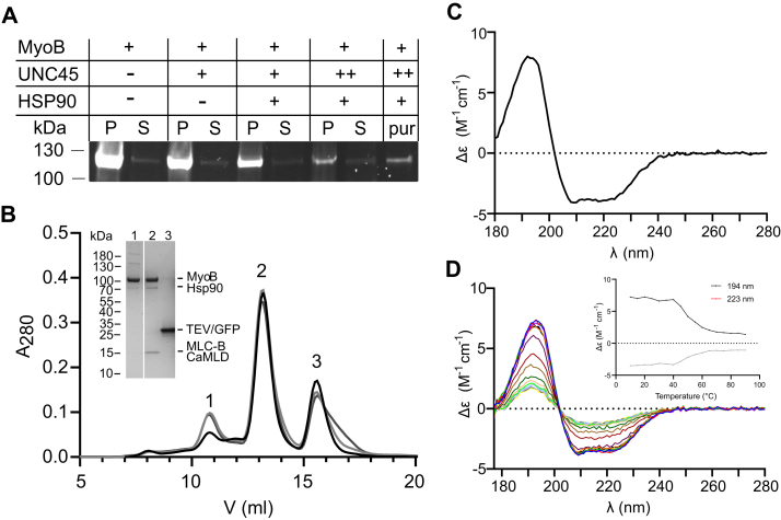
Figure 1 MyoB solubility and the MyoB–MLCB CaMLD complex purification.A, SDS-PAGE imaged under UV light for detection of eGFP-MyoB fusion protein. S: soluble fraction; P: insoluble fraction; pur: purified protein. Equal culture and buffer volumes were used for all samples, with the exception of pur that acts as a control. B, elution of MyoB–MLC-B CaMLD from a Superdex S200 10/300 column in 20 mM Tris–HCl (pH 8.5), 150 mM KCl, 0.5 mM TCEP, 5 mM MgCl2, 0.1 mM EGTA at a flow rate of 0.35 ml min−1. The different black-gray curves represent three independent purifications. The inset shows samples from the SEC peaks on SDS-PAGE. Peak 1 contains partially unfolded MyoB, peak 2 the folded MyoB–MLC-B CaMLD complex, and peak 3 Strep-tag-eGFP/TEV. C, SR-CD spectrum of the MyoB–MLC-B CaMLD complex. D, SR-CD spectra of the MyoB–MLC-B CaMLD complex at temperatures between 10 °C and 90 °C (5 °C intervals). The inset shows the change in Δε at 223 and 194 nm upon heating. CaMLD, calmodulin like-domain; MLC, myosin light chain; Myo, myosin; SEC, size-exclusion chromatography; SR-CD, synchrotron radiation-CD; TCEP, tris(2-carboxyethyl)phosphine.
Figure 1 MyoB solubility and the MyoB–MLCB CaMLD complex purification.A, SDS-PAGE imaged under UV light for detection of eGFP-MyoB fusion protein. S: soluble fraction; P: insoluble fraction; pur: purified protein. Equal culture and buffer volumes were used for all samples, with the exception of pur that acts as a control. B, elution of MyoB–MLC-B CaMLD from a Superdex S200 10/300 column in 20 mM Tris–HCl (pH 8.5), 150 mM KCl, 0.5 mM TCEP, 5 mM MgCl2, 0.1 mM EGTA at a flow rate of 0.35 ml min−1. The different black-gray curves represent three independent purifications. The inset shows samples from the SEC peaks on SDS-PAGE. Peak 1 contains partially unfolded MyoB, peak 2 the folded MyoB–MLC-B CaMLD complex, and peak 3 Strep-tag-eGFP/TEV. C, SR-CD spectrum of the MyoB–MLC-B CaMLD complex. D, SR-CD spectra of the MyoB–MLC-B CaMLD complex at temperatures between 10 °C and 90 °C (5 °C intervals). The inset shows the change in Δε at 223 and 194 nm upon heating. CaMLD, calmodulin like-domain; MLC, myosin light chain; Myo, myosin; SEC, size-exclusion chromatography; SR-CD, synchrotron radiation-CD; TCEP, tris(2-carboxyethyl)phosphine.CC BY-NC-ND · PubMed Central
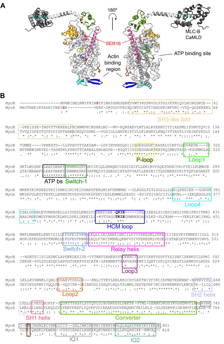
Figure 2 MyoB structure.A, predicted structure of MyoB generated using Alphafold (56) with functional regions of the motor domain and the lever arm highlighted. The Alphafold (56) predicted structure of MLC-B CaMLD was placed on MyoB IQ2 using superposition of the MTIP–MyoA complex (PDB code 6YCY (23)). B, sequence alignment of MyoA and MyoB using T-COFFEE (57) with functional regions of the motor and lever highlighted corresponding to A (2, 27, 53). The residues that influence the force and the speed at which the MyoA moves along actin are shown in red (Ser19) and brown (Lys769) (27). The dotted line box indicates the MyoB neck sequence (residues 742–801) used in this study. The residues in bold inside the HCM loop box (blue) indicate the TEDS site. IQ1 for ELC binding and IQ2 for regulatory light chain binding are shown in gray and pale green, respectively. CaMLD, calmodulin like-domain; MLC, myosin light chain; Myo, myosin; PDB, Protein Data Bank.
Figure 2 MyoB structure.A, predicted structure of MyoB generated using Alphafold (56) with functional regions of the motor domain and the lever arm highlighted. The Alphafold (56) predicted structure of MLC-B CaMLD was placed on MyoB IQ2 using superposition of the MTIP–MyoA complex (PDB code 6YCY (23)). B, sequence alignment of MyoA and MyoB using T-COFFEE (57) with functional regions of the motor and lever highlighted corresponding to A (2, 27, 53). The residues that influence the force and the speed at which the MyoA moves along actin are shown in red (Ser19) and brown (Lys769) (27). The dotted line box indicates the MyoB neck sequence (residues 742–801) used in this study. The residues in bold inside the HCM loop box (blue) indicate the TEDS site. IQ1 for ELC binding and IQ2 for regulatory light chain binding are shown in gray and pale green, respectively. CaMLD, calmodulin like-domain; MLC, myosin light chain; Myo, myosin; PDB, Protein Data Bank.CC BY-NC-ND · PubMed Central
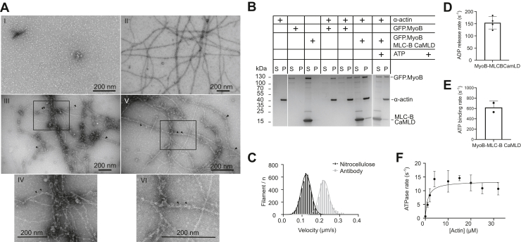
Figure 3 MyoB binding to actin.A, I - Negative-stained electron micrograph of MyoB–MLC-B CaMLD at 100 nM. II- Negative-stained electron micrograph of JAS-stabilized P. falciparum actin I at 500 nM. III and V - Negative-stained electron micrographs of MyoB–MLC-B CaMLD binding to filamentous P. falciparum actin I, at an actin:myosin ratio of 5:1. The scale bars represent 200 nm. Arrowheads indicate possible MyoB–MLC-B CaMLD particles. IV and VI – insets with magnification of represented square from II and V, respectively. B, cosedimentation of MyoB with filamentous α-actin, in presence or absence of ATP. C, motility assay of the MyoB–MLC-B CaMLD complex using nitrocellulose and antibody capture. Nitrocellulose captured data are colored black and the antibody captured data are colored gray. The antibody-capture method exhibited faster motility. D, the ADP release rate from the acto-[MyoB-MLC-B CaMLD].ADP complex was 154 ± 25 s−1, at 24 °C (four independent experiments). E, rate of ATP-induced dissociation of the acto-[MyoB-MLC-B CaMLD] rigor complex was 615 s−1, at 24 °C (two independent experiments). F, actin-activated steady-state ATPase activity of the MyoB–MLC-B CaMLD complex showed 20-fold lower Kapp but similar kcat values to the MyoA–MTIP complex (four independent experiments). All assays were performed using filamentous α-actin. CaMLD, calmodulin like-domain; MLC, myosin light chain; Myo, myosin.
Figure 3 MyoB binding to actin.A, I - Negative-stained electron micrograph of MyoB–MLC-B CaMLD at 100 nM. II- Negative-stained electron micrograph of JAS-stabilized P. falciparum actin I at 500 nM. III and V - Negative-stained electron micrographs of MyoB–MLC-B CaMLD binding to filamentous P. falciparum actin I, at an actin:myosin ratio of 5:1. The scale bars represent 200 nm. Arrowheads indicate possible MyoB–MLC-B CaMLD particles. IV and VI – insets with magnification of represented square from II and V, respectively. B, cosedimentation of MyoB with filamentous α-actin, in presence or absence of ATP. C, motility assay of the MyoB–MLC-B CaMLD complex using nitrocellulose and antibody capture. Nitrocellulose captured data are colored black and the antibody captured data are colored gray. The antibody-capture method exhibited faster motility. D, the ADP release rate from the acto-[MyoB-MLC-B CaMLD].ADP complex was 154 ± 25 s−1, at 24 °C (four independent experiments). E, rate of ATP-induced dissociation of the acto-[MyoB-MLC-B CaMLD] rigor complex was 615 s−1, at 24 °C (two independent experiments). F, actin-activated steady-state ATPase activity of the MyoB–MLC-B CaMLD complex showed 20-fold lower Kapp but similar kcat values to the MyoA–MTIP complex (four independent experiments). All assays were performed using filamentous α-actin. CaMLD, calmodulin like-domain; MLC, myosin light chain; Myo, myosin.CC BY-NC-ND · PubMed Central
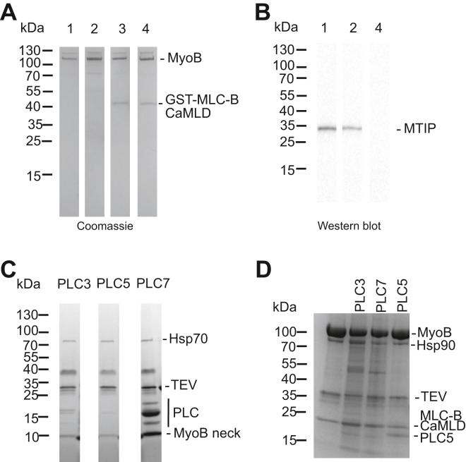
Figure 4 Screening of MyoB IQ1-binding calmodulin.A, Coomassie brilliant blue stained SDS-PAGE gel showing the binding of MTIP, ELC, and MLC-B CaMLD to MyoB by a pull-down assay. 1: MyoB-MTIP; 2: MyoB-MTIP-ELC; 3: MyoB-GST-MLC-B CaMLD; 4: MyoB-MLC-B CaMLD-ELC. B, Western blot of the same samples as panel (A). Anti-His antibody was used to verify the presence or absence of MTIP and ELC. C, binding of PLCs to MyoB neck domain. PLC3, PLC5, and PLC7 were identified as light chains interacting with MyoB. D, pull down of PLC3, PLC7, and PLC5 using the MyoB–MLC-B CaMLD complex. The bands not labeled in the figure are contaminants from insect cell expression system. CaMLD, calmodulin like-domain; ELC, essential light chain; MLC, myosin light chain; Myo, myosin.
Figure 4 Screening of MyoB IQ1-binding calmodulin.A, Coomassie brilliant blue stained SDS-PAGE gel showing the binding of MTIP, ELC, and MLC-B CaMLD to MyoB by a pull-down assay. 1: MyoB-MTIP; 2: MyoB-MTIP-ELC; 3: MyoB-GST-MLC-B CaMLD; 4: MyoB-MLC-B CaMLD-ELC. B, Western blot of the same samples as panel (A). Anti-His antibody was used to verify the presence or absence of MTIP and ELC. C, binding of PLCs to MyoB neck domain. PLC3, PLC5, and PLC7 were identified as light chains interacting with MyoB. D, pull down of PLC3, PLC7, and PLC5 using the MyoB–MLC-B CaMLD complex. The bands not labeled in the figure are contaminants from insect cell expression system. CaMLD, calmodulin like-domain; ELC, essential light chain; MLC, myosin light chain; Myo, myosin.CC BY-NC-ND · PubMed Central

Verification

Claims the paper makes

The claims and statistics of this paper haven’t been checked yet.

Run the check — free, no account needed. It reads the open-access text and checks the sources it cites.

A few free checks a day · papers already verified open instantly · results are public

Self-check

Percentages, sums and reported differences re-computed from the paper’s own numbers.

The self-check needs the paper’s full text.

Citations

Cites

Show all 1 references

Research works printed but not linked: 54

  1. 10.1016/j.semcdb.2011.09.022 · DOI
  2. 10.1016/j.jsb.2015.02.008 · DOI
  3. Frénal, K. , Dubremetz, J. F. , Lebrun, M. , and Soldati-Favre, D. (2017) Gliding motility powers invasion and egress in Apicomplexa. Nat. Rev. Microbiol. 10.1038/nrmicro.2017.86 · DOI
  4. 10.1073/pnas.0506307103 · DOI · Paper page
  5. 10.1146/annurev-cellbio-100809-151502 · DOI
  6. 10.1078/0932-4739-00015 · DOI
  7. 10.1074/jbc.M115.637694 · DOI
  8. 10.1074/jbc.M117.813972 · DOI
  9. 10.1371/journal.ppat.1008437 · DOI
  10. 10.1074/jbc.M509807200 · DOI · Paper page
  11. 10.1242/jcs.00194 · DOI
  12. 10.1016/j.jmb.2007.06.016 · DOI
  13. 10.1074/jbc.M117.802769 · DOI
  14. 10.1111/cmi.13082 · DOI
  15. 10.1007/s00436-017-5417-y · DOI
  16. 10.1007/s00436-018-6039-8 · DOI
  17. 10.1242/jcs.094300 · DOI · Paper page
  18. 10.1073/pnas.1500851112 · DOI · Paper page
  19. 10.1038/sj.emboj.7600458 · DOI · Paper page
  20. 10.1016/S0006-3495(99)77443-6 · DOI · Paper page
  21. 10.1016/S0006-3495(01)76260-1 · DOI · Paper page
  22. Moussaoui, D. , Robblee, J. P. , Auguin, D. , Krementsova, E. B. , Haase, S. , Blake, T. C. A. , Baum, J. , Robert-Paganin, J. , Trybus, K. M. , and Houdusse, A. (2020) Full-length plasmodium falciparum myosin a and essential light chain pfelc structures provide new anti- malarial targets. Elife. 10.7554/eLife.60581 · DOI
  23. 10.1038/s41467-019-11120-0 · DOI
  24. 10.1038/s42003-020-01283-8 · DOI
  25. 10.1101/2020.07.02.183871 · DOI
  26. 10.1016/S0092-8674(00)81332-X · DOI
  27. 10.1016/j.jmb.2005.11.027 · DOI
  28. 10.1074/jbc.RA119.009825 · DOI
  29. 10.1074/jbc.M114.572453 · DOI
  30. 10.1242/jcs.02774 · DOI
  31. 10.1074/jbc.M117.801456 · DOI
  32. 10.1073/pnas.96.24.13726 · DOI · Paper page
  33. 10.1038/s41579-019-0306-2 · DOI · Paper page
  34. 10.1074/jbc.M200713200 · DOI
  35. 10.1186/gb-2007-8-9-r183 · DOI
  36. 10.1073/pnas.1409459111 · DOI
  37. 10.1126/science.1066648 · DOI
  38. 10.1016/j.tcb.2008.03.007 · DOI
  39. 10.1074/jbc.M800757200 · DOI
  40. 10.1016/j.cell.2012.12.025 · DOI
  41. 10.1038/s41467-021-22093-4 · DOI
  42. 10.1080/19490992.2015.1054092 · DOI
  43. 10.1016/j.chom.2012.06.005 · DOI
  44. 10.1101/gr.129692.111 · DOI
  45. 10.1038/srep40655 · DOI
  46. 10.1107/S0021889810008289 · DOI
  47. 10.1107/S1600576717007786 · DOI · Paper page
  48. 10.1002/pro.3943 · DOI · Paper page
  49. 10.1016/S0076-6879(08)04206-7 · DOI
  50. 10.1038/s41598-017-11330-w · DOI
  51. 10.1093/nar/gkr245 · DOI · Paper page
  52. 10.1002/ar.22973 · DOI
  53. 10.1021/bi992420a · DOI
  54. 10.7554/eLife.01603 · DOI

Metadata

Authors
Pires, I., Hung, Y.-F., Bergmann, U., Molloy, J. E., & Kursula, I.
Affiliations
  • University of Oulu (FI)
  • The Francis Crick Institute (GB)
  • University of Bergen (NO)
Corresponding author
  • Pires, Isa
  • Kursula, Inari
Venue
Journal of Biological Chemistry
Publisher
openRxiv
Published
2022-05-28
Volume
298
Issue
12
Pages
102634-102634
ISSN
0021-9258 (ISSN-L)
OpenAlex
W4306958619
Cited by
1
References
1
Citations by year
2024: 1
FWCI
0.22
Type
journal article
Language
en
Open access
Licence
cc-by
Locations
  • Journal of Biological Chemistry (publishedVersion, cc-by)
  • bioRxiv (Cold Spring Harbor Laboratory) (acceptedVersion, cc-by-nc-nd)
  • PubMed Central (publishedVersion, cc-by-nc-nd)
  • PubMed (publishedVersion)
  • Desy publication database (The Deutsches Elektronen-Synchrotron) (submittedVersion, other-oa)
  • NORA - Norwegian Open Research Archives (submittedVersion, cc-by)
  • Figshare (submittedVersion, cc-by-nc-nd)
  • The Francis Crick Institute
Funders
  • Wellcome Trust (FC001119)
  • Francis Crick Institute (10119, FC001119)
  • Cancer Research UK (FC001119)
  • Academy of Finland
  • Emil Aaltosen Säätiö
  • Norges Forskningsråd
  • Sigrid Juséliuksen Säätiö
  • Medical Research Council (FC001119)
  • Deutsches Elektronen-Synchrotron
  • Biocenter, University of Oulu
Topics
  • Health Sciences › Medicine › Public Health, Environmental and Occupational Health › Malaria Research and Control
  • Health Sciences › Medicine › Cardiology and Cardiovascular Medicine › Cardiomyopathy and Myosin Studies
  • Health Sciences › Medicine › Physiology (medical) › Erythrocyte Function and Pathophysiology
Keywords
Plasmodium falciparum, myosin B, calmodulin-like domain, ATPase activity, actin binding, merozoite invasion, erythrocyte invasion, HSP90, molecular chaperones, small-angle X-ray scattering, actin filament motility, ADP release, IQ motif, parasite motility
MeSH terms
Actins, Calmodulin, Myosins, Plasmodium falciparum, Protozoan Proteins, Nonmuscle Myosin Type IIB

About these checks

Registry, retraction, Cites and statistics results are computed, not judged, with one exception: a printed reference that no index lists is read once by a model before it counts as a citation error. Claims are extracted by a model and kept only when their quote appears verbatim in the text; each cited source is then checked, with a model’s help, for whether it says what the claim relies on. Those verdicts can be wrong — read the claim’s quote and the cited source before relying on one.

Statistics and arithmetic are recomputed, not judged.

Confidence bands are shown only for fields calibrated on our annotated test set. None is calibrated yet, so no band is shown.

What counts as a claim ›How we measure confidence ›How we check this ›

Are you an author, or do you see a problem with this page? Request a review

How to cite

Cite CoChat for the checks on this page, and the paper for its own findings.

This page
CoChat. (2026). Citation check: Characterization of Plasmodium falciparum myosin B in complex with the calmodulin-like domain of its light chain MLC-B. https://app.cochat.ai/papers/10.1101/2022.05.28.493848
The paper
Pires, I., Hung, Y.-F., Bergmann, U., Molloy, J. E., & Kursula, I. (2022). Characterization of Plasmodium falciparum myosin B in complex with the calmodulin-like domain of its light chain MLC-B. Journal of Biological Chemistry. https://doi.org/10.1101/2022.05.28.493848

Data sources

Bibliographic data: Semantic Scholar, OpenAlex, Crossref.

Contains information from the Semantic Scholar Academic Graph Datasets, made available under the ODC Attribution License (ODC-BY 1.0).

Semantic Scholar’s data is described in: Kinney, R., et al. (2023). The Semantic Scholar Open Data Platform. arXiv:2301.10140.