

The DOI matches its Crossref record; 1 of the 1 references it cites were found in a registry or an index.
Assisting and Accelerating NMR Assignment with Restrained Structure Prediction
Open accessPreprint — published as 10.1038/s42003-025-08466-1 · Paper page
Abstract
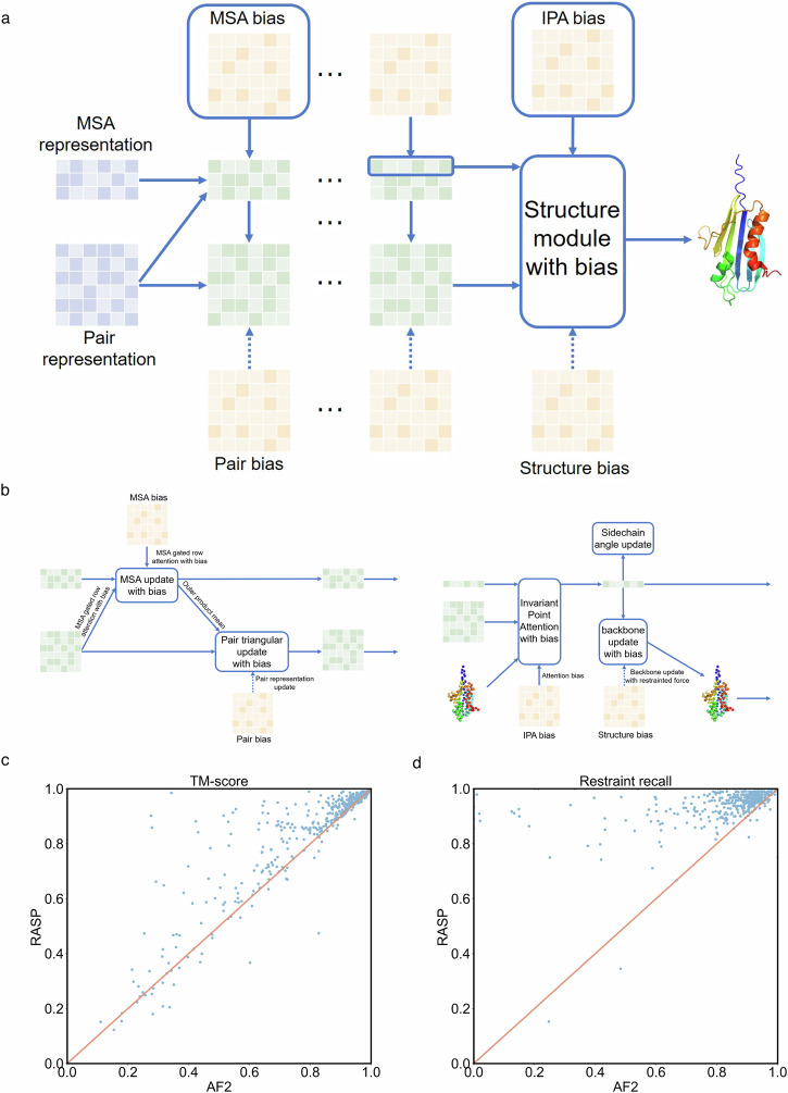
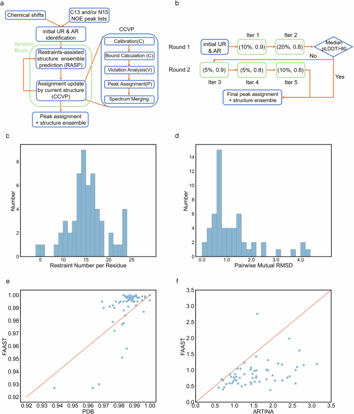
NMR experiments can detect in situ structures and dynamic interactions, but the NMR assignment process requires expertise and is time-consuming, thereby limiting its applicability. Deep learning algorithms have been employed to aid in experimental data analysis. In this work, we developed a RASP model which can enhance structure prediction with restraints. Based on the Evoformer and structure module architecture of AlphaFold, this model can predict structure based on sequence and a flexible number of input restraints. Moreover, it can evaluate the consistency between the predicted structure and the imposed restraints. Based on this model, we constructed an iterative NMR NOESY peak assignment pipeline named FAAST, to accelerate assignment process of NOESY restraints and obtaining high quality structure ensemble. The RASP model and FAAST pipeline not only allow for the leveraging of experimental restraints to improve model prediction, but can also facilitate and expedite experimental data analysis with their integrated capabilities.
Overview
The claims and statistics of this paper haven’t been checked yet.
Run the check — free, no account needed. It reads the open-access text and checks the sources it cites.
Integrity signals
Registry
Cites
Statistics
Figures
Verification
Claims the paper makes
The claims and statistics of this paper haven’t been checked yet.
Run the check — free, no account needed. It reads the open-access text and checks the sources it cites.
Self-check
Percentages, sums and reported differences re-computed from the paper’s own numbers.
The self-check needs the paper’s full text.
Citations
Cites
Highly accurate protein structure prediction with AlphaFold
Jumper, J., Evans, R., Pritzel, A., Green, T., Figurnov, M., Ronneberger, O., Tunyasuvunakool, K., Bates, R., Žídek, A., Potapenko, A., et al. (2021) · Nature · doi:10.1038/s41586-021-03819-2 · Cited by 43,764
Research works printed but not linked: 29
- 2011, NMR studies of protein structure and dynamics[J], Journal of magnetic resonance, 213, 477, 10.1016/j.jmr.2011.09.009 · DOI
- 1990, Protein structure determination in solution by NMR spectroscopy[J], Journal of Biological Chemistry, 265, 22059, 10.1016/S0021-9258(18)45665-7 · DOI
- 2022, Rapid protein assignments and structures from raw NMR spectra with the deep learning technique ARTINA[J], Nature Communications, 13, 6151, 10.1038/s41467-022-33879-5 · DOI
- 10.1007/s10858-015-9924-9 · DOI
- 10.1006/jmbi.1997.1044 · DOI
- 10.1093/bioinformatics/btl589 · DOI
- 10.1016/S0022-2836(02)00241-3 · DOI
- 2019, Protein structure prediction using sparse NOE and RDC restraints with Rosetta in CASP13[J], Proteins, 87, 1341, 10.1002/prot.25769 · DOI
- 10.1016/j.str.2019.08.012 · DOI
- 2016, Structure prediction using sparse simulated NOE restraints with Rosetta in CASP11, Proteins, 84, 181
- Evans R , O’Neill M , Pritzel A , et al. Protein complex prediction with AlphaFold-Multimer[J]. BioRxiv, 2021, biorxiv: 2021.10.04.463034. · DOI · Paper page
- 2022, Improved AlphaFold modeling with implicit experimental information[J], Nature Methods, 19, 1376, 10.1038/s41592-022-01645-6 · DOI · Paper page
- Terwilliger T C , Afonine P V , Liebschner D , et al. Accelerating crystal structure determination with iterative AlphaFold prediction[J]. Acta Crystallographica Section D: Structural Biology, 2023, 79(3). · DOI
- Stahl K , Graziadei A , Dau T , et al. Protein structure prediction with in-cell photo-crosslinking mass spectrometry and deep learning[J]. Nature Biotechnology, 2023: 1–10. · DOI
- Liu S , Zhang J , Chu H , et al. PSP: million-level protein sequence dataset for protein structure prediction[J]. arXiv preprint arXiv:2206.12240, 2022. · arXiv
- 2021, Continuous Automated Model EvaluatiOn (CAMEO)—Perspectives on the future of fully automated evaluation of structure prediction methods[J], Proteins: Structure, Function, and Bioinformatics, 89, 1977, 10.1002/prot.26213 · DOI
- 2019, Introducing “best single template” models as reference baseline for the Continuous Automated Model Evaluation (CAMEO)[J], Proteins: Structure, Function, and Bioinformatics, 87, 1378, 10.1002/prot.25815 · DOI
- 2018, Continuous Automated Model EvaluatiOn (CAMEO) complementing the critical assessment of structure prediction in CASP12[J], Proteins: Structure, Function, and Bioinformatics, 86, 387, 10.1002/prot.25431 · DOI · Paper page
- 10.1093/database/bat031 · DOI
- 10.1002/prot.20264 · DOI · Paper page
- 10.1093/bioinformatics/btq066 · DOI · Paper page
- 2022, AlphaFold Models of Small Proteins Rival the Accuracy of Solution NMR Structures[J], Frontiers in Molecular Biosciences, 9, 877000, 10.3389/fmolb.2022.877000 · DOI · Paper page
- Li E H , Spaman L , Tejero R , et al. Blind Assessment of Monomeric AlphaFold2 Protein Structure Models with Experimental NMR Data[J]. BioRxiv preprint, 2023, biorxiv: 2023.01.22.525096. · DOI
- 2020, A method for validating the accuracy of NMR protein structures[J], Nature Communications, 11, 6321, 10.1038/s41467-020-20177-1 · DOI
- 10.1007/s10858-015-9955-2 · DOI
- 2017, Openmm 7: Rapid development of high performance algorithms for molecular dynamics[J], PLOS Computational Biology, 13, 1
- 10.1093/nar/28.1.235 · DOI · Paper page
- 2023, Biological Magnetic Resonance Data Bank[J], Nucleic Acids Research, 51, D368, 10.1093/nar/gkac1050 · DOI
- 10.1093/nar/gki524 · DOI · Paper page
Metadata
- Authors
- Liu, S., Chu, H., Xie, Y., Wu, F., Ni, N., Wang, C., Mu, F., Wei, J., Zhang, J., Chen, M., Li, J., Yu, F., Fu, H., Wang, S., Tian, C., Wang, Z., & Gao, Y. Q.
- Affiliations
- Changping Laboratory (CN)
- Huawei Technologies (China) (CN)
- Chinese Academy of Sciences (CN)
- Peking University (CN)
- East China University of Science and Technology (CN)
- University of Science and Technology of China (CN)
- Corresponding author
- Liu, Sirui
- Wang, Zidong
- Gao, Yi Qin
- ORCID
- Venue
- Communications Biology
- Publisher
- openRxiv
- Published
- 2023-04-17
- Volume
- 8
- Issue
- 1
- Pages
- 1067-1067
- ISSN
- 2399-3642 (ISSN-L)
- PMC
- PMC12274491
- OpenAlex
- W4412521537
- Cited by
- 10
- References
- 1
- Citations by year
- 2026: 5 · 2025: 4 · 2024: 1
- FWCI
- 2.69
- Type
- journal article
- Language
- en
- Open access
- gold
- doi.org
- Licence
- cc-by-nc-nd
- Locations
- Communications Biology (publishedVersion, cc-by-nc-nd)
- bioRxiv (Cold Spring Harbor Laboratory) (acceptedVersion, cc-by-nc-nd)
- PubMed Central (publishedVersion, cc-by-nc-nd)
- PubMed (publishedVersion)
- DOAJ (DOAJ: Directory of Open Access Journals) (submittedVersion, cc-by-sa)
- Funders
- National Natural Science Foundation of China (92053202)
- National Science and Technology Major Project
- Topics
- Life Sciences › Biochemistry, Genetics and Molecular Biology › Molecular Biology › Protein Structure and Dynamics
- Physical Sciences › Chemistry › Spectroscopy › Advanced NMR Techniques and Applications
- Life Sciences › Biochemistry, Genetics and Molecular Biology › Structural Biology › Enzyme Structure and Function
- Keywords
- NMR resonance assignment, NOESY, protein dynamics, multidomain proteins, structure ensemble, deep learning
- MeSH terms
- Models, Molecular, Magnetic Resonance Spectroscopy, Protein Conformation, Proteins, Protein Folding, Nuclear Magnetic Resonance, Biomolecular
About these checks
Registry, retraction, Cites and statistics results are computed, not judged, with one exception: a printed reference that no index lists is read once by a model before it counts as a citation error. Claims are extracted by a model and kept only when their quote appears verbatim in the text; each cited source is then checked, with a model’s help, for whether it says what the claim relies on. Those verdicts can be wrong — read the claim’s quote and the cited source before relying on one.
Statistics and arithmetic are recomputed, not judged.
Confidence bands are shown only for fields calibrated on our annotated test set. None is calibrated yet, so no band is shown.
What counts as a claim ›How we measure confidence ›How we check this ›
Are you an author, or do you see a problem with this page? Request a review
How to cite
Cite CoChat for the checks on this page, and the paper for its own findings.
- This page
- CoChat. (2026). Citation check: Assisting and Accelerating NMR Assignment with Restrained Structure Prediction. https://app.cochat.ai/papers/10.1101/2023.04.14.536890
- The paper
- Liu, S., Chu, H., Xie, Y., Wu, F., Ni, N., Wang, C., Mu, F., Wei, J., Zhang, J., Chen, M., Li, J., Yu, F., Fu, H., Wang, S., Tian, C., Wang, Z., & Gao, Y. Q. (2023). Assisting and Accelerating NMR Assignment with Restrained Structure Prediction. Communications Biology. https://doi.org/10.1101/2023.04.14.536890
