PreprintCited by 5Force Microscopy Techniques and ApplicationsAdvanced biosensing and bioanalysis techniquesAdvanced Fluorescence Microscopy Techniques

The DOI matches its Crossref record; 1 of the 1 references it cites were found in a registry or an index.

Mapping Single-molecule Protein Complexes in 3D with DNA Nanoswitch Calipers

Open access
Shrestha, Prakash, Yang, Darren, Shih, William M., Wong, Wesley P.·2023·Journal of the American Chemical Society

Preprint — published as 10.1021/jacs.3c10262

Abstract

The ability to accurately map the 3D geometry of single-molecule complexes in trace samples would lead to new insights into molecular mechanics and provide an approach for single-molecule structural proteomics. To enable this, we have developed a high-resolution force-spectroscopy method capable of measuring multiple distances between labeled sites in natively folded protein complexes. Our approach combines reconfigurable nanoscale devices we call DNA Nanoswitch Calipers, which we have previously introduced, with a force-based barcoding system to distinguish each measurement location. We demonstrate our approach by reconstructing the tetrahedral geometry of biotin-binding sites in natively folded streptavidin, with 1.5-2.5 Å agreement to previously reported structures.

Overview

The claims and statistics of this paper haven’t been checked yet.

Run the check — free, no account needed. It reads the open-access text and checks the sources it cites.

A few free checks a day · papers already verified open instantly · results are public

Integrity signals

Registry

Crossref · 10.1101/2023.07.10.548386

Cites

1 of 1 references verified0 citation errors · 0 not checkable

Statistics

Figures

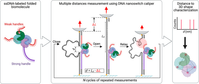
Figure 1 Overview of single-molecule 3D mapping with DNA nanoswitch calipers (DNC). (Left) Natively folded biomolecular complex labeled with ssDNA handles: a strong handle (purple) that serves as an anchoring site for DNC and shorter weak handles (red). (Middle) Multiple distances are measured between red and purple handles using a DNC actuated by mechanical force. Each DNC has one “grabber” complementary to the weak handles and one complementary to the strong handle. As force is cycled between low and high forces, the caliper alternates between looped and linear states, due to engagement then release of the weak handle. (Top right) Distribution of measured distances d calculated by subtracting the change in length ΔL from the effective loop length L0, which serves as a reference to convert changes in length to absolute distance. (Bottom right) Reconstructed geometry for positions of DNA-labeled sites in the folded biomolecular complex.
Figure 1 Overview of single-molecule 3D mapping with DNA nanoswitch calipers (DNC). (Left) Natively folded biomolecular complex labeled with ssDNA handles: a strong handle (purple) that serves as an anchoring site for DNC and shorter weak handles (red). (Middle) Multiple distances are measured between red and purple handles using a DNC actuated by mechanical force. Each DNC has one “grabber” complementary to the weak handles and one complementary to the strong handle. As force is cycled between low and high forces, the caliper alternates between looped and linear states, due to engagement then release of the weak handle. (Top right) Distribution of measured distances d calculated by subtracting the change in length ΔL from the effective loop length L0, which serves as a reference to convert changes in length to absolute distance. (Bottom right) Reconstructed geometry for positions of DNA-labeled sites in the folded biomolecular complex.CC BY-NC-ND · PubMed Central
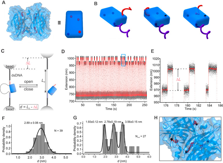
Figure 2 Single-molecule distance measurements of streptavidin–biotin complexes with only two biotin sites occupied. (A) Structure of streptavidin–biotin complex (PDB entry 6M9B) (left) with a schematic representation (right) where spheres indicate biotins bound to streptavidin. (B) Schematic of streptavidin bound to two biotinylated ssDNA handles illustrating three distinct configurations. The longer (purple) strong handle provides strong attachment to the DNC, while the shorter (red) weak handle shears off at lower force. Biotinylated handles are hybridized to the DNC construct prior to the introduction of streptavidin to yield one pair of handles per molecule (Figure S1). (C) DNC tethered between two optically trapped microbeads and actuated by force to measure distances between biotin-binding sites in streptavidin. DNC looping occurs when both handles are engaged; unlooping occurs when the weak handle shears off at high force; relooping occurs with weak handle rebinding at low force. (D) Data showing DNC extension over cycles of low and high force (raw data in black, 200-point sliding window average in red). (E) Zoom-in of blue rectangle in D, showing change in extension due to DNC unlooping. (F) Distance histogram calculated by subtracting ΔL from effective loop length (L0). (G) Histogram of per-molecule average distances for 27 molecular complexes. Plus–minus values indicate sigma fitting parameters from the multipeak Gaussian fit. (H) DNC determined positions of carbon in the carboxylic group of each biotin (black spheres) superimposed on the crystal structure (PDB 6M9B).
Figure 2 Single-molecule distance measurements of streptavidin–biotin complexes with only two biotin sites occupied. (A) Structure of streptavidin–biotin complex (PDB entry 6M9B) (left) with a schematic representation (right) where spheres indicate biotins bound to streptavidin. (B) Schematic of streptavidin bound to two biotinylated ssDNA handles illustrating three distinct configurations. The longer (purple) strong handle provides strong attachment to the DNC, while the shorter (red) weak handle shears off at lower force. Biotinylated handles are hybridized to the DNC construct prior to the introduction of streptavidin to yield one pair of handles per molecule (Figure S1). (C) DNC tethered between two optically trapped microbeads and actuated by force to measure distances between biotin-binding sites in streptavidin. DNC looping occurs when both handles are engaged; unlooping occurs when the weak handle shears off at high force; relooping occurs with weak handle rebinding at low force. (D) Data showing DNC extension over cycles of low and high force (raw data in black, 200-point sliding window average in red). (E) Zoom-in of blue rectangle in D, showing change in extension due to DNC unlooping. (F) Distance histogram calculated by subtracting ΔL from effective loop length (L0). (G) Histogram of per-molecule average distances for 27 molecular complexes. Plus–minus values indicate sigma fitting parameters from the multipeak Gaussian fit. (H) DNC determined positions of carbon in the carboxylic group of each biotin (black spheres) superimposed on the crystal structure (PDB 6M9B).CC BY-NC-ND · PubMed Central
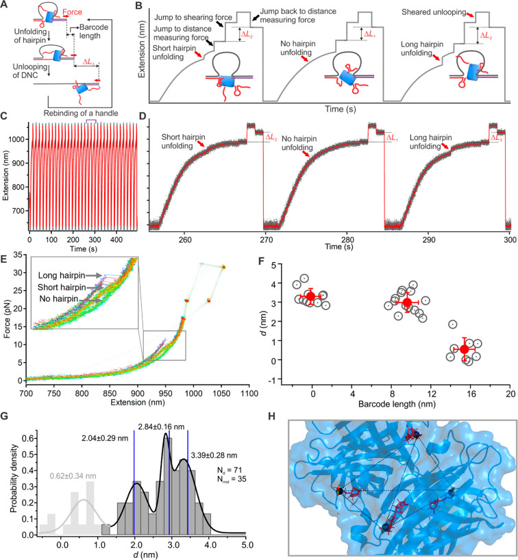
Figure 3 3D mapping of streptavidin–biotin complex with force-based barcodes to resolve multiple distance measurements per molecule. (A) Schematic showing incorporation and readout of hairpin barcodes incorporated into weak handles (red). First, hairpin opening enables handle identification from the change in length, and then loop opening at a higher force enables the distance between handles to be measured as before. Dropping the force enables rebinding to another handle. (B) Outline of distance measurement strategy consisting of a force ramp for hairpin readout, followed by jumps in force to ∼20 pN to measure the distance, ∼30 pN to shear the weak handle, ∼20 pN to measure the change in length, and then ∼0 pN to enable rebinding. Three cycles are shown, illustrating unfolding signatures of the three barcodes. (C) Extension vs time data for a DNC over multiple force cycles (raw data in gray, smoothed in red). (D) Zoom-in of a section (indicated by purple arrows) from C depicting the sequential readout of all three barcodes. (E) Force vs extension curves of repeated cycles depicting the measurement of all three barcode lengths in one streptavidin molecule. (F) Scatter plot of measured barcode lengths vs handle distances in a single molecule. For each cluster, red circles indicate mean, and error bars indicate standard deviation. (G) Histogram of per-molecule distances aggregated over multiple molecules. Plus–minus values indicate sigma fitting parameters from the multipeak Gaussian fit. Blue vertical lines indicate expected distances between biotin-binding sites based on the crystal structure (PDB 6M9B). (H) Superposition of the DNC determined the position of the carbon in the carboxylic group of each biotin (black spheres) on the crystal structure.
Figure 3 3D mapping of streptavidin–biotin complex with force-based barcodes to resolve multiple distance measurements per molecule. (A) Schematic showing incorporation and readout of hairpin barcodes incorporated into weak handles (red). First, hairpin opening enables handle identification from the change in length, and then loop opening at a higher force enables the distance between handles to be measured as before. Dropping the force enables rebinding to another handle. (B) Outline of distance measurement strategy consisting of a force ramp for hairpin readout, followed by jumps in force to ∼20 pN to measure the distance, ∼30 pN to shear the weak handle, ∼20 pN to measure the change in length, and then ∼0 pN to enable rebinding. Three cycles are shown, illustrating unfolding signatures of the three barcodes. (C) Extension vs time data for a DNC over multiple force cycles (raw data in gray, smoothed in red). (D) Zoom-in of a section (indicated by purple arrows) from C depicting the sequential readout of all three barcodes. (E) Force vs extension curves of repeated cycles depicting the measurement of all three barcode lengths in one streptavidin molecule. (F) Scatter plot of measured barcode lengths vs handle distances in a single molecule. For each cluster, red circles indicate mean, and error bars indicate standard deviation. (G) Histogram of per-molecule distances aggregated over multiple molecules. Plus–minus values indicate sigma fitting parameters from the multipeak Gaussian fit. Blue vertical lines indicate expected distances between biotin-binding sites based on the crystal structure (PDB 6M9B). (H) Superposition of the DNC determined the position of the carbon in the carboxylic group of each biotin (black spheres) on the crystal structure.CC BY-NC-ND · PubMed Central

Verification

Claims the paper makes

The claims and statistics of this paper haven’t been checked yet.

Run the check — free, no account needed. It reads the open-access text and checks the sources it cites.

A few free checks a day · papers already verified open instantly · results are public

Self-check

Percentages, sums and reported differences re-computed from the paper’s own numbers.

The self-check needs the paper’s full text.

Citations

Cites

Research works printed but not linked: 37

  1. 2017, Structural Biology and the Design of New Therapeutics: From HIV and Cancer to Mycobacterial Infections: A Paper Dedicated to John Kendrew, Journal of Molecular Biology, 429, 2677, 10.1016/j.jmb.2017.06.014 · DOI
  2. 10.1038/nrmicro2747 · DOI
  3. 2021, Highly accurate protein structure prediction for the human proteome, Nature, 596, 590, 10.1038/s41586-021-03828-1 · DOI · Paper page
  4. 10.1126/science.1113801 · DOI
  5. 10.1126/science.aau1044 · DOI
  6. 10.1038/nrm.2017.71 · DOI
  7. 10.1021/nl103427w · DOI
  8. 10.1126/science.1127344 · DOI
  9. 10.1126/science.aat4346 · DOI
  10. 2018, Cryo-electron microscopy for structural analysis of dynamic biological macromolecules, Biochimica et Biophysica Acta (BBA) - General Subjects, 1862, 324, 10.1016/j.bbagen.2017.07.020 · DOI
  11. 10.1038/nrd.2018.77 · DOI
  12. Lerner, E. et al. Toward dynamic structural biology: Two decades of single-molecule Förster resonance energy transfer. Science 359, eaan1133 (2018). · DOI
  13. 10.1016/j.sbi.2016.11.012 · DOI
  14. 2017, Cool and dynamic: single-molecule fluorescence-based structural biology, Nat Methods, 14, 123, 10.1038/nmeth.4159 · DOI
  15. 10.7554/eLife.60416 · DOI
  16. 10.1073/pnas.2206513119 · DOI
  17. 2017, Confined space facilitates G-quadruplex formation, Nature Nanotech, 12, 582, 10.1038/nnano.2017.29 · DOI
  18. 10.1126/science.1207598 · DOI · Paper page
  19. 10.1016/j.jmb.2021.167207 · DOI
  20. 2022, Nanopore-based technologies beyond DNA sequencing, Nat. Nanotechnol, 17, 1136, 10.1038/s41565-022-01193-2 · DOI
  21. 10.1073/pnas.0509217103 · DOI
  22. 2015, Imaging and three-dimensional reconstruction of chemical groups inside a protein complex using atomic force microscopy, Nature Nanotech, 10, 264, 10.1038/nnano.2014.335 · DOI
  23. 2021, Single-molecule mechanical fingerprinting with DNA nanoswitch calipers, Nat. Nanotechnol, 16, 1362, 10.1038/s41565-021-00979-0 · DOI
  24. 10.1016/0003-9861(64)90150-X · DOI
  25. 10.1073/pnas.86.7.2190 · DOI
  26. 10.1107/S0907444911027806 · DOI
  27. 10.1021/bi992715o · DOI
  28. 10.1021/la00014a003 · DOI
  29. Sedlak, S. M. , Schendel, L. C. , Gaub, H. E. & Bernardi, R. C. Streptavidin/biotin: Tethering geometry defines unbinding mechanics. Science Advances 6, eaay5999 (2020). · DOI
  30. 2020, Designed anchoring geometries determine lifetimes of biotin–streptavidin bonds under constant load and enable ultra-stable coupling, Nanoscale, 12, 21131, 10.1039/D0NR03665J · DOI
  31. 2019, Making routine native SAD a reality: lessons from beamline X06DA at the Swiss Light Source, Acta Cryst D, 75, 262, 10.1107/S2059798319003103 · DOI
  32. 10.1529/biophysj.106.091116 · DOI
  33. 10.1038/nmeth929 · DOI
  34. 10.1126/science.2911722 · DOI
  35. 10.1088/0957-4484/22/49/494005 · DOI
  36. 10.1038/s41467-021-21033-6 · DOI
  37. Bustamante, C. , Chemla, Y. R. & Moffitt, J. R. High-resolution dual-trap optical tweezers with differential detection: instrument design. Cold Spring Harb Protoc 2009, pdb.ip73 (2009). · DOI

Metadata

Authors
Shrestha, P., Yang, D., Shih, W. M., & Wong, W. P.
Affiliations
  • Boston Children's Hospital (US)
  • Harvard University (US)
Corresponding author
Wong, Wesley P.
Venue
Journal of the American Chemical Society
Publisher
openRxiv
Published
2023-07-11
Volume
145
Issue
51
Pages
27916-27921
ISSN
0002-7863 (ISSN-L)
OpenAlex
W4389764840
Cited by
5
References
1
Citations by year
2026: 1 · 2025: 2 · 2024: 2
FWCI
0.58
Type
journal article
Language
en
Open access
Licence
cc-by-nc-nd
Locations
  • Journal of the American Chemical Society (publishedVersion, cc-by-nc-nd)
  • bioRxiv (Cold Spring Harbor Laboratory) (acceptedVersion)
  • PubMed Central (publishedVersion, cc-by-nc-nd)
  • PubMed (publishedVersion)
  • PubMed Central (submittedVersion)
Funders
  • Alfred P. Sloan Foundation (G-2021-169145)
  • Harvard University
  • National Institutes of Health
  • Hansjörg Wyss Institute for Biologically Inspired Engineering, Harvard University
  • Office of Naval Research (N00014-15-1-0073)
  • National Institute of General Medical Sciences (GM119537, R35GM119537)
Topics
  • Physical Sciences › Physics and Astronomy › Atomic and Molecular Physics, and Optics › Force Microscopy Techniques and Applications
  • Life Sciences › Biochemistry, Genetics and Molecular Biology › Molecular Biology › Advanced biosensing and bioanalysis techniques
  • Life Sciences › Biochemistry, Genetics and Molecular Biology › Biophysics › Advanced Fluorescence Microscopy Techniques
Keywords
single-molecule force spectroscopy, streptavidin, biotin binding site, tetrahedral geometry, nanodevices

About these checks

Registry, retraction, Cites and statistics results are computed, not judged, with one exception: a printed reference that no index lists is read once by a model before it counts as a citation error. Claims are extracted by a model and kept only when their quote appears verbatim in the text; each cited source is then checked, with a model’s help, for whether it says what the claim relies on. Those verdicts can be wrong — read the claim’s quote and the cited source before relying on one.

Statistics and arithmetic are recomputed, not judged.

Confidence bands are shown only for fields calibrated on our annotated test set. None is calibrated yet, so no band is shown.

What counts as a claim ›How we measure confidence ›How we check this ›

Are you an author, or do you see a problem with this page? Request a review