Student or educator? Sign up with your school email and get 6 months of Pro free.
PreprintCited by 3Protein Kinase Regulation and GTPase SignalingChemical Synthesis and AnalysisProtein Structure and Dynamics

The DOI matches its Crossref record; 1 of the 1 references it cites were found in a registry or an index.

Molecularly imprinted nanoparticles reveal regulatory scaffolding features in Pyk2 tyrosine kinase

Open access
Zanela, Tania M. Palhano, Zangiabadi, Milad, Zhao, Yan, Underbakke, Eric S.·2023·RSC Chemical Biology

Preprint — published as 10.1039/d3cb00228d · Paper page

Abstract

Pyk2 is a multi-domain non-receptor tyrosine kinase that serves dual roles as signaling enzyme and scaffold. Pyk2 activation involves a multi-stage cascade of conformational rearrangements and protein interactions initiated by autophosphorylation of a linker site. Linker phosphorylation recruits Src kinase, and Src-mediated phosphorylation of the Pyk2 activation loop confers full activation. The regulation and accessibility of the initial Pyk2 autophosphorylation site remains unclear. We employed peptide-binding molecularly imprinted nanoparticles (MINPs) to probe the regulatory conformations controlling Pyk2 activation. MINPs differentiating local structure and phosphorylation state revealed that the Pyk2 autophosphorylation site is protected in the autoinhibited state. Activity profiling of Pyk2 variants implicated FERM and linker residues responsible for constraining the autophosphorylation site. MINPs targeting each Src docking site disrupt the higher-order kinase interactions critical for activation complex maturation. Ultimately, MINPs targeting key regulatory motifs establish a useful toolkit for probing successive activational stages in the higher-order Pyk2 signaling complex. Abstract Figure:

Overview

The claims and statistics of this paper haven’t been checked yet.

Run the check — free, no account needed. It reads the open-access text and checks the sources it cites.

A few free checks a day · papers already verified open instantly · results are public

Integrity signals

Registry

Crossref · 10.1101/2023.08.05.552112

Cites

1 of 1 references verified0 citation errors · 0 not checkable

Statistics

Figures

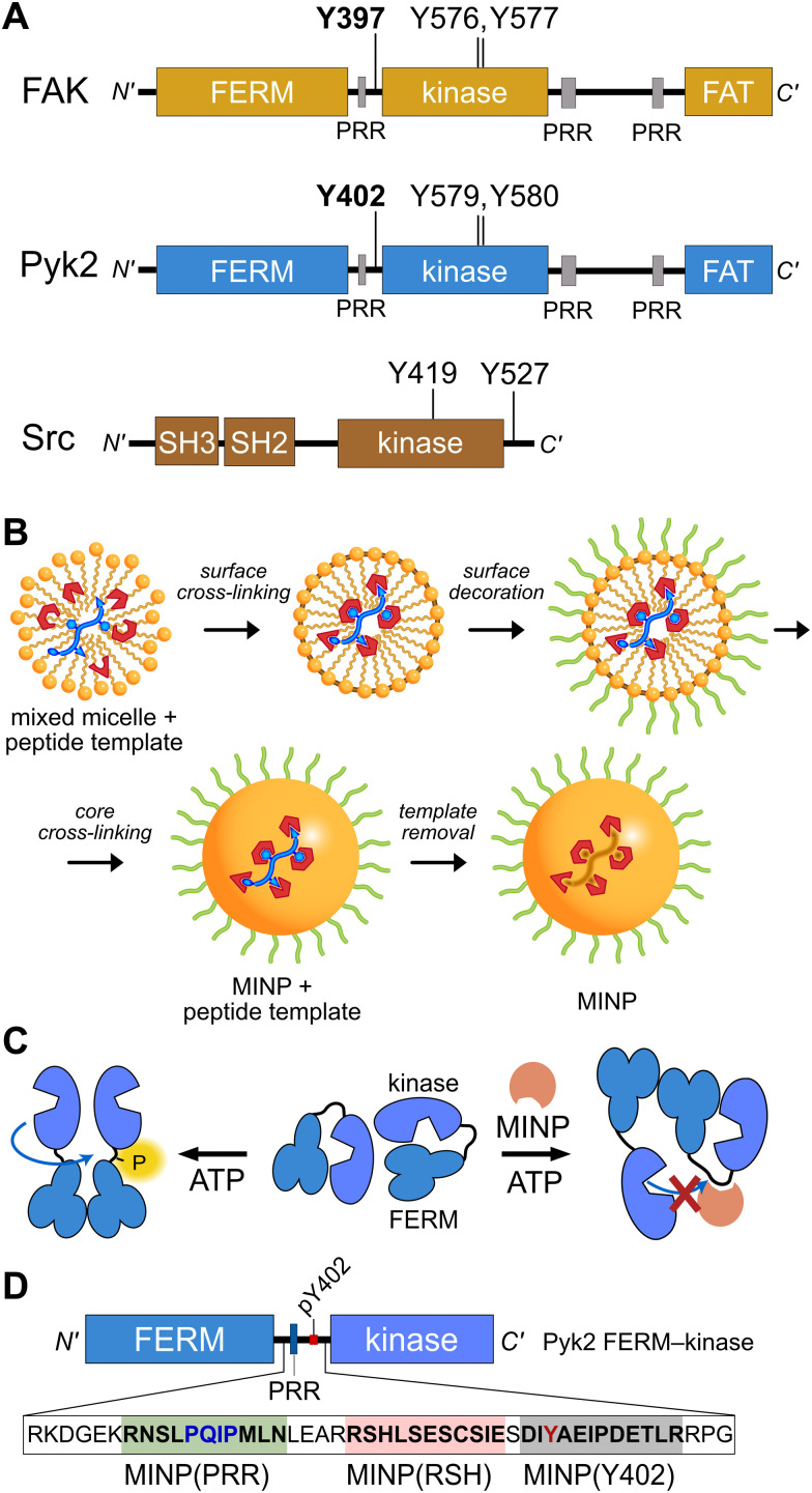
Fig. 1 Tyrosine kinase regulatory features probed by MINPs (A) Domain organization of Pyk2, FAK, and Src kinase. Proline-rich regions (PRR) and phosphoacceptor tyrosines are annotated with linker autophosphorylation sites in bold. (B) Cartoon representation of peptide templated MINP preparation. Mixed micelles with functional monomers are cross-linked around peptide (yellow and blue) to afford a binding pocket with shape and functional group complementarity.8 (C) Schematic model for MINP-mediated inhibition of Pyk2 autophosphorylation. (D) Pyk2 FERM-kinase construct highlighting linker sequences used for MINP imprinting.
Fig. 1 Tyrosine kinase regulatory features probed by MINPs (A) Domain organization of Pyk2, FAK, and Src kinase. Proline-rich regions (PRR) and phosphoacceptor tyrosines are annotated with linker autophosphorylation sites in bold. (B) Cartoon representation of peptide templated MINP preparation. Mixed micelles with functional monomers are cross-linked around peptide (yellow and blue) to afford a binding pocket with shape and functional group complementarity.8 (C) Schematic model for MINP-mediated inhibition of Pyk2 autophosphorylation. (D) Pyk2 FERM-kinase construct highlighting linker sequences used for MINP imprinting.CC BY · PubMed Central
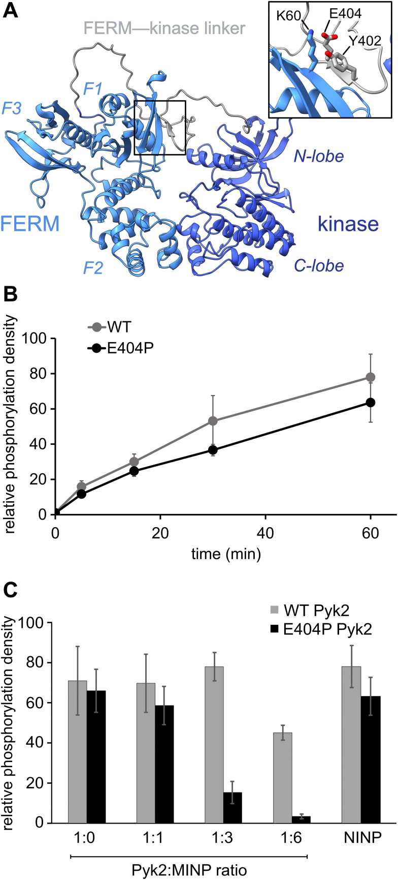
Fig. 2 Pyk2 kinase regulatory conformations probed by molecularly imprinted nanoparticles. (A) AlphaFold-derived structural model of Pyk2 FERM-kinase with inset detailing the small β-sheet constraining Y402 in the Pyk2 linker segment colored in gray. (B) Kinase activity time course of WT and E404P Pyk2 FERM-kinase (0.5 μM). Tyrosine phosphorylation was detected via Western blotting with site-specific anti-phospho-Y402 primary antibody. (C) Inhibition of Pyk2 autophosphorylation by MINP(Y402) at protein : MINP ratios of 1 : 1, 1 : 3, and 1 : 6, with the NINP as the control at 1 : 6 ratio. Pyk2 concentration was fixed at 0.5 μM, and autophosphorylation was assessed 5 min after reaction initiation with ATP. Tyrosine phosphorylation was detected by dot immunoblotting with anti-phosphotyrosine primary antibody (PY20). Activity assay replicates (n = 3–5) represent independent reactions with error bars signifying standard deviation.
Fig. 2 Pyk2 kinase regulatory conformations probed by molecularly imprinted nanoparticles. (A) AlphaFold-derived structural model of Pyk2 FERM-kinase with inset detailing the small β-sheet constraining Y402 in the Pyk2 linker segment colored in gray. (B) Kinase activity time course of WT and E404P Pyk2 FERM-kinase (0.5 μM). Tyrosine phosphorylation was detected via Western blotting with site-specific anti-phospho-Y402 primary antibody. (C) Inhibition of Pyk2 autophosphorylation by MINP(Y402) at protein : MINP ratios of 1 : 1, 1 : 3, and 1 : 6, with the NINP as the control at 1 : 6 ratio. Pyk2 concentration was fixed at 0.5 μM, and autophosphorylation was assessed 5 min after reaction initiation with ATP. Tyrosine phosphorylation was detected by dot immunoblotting with anti-phosphotyrosine primary antibody (PY20). Activity assay replicates (n = 3–5) represent independent reactions with error bars signifying standard deviation.CC BY · PubMed Central
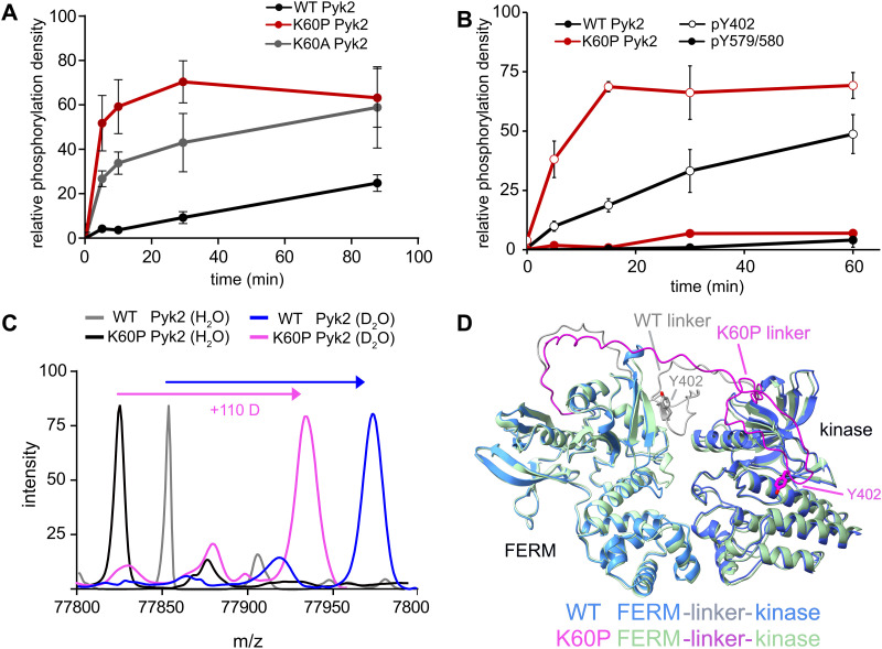
Fig. 3 FERM domain variants modulate Pyk2 autophosphorylation. (A) Kinase activity time courses of WT Pyk2 FERM-kinase (0.5 μM) and variants K60P and K60A. Tyrosine phosphorylation was detected via Western blotting with pan-specific anti-phosphotyrosine primary antibody (PY20). (B) Site-specific phosphorylation was monitored by blotting WT Pyk2 (black) and K60P Pyk2 FERM-kinase (red) with anti-phospho-pY579/pY580 (filled circles) or anti-phospho-Y402 primary antibody (hollow circles). Phosphorylation levels were quantified by densitometry. Activity assay replicates (n = 3) represent independent reactions with error bars signifying standard deviation. (C) Global HDX-MS spectra of WT and K60P Pyk2 FERM-kinase. Spectra include unlabeled (H2O) WT (grey) and K60P (black) superimposed with 2.5 min D2O exposure of WT (blue) and K60P Pyk2 (magenta). (D) AlphaFold (ver. 2.3.1) generated models of WT (blue) and K60P (green) Pyk2 autoinhibitory conformation. The FERM-kinase linker is highlighted with the Y402 site for WT (grey) and K60P (magenta).
Fig. 3 FERM domain variants modulate Pyk2 autophosphorylation. (A) Kinase activity time courses of WT Pyk2 FERM-kinase (0.5 μM) and variants K60P and K60A. Tyrosine phosphorylation was detected via Western blotting with pan-specific anti-phosphotyrosine primary antibody (PY20). (B) Site-specific phosphorylation was monitored by blotting WT Pyk2 (black) and K60P Pyk2 FERM-kinase (red) with anti-phospho-pY579/pY580 (filled circles) or anti-phospho-Y402 primary antibody (hollow circles). Phosphorylation levels were quantified by densitometry. Activity assay replicates (n = 3) represent independent reactions with error bars signifying standard deviation. (C) Global HDX-MS spectra of WT and K60P Pyk2 FERM-kinase. Spectra include unlabeled (H2O) WT (grey) and K60P (black) superimposed with 2.5 min D2O exposure of WT (blue) and K60P Pyk2 (magenta). (D) AlphaFold (ver. 2.3.1) generated models of WT (blue) and K60P (green) Pyk2 autoinhibitory conformation. The FERM-kinase linker is highlighted with the Y402 site for WT (grey) and K60P (magenta).CC BY · PubMed Central
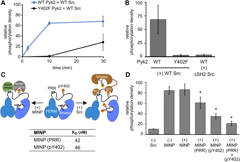
Fig. 4 Scaffolding sites in the Pyk2–Src complex probed by MINPs. (A) Kinase activity time course of WT or Y402F Pyk2 FERM-kinase (0.5 μM) in the presence of Src (0.5 μM). Site-specific phosphorylation was detected by blotting with anti-phospho-pY579/pY580 primary antibody. (B) WT or Y402F Pyk2 FERM-kinase (1 μM) was preincubated with ATP for 10 min. Activation loop phosphorylation (pY579/pY580) was measured following addition of WT Src or ΔSH2 Src [1-142,166-536] (0.5 μM). (C) Schematic illustrating MINP-mediated inhibition of Pyk2 activation loop phosphorylation. Dissociation constants were measured via isothermal titration calorimetry for MINP(pY402) and MINP(PRR) with the corresponding peptide template. (D) Activation loop phosphorylation (Y579/Y580) of phospho-Y402 Pyk2 FERM-kinase (1 μM) preincubated with MINP. Src (0.5 μM) was added 3 min after MINP equilibration. Reactions were quenched 5 min after Src addition. Activation loop phosphorylation was assessed by blotting with anti-phospho-Y579/Y580 primary antibody and quantified by densitometry. Activity assay replicates (n = 3) represent independent reactions with error bars representing standard deviation. *p < 0.05 vs. the (+) NIMP treatment.
Fig. 4 Scaffolding sites in the Pyk2–Src complex probed by MINPs. (A) Kinase activity time course of WT or Y402F Pyk2 FERM-kinase (0.5 μM) in the presence of Src (0.5 μM). Site-specific phosphorylation was detected by blotting with anti-phospho-pY579/pY580 primary antibody. (B) WT or Y402F Pyk2 FERM-kinase (1 μM) was preincubated with ATP for 10 min. Activation loop phosphorylation (pY579/pY580) was measured following addition of WT Src or ΔSH2 Src [1-142,166-536] (0.5 μM). (C) Schematic illustrating MINP-mediated inhibition of Pyk2 activation loop phosphorylation. Dissociation constants were measured via isothermal titration calorimetry for MINP(pY402) and MINP(PRR) with the corresponding peptide template. (D) Activation loop phosphorylation (Y579/Y580) of phospho-Y402 Pyk2 FERM-kinase (1 μM) preincubated with MINP. Src (0.5 μM) was added 3 min after MINP equilibration. Reactions were quenched 5 min after Src addition. Activation loop phosphorylation was assessed by blotting with anti-phospho-Y579/Y580 primary antibody and quantified by densitometry. Activity assay replicates (n = 3) represent independent reactions with error bars representing standard deviation. *p < 0.05 vs. the (+) NIMP treatment.CC BY · PubMed Central

Verification

Claims the paper makes

The claims and statistics of this paper haven’t been checked yet.

Run the check — free, no account needed. It reads the open-access text and checks the sources it cites.

A few free checks a day · papers already verified open instantly · results are public

Self-check

Percentages, sums and reported differences re-computed from the paper’s own numbers.

The self-check needs the paper’s full text.

Citations

Cites

Show all 1 references

Research works printed but not linked: 27

  1. 10.1038/376737a0 · DOI · Paper page
  2. 10.3389/fnsyn.2021.749001 · DOI
  3. 2018, Cancers (Basel), 10, 196, 10.3390/cancers10060196 · DOI
  4. 2011, Am. J. of Physiol. and Circ. Physiol, 301, H975, 10.1152/ajpheart.00241.2011 · DOI
  5. 10.1073/pnas.1317022111 · DOI
  6. 10.1523/JNEUROSCI.4992-08.2010 · DOI
  7. 2022, Commun. Biol, 5, 1, 10.1038/s42003-021-02997-z · DOI
  8. 10.1021/jacs.0c11566 · DOI
  9. 10.1016/j.cell.2007.05.041 · DOI · Paper page
  10. 10.1021/acs.biochem.9b00541 · DOI
  11. 10.1074/jbc.273.1.577 · DOI · Paper page
  12. 10.1128/MCB.14.3.1680 · DOI
  13. 10.1074/jbc.M313527200 · DOI
  14. 10.1371/journal.pone.0149231 · DOI
  15. 10.1186/s12860-019-0193-4 · DOI
  16. 10.1074/jbc.M412021200 · DOI
  17. 10.1021/jacs.6b12949 · DOI
  18. 10.1021/acsnano.2c11186 · DOI
  19. 2018, Chem. - A Euro. J, 24, 150, 10.1002/chem.201703760 · DOI
  20. 10.1021/acs.chemmater.9b01613 · DOI
  21. 10.1021/acs.nanolett.3c01374 · DOI
  22. 10.1021/jacs.0c11566 · DOI
  23. 10.1093/NAR/GKAB1061 · DOI · Paper page
  24. 10.1016/j.pbiomolbio.2015.06.001 · DOI
  25. 2014, J. Cell. Biochem, 115, 596, 10.1002/jcb.24697 · DOI
  26. 10.1016/S0896-6273(01)00220-3 · DOI · Paper page
  27. X. Zhu , Y. Bao , Y. Guo and W. Yang , Cancers (Basel), 2018, 10. · DOI

Metadata

Authors
Zanela, T. M. P., Zangiabadi, M., Zhao, Y., & Underbakke, E. S.
Affiliations
Iowa State University (US)
Corresponding author
Underbakke, Eric S.
Venue
RSC Chemical Biology
Publisher
openRxiv
Published
2023-08-05
Volume
5
Issue
5
Pages
447-453
ISSN
2633-0679 (ISSN-L)
OpenAlex
W4392776802
Cited by
3
References
1
Citations by year
2026: 1 · 2025: 1 · 2024: 1
FWCI
0.41
Type
journal article
Language
en
Open access
Licence
cc-by
Locations
  • RSC Chemical Biology (publishedVersion, cc-by)
  • bioRxiv (Cold Spring Harbor Laboratory) (acceptedVersion, cc-by)
  • PubMed Central (publishedVersion, cc-by)
  • PubMed (publishedVersion)
  • DOAJ (DOAJ: Directory of Open Access Journals) (submittedVersion)
Funders
  • National Science Foundation (1715411, 2002659, 2308625)
  • Iowa State University
  • Division of Materials Research (2308625, 2002659)
  • Division of Molecular and Cellular Biosciences (1715411)
Topics
  • Life Sciences › Biochemistry, Genetics and Molecular Biology › Molecular Biology › Protein Kinase Regulation and GTPase Signaling
  • Physical Sciences › Chemistry › Organic Chemistry › Chemical Synthesis and Analysis
  • Life Sciences › Biochemistry, Genetics and Molecular Biology › Molecular Biology › Protein Structure and Dynamics
Keywords
Pyk2, non-receptor tyrosine kinase, molecularly imprinted nanoparticles, autophosphorylation sites, kinase activation, Src kinase, FERM domain, autoinhibition, protein conformational regulation

About these checks

Registry, retraction, Cites and statistics results are computed, not judged, with one exception: a printed reference that no index lists is read once by a model before it counts as a citation error. Claims are extracted by a model and kept only when their quote appears verbatim in the text; each cited source is then checked, with a model’s help, for whether it says what the claim relies on. Those verdicts can be wrong — read the claim’s quote and the cited source before relying on one.

Statistics and arithmetic are recomputed, not judged.

Confidence bands are shown only for fields calibrated on our annotated test set. None is calibrated yet, so no band is shown.

What counts as a claim ›How we measure confidence ›How we check this ›

Are you an author, or do you see a problem with this page? Request a review

How to cite

Cite CoChat for the checks on this page, and the paper for its own findings.

This page
CoChat. (2026). Citation check: Molecularly imprinted nanoparticles reveal regulatory scaffolding features in Pyk2 tyrosine kinase. https://app.cochat.ai/papers/10.1101/2023.08.05.552112
The paper
Zanela, T. M. P., Zangiabadi, M., Zhao, Y., & Underbakke, E. S. (2023). Molecularly imprinted nanoparticles reveal regulatory scaffolding features in Pyk2 tyrosine kinase. RSC Chemical Biology. https://doi.org/10.1101/2023.08.05.552112

Data sources

Bibliographic data: Semantic Scholar, OpenAlex, Crossref.

Contains information from the Semantic Scholar Academic Graph Datasets, made available under the ODC Attribution License (ODC-BY 1.0).

Semantic Scholar’s data is described in: Kinney, R., et al. (2023). The Semantic Scholar Open Data Platform. arXiv:2301.10140.