

The DOI matches its Crossref record; 1 of the 1 references it cites were found in a registry or an index.
Design of Cyclic Peptides Targeting Protein-Protein Interactions using AlphaFold
Open accessPreprint — published as 10.3390/ijms241713257
Abstract
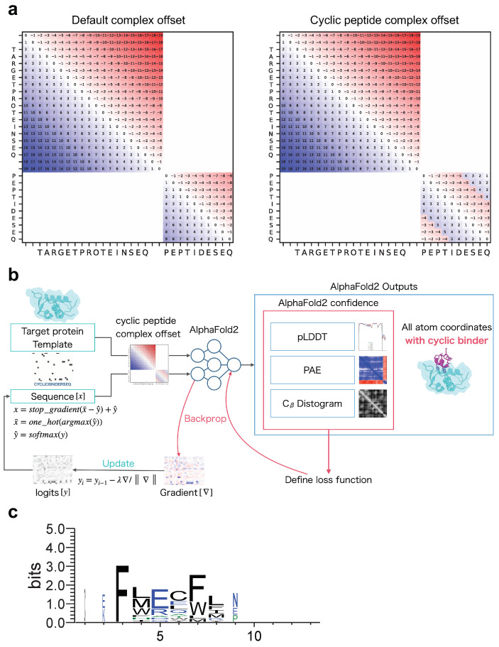
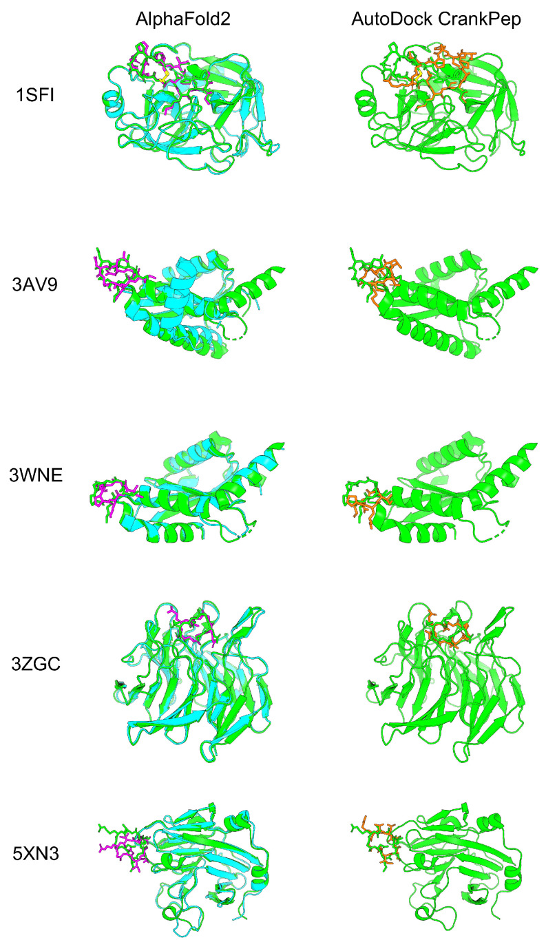
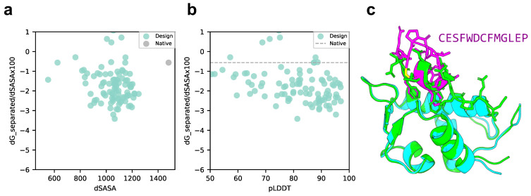
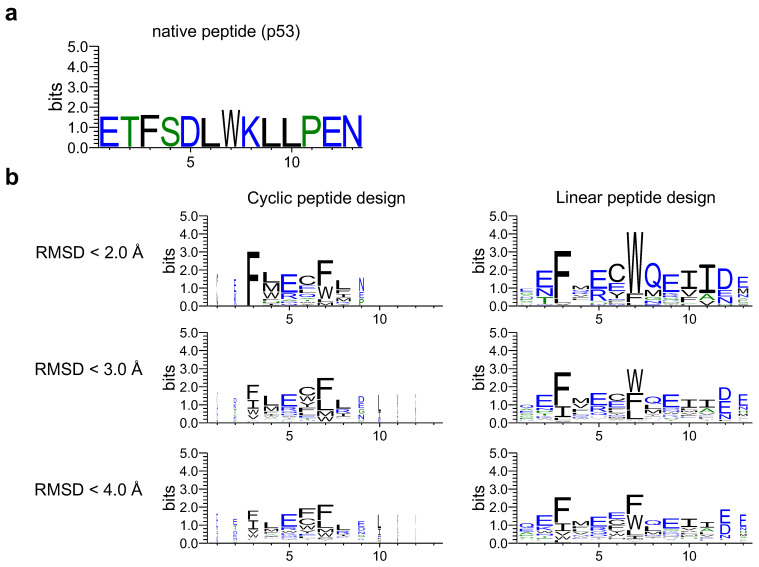
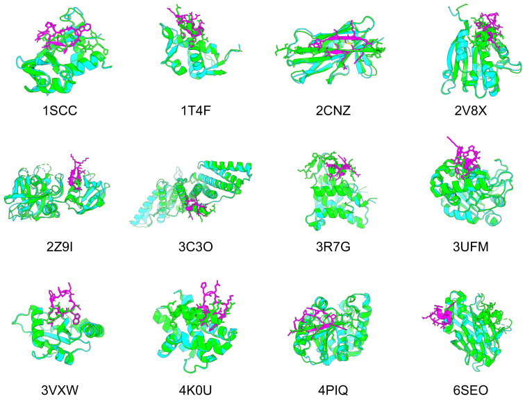
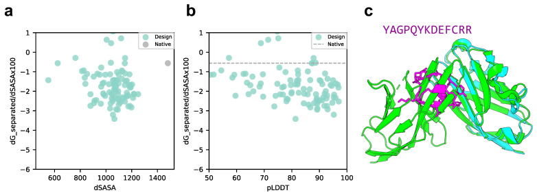
More than 930,000 protein-protein interactions (PPIs) have been identified in recent years, but their physicochemical properties differ from conventional drug targets, complicating the use of conventional small molecules as modalities. Cyclic peptides are a promising modality for targeting protein-protein interactions (PPIs), but it is difficult to predict the structure of a target protein-cyclic peptide complex or to design a cyclic peptide sequence that binds to the target protein using computational methods. Recently, AlphaFold with a cyclic offset has enabled predicting the structure of cyclic peptides, thereby enabling de novo cyclic peptide designs. We developed a cyclic peptide complex offset to enable the structural prediction of target proteins and cyclic peptide complexes and found AlphaFold2 with a cyclic peptide complex offset can predict structures with high accuracy. We also applied the cyclic peptide complex offset to the binder hallucination protocol of AfDesign, a de novo protein design method using AlphaFold, and we could design a high predicted local-distance difference test and lower separated binding energy per unit interface area than the native MDM2/p53 structure. Furthermore, the method was applied to 12 other protein-peptide complexes and one protein-protein complex. Our approach shows that it is possible to design putative cyclic peptide sequences targeting PPI.
Overview
The claims and statistics of this paper haven’t been checked yet.
Run the check — free, no account needed. It reads the open-access text and checks the sources it cites.
Integrity signals
Registry
Cites
Statistics
Figures
Verification
Claims the paper makes
The claims and statistics of this paper haven’t been checked yet.
Run the check — free, no account needed. It reads the open-access text and checks the sources it cites.
Self-check
Percentages, sums and reported differences re-computed from the paper’s own numbers.
The self-check needs the paper’s full text.
Citations
Cites
Highly accurate protein structure prediction with AlphaFold
Jumper, J., Evans, R., Pritzel, A., Green, T., Figurnov, M., Ronneberger, O., Tunyasuvunakool, K., Bates, R., Žídek, A., Potapenko, A., et al. (2021) · Nature · doi:10.1038/s41586-021-03819-2 · Cited by 43,764
Research works printed but not linked: 63
- 10.1038/468851a · DOI
- 10.1002/PRO.3978 · DOI · Paper page
- 10.1021/jm010468s · DOI
- 10.1038/nrd1343 · DOI · Paper page
- 10.1038/nrd1578 · DOI
- 10.1146/annurev-pharmtox-011613-140028 · DOI · Paper page
- 10.1016/j.tips.2013.04.007 · DOI · Paper page
- 2020, Current Challenges and Opportunities in Designing Protein–Protein Interaction Targeted Drugs, Adv. Appl. Bioinform. Chem, 13, 11
- 2021, Quantitative Estimate Index for Early-Stage Screening of Compounds Targeting Protein-Protein Interactions, Int. J. Mol. Sci, 22, 10925, 10.3390/ijms222010925 · DOI
- 10.1021/jacs.8b13178 · DOI · Paper page
- 10.1038/s41573-020-00135-8 · DOI
- 2015, Peptide Therapeutics: Targeting the Undruggable Space, Eur. J. Med. Chem, 94, 459, 10.1016/j.ejmech.2015.01.014 · DOI
- 2016, Quantifying the Chameleonic Properties of Macrocycles and Other High-Molecular-Weight Drugs, Drug Discov. Today, 21, 712, 10.1016/j.drudis.2016.02.005 · DOI
- 2021, Interplay among Conformation, Intramolecular Hydrogen Bonds, and Chameleonicity in the Membrane Permeability and Cyclophilin A Binding of Macrocyclic Peptide Cyclosporin O Derivatives, J. Med. Chem, 64, 8272, 10.1021/acs.jmedchem.1c00211 · DOI
- 2021, Large-scale membrane permeability prediction of cyclic peptides crossing a lipid bilayer based on enhanced sampling molecular dynamics simulations, J. Chem. Inf. Model, 61, 3681, 10.1021/acs.jcim.1c00380 · DOI
- 2022, Lipid composition is critical for accurate membrane permeability prediction of cyclic peptides by molecular dynamics simulations, J. Chem. Inf. Model, 62, 4549, 10.1021/acs.jcim.2c00931 · DOI
- 10.1186/s12929-016-0223-x · DOI
- 2021, The RaPID Platform for the Discovery of Pseudo-Natural Macrocyclic Peptides, Acc. Chem. Res, 54, 3604, 10.1021/acs.accounts.1c00391 · DOI
- 10.1093/nar/gkp514 · DOI
- 2021, Accelerated Antimicrobial Discovery via Deep Generative Models and Molecular Dynamics Simulations, Nat. Biomed. Eng, 5, 613, 10.1038/s41551-021-00689-x · DOI
- Cardoso, M.H. ; Orozco, R.Q. ; Rezende, S.B. ; Rodrigues, G. ; Oshiro, K.G.N. ; Cândido, E.S. ; Franco, O.L. Computer-Aided Design of Antimicrobial Peptides: Are We Generating Effective Drug Candidates? Front. Microbiol. 2020, 10, https://doi.org/10.3389/fmicb.2019.03097. · DOI
- 10.1021/acs.jcim.9b01014 · DOI
- 2020, Recent Advances in the Development of Protein–Protein Interactions Modulators: Mechanisms and Clinical Trials, Signal Transduct. Target Ther, 5, 213, 10.1038/s41392-020-00315-3 · DOI · Paper page
- 10.1016/j.drudis.2006.05.012 · DOI
- 10.1021/ci200528d · DOI · Paper page
- 2022, Virtual Screening Expands the Non-Natural Amino Acid Palette for Peptide Optimization, J. Chem. Inf. Model, 62, 2999, 10.1021/acs.jcim.2c00193 · DOI
- 10.1021/jm0306430 · DOI · Paper page
- 10.1021/ci400128m · DOI · Paper page
- 2017, Docking of Macrocycles: Comparing Rigid and Flexible Docking in Glide, J. Chem. Inf. Model, 57, 190, 10.1021/acs.jcim.6b00443 · DOI
- 10.1093/nar/gkv456 · DOI
- 2019, CABS-Dock Standalone: A Toolbox for Flexible Protein–Peptide Docking, Bioinformatics, 35, 4170, 10.1093/bioinformatics/btz185 · DOI
- 10.1093/bioinformatics/btx216 · DOI
- 10.1371/journal.pcbi.1005905 · DOI
- 10.1093/nar/gkw329 · DOI
- 10.1158/0008-5472.CAN-17-0511 · DOI
- 2022, Cyclization and Docking Protocol for Cyclic Peptide–Protein Modeling Using HADDOCK2.4, J. Chem. Theory Comput, 18, 4027, 10.1021/acs.jctc.2c00075 · DOI
- 10.1093/bioinformatics/btz459 · DOI
- 2022, High Throughput Virtual Screening (HTVS) of Peptide Library: Technological Advancement in Ligand Discovery, Eur. J. Med. Chem, 243, 114766, 10.1016/j.ejmech.2022.114766 · DOI
- 10.1126/science.abj8754 · DOI · Paper page
- Baek, M. ; Anishchenko, I. ; Humphreys, I.R. ; Cong, Q. ; Baker, D. ; DiMaio, F. Efficient and Accurate Prediction of Protein Structure Using RoseTTAFold2. bioRxiv 2023, https://doi.org/10.1101/2023.05.24.542179. · DOI
- Evans, R. ; O’Neill, M. ; Pritzel, A. ; Antropova, N. ; Senior, A. ; Green, T. ; Žídek, A. ; Bates, R. ; Blackwell, S. ; Yim, J. ; et al. Protein Complex Prediction with AlphaFold-Multimer. bioRxiv 2022, https://doi.org/10.1101/2021.10.04.463034. · DOI · Paper page
- 2021, Computed Structures of Core Eukaryotic Protein Complexes, Science, 374, eabm4805, 10.1126/science.abm4805 · DOI · Paper page
- Gulsevin, A. ; Meiler, J. Benchmarking Peptide Structure Prediction with AlphaFold2. bioRxiv 2022. https://doi.org/10.1101/2022. 02.17.480937. · DOI
- 10.1038/s41467-021-27838-9 · DOI · Paper page
- 10.1103/PhysRevLett.129.238101 · DOI · Paper page
- 2021, De Novo Protein Design by Deep Network Hallucination, Nature, 600, 547, 10.1038/s41586-021-04184-w · DOI · Paper page
- 10.1073/pnas.2017228118 · DOI · Paper page
- 2023, De Novo Protein Design by Inversion of the AlphaFold Structure Prediction Network, Protein Sci, 32, e4653, 10.1002/pro.4653 · DOI · Paper page
- Frank, C. ; Khoshouei, A. ; Stigter, Y. de ; Schiewitz, D. ; Feng, S. ; Ovchinnikov, S. ; Dietz, H. Efficient and Scalable de Novo Protein Design Using a Relaxed Sequence Space. bioRxiv 2023, https://doi.org/10.1101/2023.02.24.529906. · DOI
- 2022, Solubility-Aware Protein Binding Peptide Design Using AlphaFold, Biomedicines, 10, 1626, 10.3390/biomedicines10071626 · DOI
- Rettie, S.A. ; Campbell, K.V. ; Bera, A.K. ; Kang, A. ; Kozlov, S. ; De La Cruz, J. ; Adebomi, V. ; Zhou, G. ; DiMaio, F. ; Ovchinnikov, S. ; Bhardwaj, G. Cyclic Peptide Structure Prediction and Design Using AlphaFold. bioRxiv 2023, https://doi.org/10.1101/2023.02.25.529 · DOI
- 2022, ColabFold - Making Protein Folding Accessible to All, Nat. Methods, 19, 679, 10.1038/s41592-022-01488-1 · DOI · Paper page
- 2019, Docking Flexible Cyclic Peptides with AutoDock CrankPep, J. Chem. Theory Comput, 15, 5161, 10.1021/acs.jctc.9b00557 · DOI
- 10.1006/jmbi.1998.2401 · DOI
- 10.1002/jcc.21256 · DOI
- 10.1101/gr.849004 · DOI
- 10.1002/pro.2187 · DOI
- 10.1093/NAR/GKAB294 · DOI
- Banhos Danneskiold-Samøe, N. ; Kavi, D. ; Jude, K.M. ; Nissen, S.B. ; Wat, L.W. ; Coassolo, L. ; Zhao, M. ; Asae Santana-Oikawa, G. ; Broido, B.B. ; Garcia, K.C. ; Svensson, K.J. Rapid and Accurate Deorphanization of Ligand-Receptor Pairs Using AlphaFold. bioRxiv 2023, https://doi.org/10.1101/2023.03.16.531341. · DOI · Paper page
- 10.1021/acs.jcim.2c01083 · DOI
- Bryant, A. ; Elofsson, A. EvoBind: in Silico Directed Evolution of Peptide Binders with AlphaFold. bioRxiv 2022, https://doi.org/10.1101/2022.07.23.501214. · DOI
- Liu, J. ; Chen, Z. ; Li, Y. ; Zhao, W. ; Wu, J. ; Zhang, Z. PD-1/PD-L1 Checkpoint Inhibitors in Tumor Immunotherapy. Front. Pharmacol. 2021, 12, https://doi.org/10.3389/fphar.2021.731798. · DOI
- 2021, Rational Design of Potent Peptide Inhibitors of the PD-1:PD-L1 Interaction for Cancer Immunotherapy, J. Am. Chem. Soc, 143, 18536, 10.1021/jacs.1c08132 · DOI · Paper page
Metadata
- Authors
- Kosugi, T., & Ohue, M.
- Affiliations
- Tokyo Institute of Technology (JP)
- Corresponding author
- Ohue, Masahito
- Venue
- International Journal of Molecular Sciences
- Publisher
- openRxiv
- Published
- 2023-08-21
- Volume
- 24
- Issue
- 17
- Pages
- 13257-13257
- ISSN
- 1422-0067 (ISSN-L)
- PMC
- PMC10487914
- OpenAlex
- W4386222188
- Cited by
- 51
- References
- 1
- Citations by year
- 2026: 14 · 2025: 26 · 2024: 11
- FWCI
- 4.54
- Type
- journal article
- Language
- en
- Open access
- gold
- doi.org
- Licence
- cc-by
- Locations
- International Journal of Molecular Sciences (publishedVersion, cc-by)
- bioRxiv (Cold Spring Harbor Laboratory) (acceptedVersion)
- PubMed Central (publishedVersion, cc-by)
- PubMed (publishedVersion)
- Funders
- Japan Agency for Medical Research and Development (BINDS JP22ama121026)
- DeepMind
- Japan Society for the Promotion of Science (23H04880, JPMJAX20A3, KAKENHI 23H03496, 23K28186, 23H04887)
- Japan Science and Technology Agency (23H04887, JPMJAX20A3, 23H03496, JP23ama121026, 23H04880, JPMJFR216J)
- ACT-X
- Topics
- Life Sciences › Biochemistry, Genetics and Molecular Biology › Molecular Biology › Protein Structure and Dynamics
- Physical Sciences › Chemistry › Organic Chemistry › Chemical Synthesis and Analysis
- Life Sciences › Pharmacology, Toxicology and Pharmaceutics › Drug Discovery › Computational Drug Discovery Methods
- Keywords
- cyclic peptides, protein-protein interaction, de novo peptide design, AlphaFold2
- MeSH terms
- Excipients, Orthotic Devices, Peptides, Cyclic, Drug Delivery Systems
About these checks
Registry, retraction, Cites and statistics results are computed, not judged, with one exception: a printed reference that no index lists is read once by a model before it counts as a citation error. Claims are extracted by a model and kept only when their quote appears verbatim in the text; each cited source is then checked, with a model’s help, for whether it says what the claim relies on. Those verdicts can be wrong — read the claim’s quote and the cited source before relying on one.
Statistics and arithmetic are recomputed, not judged.
Confidence bands are shown only for fields calibrated on our annotated test set. None is calibrated yet, so no band is shown.
What counts as a claim ›How we measure confidence ›How we check this ›
Are you an author, or do you see a problem with this page? Request a review
