Student or educator? Sign up with your school email and get 6 months of Pro free.
PreprintCited by 4Redox biology and oxidative stressEnzyme Structure and FunctionBioactive Compounds and Antitumor Agents

The DOI matches its Crossref record; 1 of the 1 references it cites were found in a registry or an index.

Warfarin analogs target disulfide bond-forming enzymes and suggest a residue important for quinone and coumarin binding

Open access
Chavez, Dariana, Amarquaye, Gwendolyn Nita, Mejia-Santana, Adrian, Dyotima, Ryan, Kayley, Zeng, Lifan, Landeta, Cristina·2024·Journal of Biological Chemistry

Preprint — published as 10.1016/j.jbc.2024.107383 · Paper page

Abstract

Disulfide bond formation has a central role in protein folding of both eukaryotes and prokaryotes. DsbB and VKOR enzymes catalyze the oxidation of the oxidoreductase partner and the formation of de novo disulfide bonds using quinone as cofactor. We have used E. coli and a family of warfarin analogs to study de novo disulfide bond formation. We found that human VKORc1 can function in E. coli by removing two positive residues, allowing the search for novel anticoagulants. One analog was capable of inhibiting both bacterial DsbB and VKOR, and a second one antagonized only the mammalian enzymes. We identified the two amino acid residues responsible for binding. One of these is also essential for quinone binding in both DsbB and VKOR. Our studies highlight a conserved role of this residue in de novo disulfide-generating enzymes and enable the design of novel anticoagulants or antibacterials using coumarin as a scaffold.

Overview

The claims and statistics of this paper haven’t been checked yet.

Run the check — free, no account needed. It reads the open-access text and checks the sources it cites.

A few free checks a day · papers already verified open instantly · results are public

Integrity signals

Registry

Crossref · 10.1101/2024.02.18.580799

Cites

1 of 1 references verified0 citation errors · 0 not checkable

Statistics

Figures

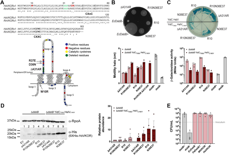
Figure 1 HsVKOR is functional in E. coli by mutating two positive residues in the extracytoplasmic loop.A, alignment of Human (Q9BQB6) and rat (Q6TEK4) VKOR using clustal omega (https://www.ebi.ac.uk/jdispatcher/msa/clustalo) membrane protein visualization was done using Protter (https://wlab.ethz.ch/protter/start). Residues highlighted in red were analyzed in this study. Residues highlighted in green were deleted based on a previous spontaneous mutation. B, HsVKOR variants complement motility of E. coli ΔdsbB mutant. Swarming halos of HsVKOR mutants expressed in E. coli ΔdsbB or ΔdsbB YidCT362I HslVC160Y. Top, a representative image. Bottom, halos measured after 48 h incubation at 30 °C. Data represents average ± SD of at least three independent experiments. C, HsVKOR variants complement the β-Galdbs phenotype of E. coli ΔdsbB YidCT362I HslVC160Y strain. Top, a representative image. Bottom, β-Gal activity was quantified after 18 h growth at 30 °C. Data represents average ± SD of three independent experiments. D, mutations in residues 36–37 of HsVKOR help stabilize the protein. Western blotting using anti-His antibody was used to detect HsVKOR variants and anti-RpoA as a loading control. A representative image is shown. The adjusted total band volume of His and RpoA bands was done using ChemiDoc MP. Data represents average ± SD of six independent experiments. E, HsVKORN36E37 complements anaerobic growth of E. coli ΔdsbB. Aerobically grown cells were diluted to ∼9 × 10⁶ CFU/ml (gray dotted line) into anaerobic minimal medium containing 100 mM potassium nitrate. Bacteria were enumerated after 24 h of anaerobic incubation at 37 °C. Data represents the average ± SEM of at least three independent experiments.
Figure 1 HsVKOR is functional in E. coli by mutating two positive residues in the extracytoplasmic loop.A, alignment of Human (Q9BQB6) and rat (Q6TEK4) VKOR using clustal omega (https://www.ebi.ac.uk/jdispatcher/msa/clustalo) membrane protein visualization was done using Protter (https://wlab.ethz.ch/protter/start). Residues highlighted in red were analyzed in this study. Residues highlighted in green were deleted based on a previous spontaneous mutation. B, HsVKOR variants complement motility of E. coli ΔdsbB mutant. Swarming halos of HsVKOR mutants expressed in E. coli ΔdsbB or ΔdsbB YidCT362I HslVC160Y. Top, a representative image. Bottom, halos measured after 48 h incubation at 30 °C. Data represents average ± SD of at least three independent experiments. C, HsVKOR variants complement the β-Galdbs phenotype of E. coli ΔdsbB YidCT362I HslVC160Y strain. Top, a representative image. Bottom, β-Gal activity was quantified after 18 h growth at 30 °C. Data represents average ± SD of three independent experiments. D, mutations in residues 36–37 of HsVKOR help stabilize the protein. Western blotting using anti-His antibody was used to detect HsVKOR variants and anti-RpoA as a loading control. A representative image is shown. The adjusted total band volume of His and RpoA bands was done using ChemiDoc MP. Data represents average ± SD of six independent experiments. E, HsVKORN36E37 complements anaerobic growth of E. coli ΔdsbB. Aerobically grown cells were diluted to ∼9 × 10⁶ CFU/ml (gray dotted line) into anaerobic minimal medium containing 100 mM potassium nitrate. Bacteria were enumerated after 24 h of anaerobic incubation at 37 °C. Data represents the average ± SEM of at least three independent experiments.CC BY · PubMed Central
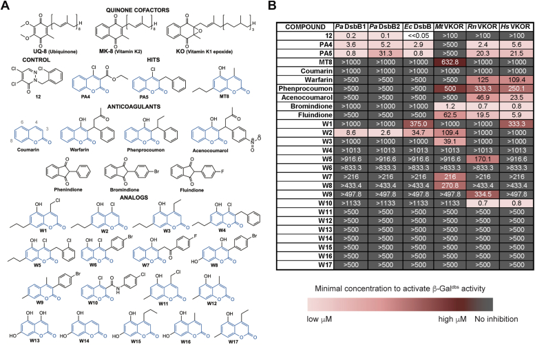
Figure 2 In vivo inhibition of DsbB and VKOR enzymes with warfarin analogs using a heterologous E. coli platform expressing β-Galdbs.A, chemical structures of cofactors, anticoagulants, and warfarin analogs tested against DsbB and VKOR. B, structure-activity relationship analysis of warfarin analogs. E. coli ΔdsbB strain expressing the β-Galdbs carrying P. aeruginosa (Pa) DsbB1, E. coli (Ec) DsbB, M. tuberculosis (Mt), rat (Rn), or human (Hs) VKOR proteins. Strains were grown for 24 h at 30 °C in 384-well minimal media plates containing X-Gal and a serial dilution of drugs (often 0–500 μM). The minimal concentration of drug to produce a pale blue color was reported as the minimal concentration to activate β-Galdbs. The results represent the average of at least three independent experiments using color coding.
Figure 2 In vivo inhibition of DsbB and VKOR enzymes with warfarin analogs using a heterologous E. coli platform expressing β-Galdbs.A, chemical structures of cofactors, anticoagulants, and warfarin analogs tested against DsbB and VKOR. B, structure-activity relationship analysis of warfarin analogs. E. coli ΔdsbB strain expressing the β-Galdbs carrying P. aeruginosa (Pa) DsbB1, E. coli (Ec) DsbB, M. tuberculosis (Mt), rat (Rn), or human (Hs) VKOR proteins. Strains were grown for 24 h at 30 °C in 384-well minimal media plates containing X-Gal and a serial dilution of drugs (often 0–500 μM). The minimal concentration of drug to produce a pale blue color was reported as the minimal concentration to activate β-Galdbs. The results represent the average of at least three independent experiments using color coding.CC BY · PubMed Central
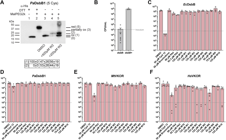
Figure 3 In vivo inhibition of DsbB and VKOR enzymes with warfarin analogs using a heterologous E. coli platform.A, in vivo alkylation was performed in E. coli cells expressing PaDsbB1 grown aerobically in minimal medium with DMSO or compound W2. TCA-precipitated proteins were alkylated with 12.5 mM of MalPEG-2 kDa. Samples were run by SDS-PAGE and immunoblotted with anti-His antibody. 100 mM dithiothreitol (DTT) was used for reducing DSBs. There are four catalytic cysteines in PaDsbB1 plus one non-catalytic. Ox, oxidized the four catalytic cysteines are forming two DSB; red, reduced all cysteines are in the thiol form; partially ox, two catalytic cysteines reduced and two oxidized. The number in parenthesis indicates the cysteines that are in the free thiol form and were derivatized with MalPEG2k (2 kDa addition per cysteine). B, strains were grown aerobically to mid-log phase and diluted to ∼1 × 10⁵ CFU/ml (gray dotted line) into minimal medium containing 100 mM potassium nitrate. Bacteria were enumerated after 24 h of anaerobic incubation at 37 °C. Data represents the average ± SEM of at least three independent experiments. C–F, strains were grown as in (B) but in the presence of DMSO-diluted drugs which were added to one final concentration (indicated in X-axis) with a 0.25% final concentration of DMSO. Bacteria were enumerated after 24 h of anaerobic incubation at 37 °C. Data represents the average ± SEM of at least three independent experiments.
Figure 3 In vivo inhibition of DsbB and VKOR enzymes with warfarin analogs using a heterologous E. coli platform.A, in vivo alkylation was performed in E. coli cells expressing PaDsbB1 grown aerobically in minimal medium with DMSO or compound W2. TCA-precipitated proteins were alkylated with 12.5 mM of MalPEG-2 kDa. Samples were run by SDS-PAGE and immunoblotted with anti-His antibody. 100 mM dithiothreitol (DTT) was used for reducing DSBs. There are four catalytic cysteines in PaDsbB1 plus one non-catalytic. Ox, oxidized the four catalytic cysteines are forming two DSB; red, reduced all cysteines are in the thiol form; partially ox, two catalytic cysteines reduced and two oxidized. The number in parenthesis indicates the cysteines that are in the free thiol form and were derivatized with MalPEG2k (2 kDa addition per cysteine). B, strains were grown aerobically to mid-log phase and diluted to ∼1 × 10⁵ CFU/ml (gray dotted line) into minimal medium containing 100 mM potassium nitrate. Bacteria were enumerated after 24 h of anaerobic incubation at 37 °C. Data represents the average ± SEM of at least three independent experiments. C–F, strains were grown as in (B) but in the presence of DMSO-diluted drugs which were added to one final concentration (indicated in X-axis) with a 0.25% final concentration of DMSO. Bacteria were enumerated after 24 h of anaerobic incubation at 37 °C. Data represents the average ± SEM of at least three independent experiments.CC BY · PubMed Central
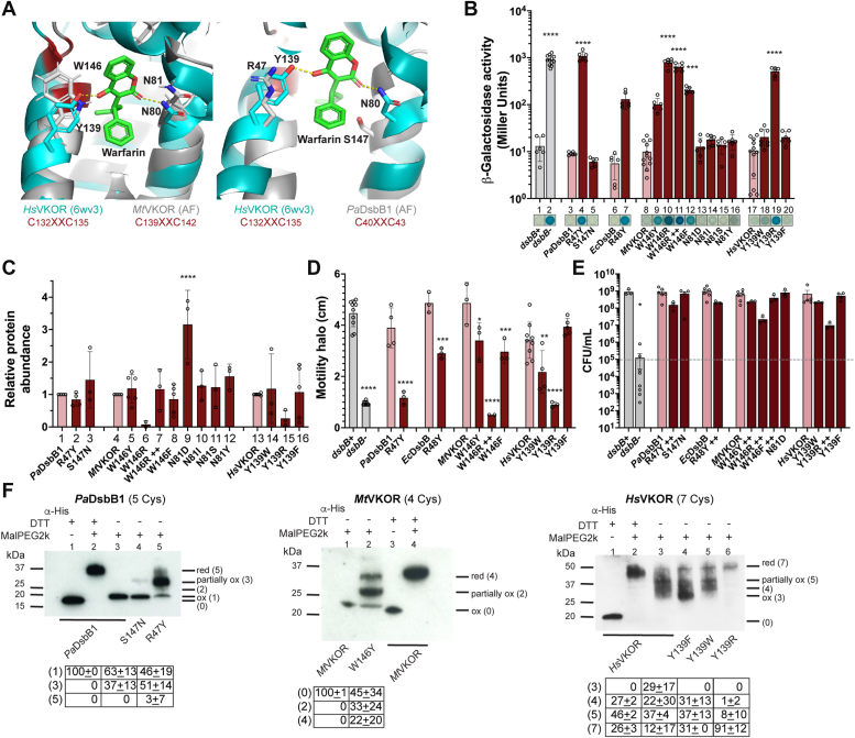
Figure 4 The fourth residue downstream the catalytic CXXC motif is required for de novo oxidation of these cysteines in DsbB and VKOR.A, structural alignment of HsVKOR-warfarin (6wv3) with the AlphaFold (36) predicted structure of MtVKOR (left, I6X5W1) using PyMOL (The PyMOL Molecular Graphics System, Version 2.0 Schrödinger, LLC). Structural alignment of HsVKOR-warfarin (6wv3, residues 97–155) with the AlphaFold predicted structure of PaDsbB1 (right, Q02TM7, residues 1–60). Catalytic cysteines are highlighted in red, and residues that align with N80 and Y139 of HsVKOR are indicated in both alignments. B, mutants in the CXXC+4 in both DsbB and VKOR display high β-Gal activity. β-Gal activity in these strains was quantified after 18 h growth at 30 °C. A representative image is shown on the X-axis. ++ indicates more IPTG. Data represents average ± SD of at least three independent experiments. C, protein abundance across CXXC+4 mutants was determined by Western blot using anti-His antibody and anti-RpoA as loading control compared to their wildtype. Data represents average ± SD of at least three independent experiments. D, CXXC+4 mutants are less motile than the wildtype enzymes. Data represents average ± SD of at least three independent experiments. E, CXXC+4 mutants grow anaerobically. Log-phase aerobically grown cells were diluted to ∼1 × 10⁵ CFU/ml (gray dotted line) into anaerobic minimal medium containing 100 mM potassium nitrate. Bacteria were enumerated after 24 h of anaerobic incubation at 37 °C. Data represents the average ± SEM of at least three independent experiments. Statistical tests were done using Ordinary one-way ANOVA multiple comparisons. p-value ≤ 0.0001 (∗∗∗∗), 0.0002 (∗∗∗), 0.021 (∗∗), 0.0332 (∗), and ns ( ). F, CXXC+4 mutants are found more predominantly in partially reduced states. In vivo alkylation was performed in cells grown aerobically in minimal medium. TCA-precipitated proteins were alkylated with 12.5 mM of MalPEG2k. Samples were run by SDS-PAGE and immunoblotted with anti-His antibody. 100 mM dithiothreitol (DTT) was used for reducing DSBs. Images are representative of at least two independent experiments. There are four catalytic cysteines in each protein plus one non-catalytic in PaDsbB1 or three non-catalytic cysteines in HsVKOR. Ox, oxidized the four catalytic cysteines are forming two DSB (which for MtVKOR the mobility of the oxidized form is the same as that of the reduced protein with no alkylating agent present); red, reduced all cysteines (catalytic and non-catalytic) are in the thiol form; partially ox, two catalytic cysteines reduced and two oxidized. The number in parenthesis indicates the cysteines that are in the free thiol form and were derivatized with MalPEG (2 kDa addition per cysteine).
Figure 4 The fourth residue downstream the catalytic CXXC motif is required for de novo oxidation of these cysteines in DsbB and VKOR.A, structural alignment of HsVKOR-warfarin (6wv3) with the AlphaFold (36) predicted structure of MtVKOR (left, I6X5W1) using PyMOL (The PyMOL Molecular Graphics System, Version 2.0 Schrödinger, LLC). Structural alignment of HsVKOR-warfarin (6wv3, residues 97–155) with the AlphaFold predicted structure of PaDsbB1 (right, Q02TM7, residues 1–60). Catalytic cysteines are highlighted in red, and residues that align with N80 and Y139 of HsVKOR are indicated in both alignments. B, mutants in the CXXC+4 in both DsbB and VKOR display high β-Gal activity. β-Gal activity in these strains was quantified after 18 h growth at 30 °C. A representative image is shown on the X-axis. ++ indicates more IPTG. Data represents average ± SD of at least three independent experiments. C, protein abundance across CXXC+4 mutants was determined by Western blot using anti-His antibody and anti-RpoA as loading control compared to their wildtype. Data represents average ± SD of at least three independent experiments. D, CXXC+4 mutants are less motile than the wildtype enzymes. Data represents average ± SD of at least three independent experiments. E, CXXC+4 mutants grow anaerobically. Log-phase aerobically grown cells were diluted to ∼1 × 10⁵ CFU/ml (gray dotted line) into anaerobic minimal medium containing 100 mM potassium nitrate. Bacteria were enumerated after 24 h of anaerobic incubation at 37 °C. Data represents the average ± SEM of at least three independent experiments. Statistical tests were done using Ordinary one-way ANOVA multiple comparisons. p-value ≤ 0.0001 (∗∗∗∗), 0.0002 (∗∗∗), 0.021 (∗∗), 0.0332 (∗), and ns ( ). F, CXXC+4 mutants are found more predominantly in partially reduced states. In vivo alkylation was performed in cells grown aerobically in minimal medium. TCA-precipitated proteins were alkylated with 12.5 mM of MalPEG2k. Samples were run by SDS-PAGE and immunoblotted with anti-His antibody. 100 mM dithiothreitol (DTT) was used for reducing DSBs. Images are representative of at least two independent experiments. There are four catalytic cysteines in each protein plus one non-catalytic in PaDsbB1 or three non-catalytic cysteines in HsVKOR. Ox, oxidized the four catalytic cysteines are forming two DSB (which for MtVKOR the mobility of the oxidized form is the same as that of the reduced protein with no alkylating agent present); red, reduced all cysteines (catalytic and non-catalytic) are in the thiol form; partially ox, two catalytic cysteines reduced and two oxidized. The number in parenthesis indicates the cysteines that are in the free thiol form and were derivatized with MalPEG (2 kDa addition per cysteine).CC BY · PubMed Central
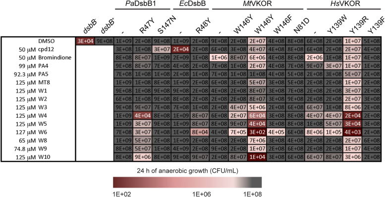
Figure 5 CXXC+4 mutants are sensitized to 4-chloro-coumarin analogs with substitutions at position 3. Log-phase aerobically grown cells were diluted to ∼1 × 10⁵ CFU/ml into anaerobic minimal medium containing 100 mM potassium nitrate. DMSO-diluted drugs were added to one final concentration indicated (final 0.25% DMSO). Bacteria were enumerated after 24 h of anaerobic incubation at 37 °C. The results represent the average of at least three independent experiments using color coding. See Fig. S3 for complete data.
Figure 5 CXXC+4 mutants are sensitized to 4-chloro-coumarin analogs with substitutions at position 3. Log-phase aerobically grown cells were diluted to ∼1 × 10⁵ CFU/ml into anaerobic minimal medium containing 100 mM potassium nitrate. DMSO-diluted drugs were added to one final concentration indicated (final 0.25% DMSO). Bacteria were enumerated after 24 h of anaerobic incubation at 37 °C. The results represent the average of at least three independent experiments using color coding. See Fig. S3 for complete data.CC BY · PubMed Central
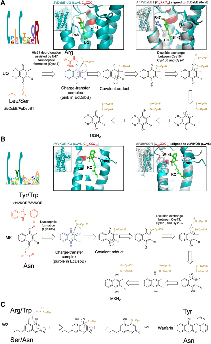
Figure 6 Role of CXXC+4 residue in both DsbB and VKOR enzymes is conserved and stabilizes the quinone intermediates. See text for details. HMM logo of the Pfam seed models for DsbB (PF02600) and VKOR (PF07884) visualized with Skylign (https://www.ebi.ac.uk/interpro/) (46). A, structural alignment of EcDsbB-UQ (6wvf) with the AlphaFold (36) predicted structure of PaDsbB1 (Q02TM7). B, structural alignment of HsVKOR-KO (6wv5) with the AlphaFold (36) predicted structure of MtVKOR (I6X5W1). Alignments were done using PyMOL (The PyMOL Molecular Graphics System, Version 2.0 Schrödinger, LLC). Residues important for quinone and warfarin-like molecules are depicted. Quinone intermediates are presented with the residues that stabilize them at the top and bottom (this does not reflect the structural position). Models were generated with ChemDraw 22.2.0 (47) using models from Ref. (25, 48). C, proposed mechanism of binding of DsbB and VKOR enzymes to compound W2 (left) based on HsVKOR with warfarin (right) as previously reported (23).
Figure 6 Role of CXXC+4 residue in both DsbB and VKOR enzymes is conserved and stabilizes the quinone intermediates. See text for details. HMM logo of the Pfam seed models for DsbB (PF02600) and VKOR (PF07884) visualized with Skylign (https://www.ebi.ac.uk/interpro/) (46). A, structural alignment of EcDsbB-UQ (6wvf) with the AlphaFold (36) predicted structure of PaDsbB1 (Q02TM7). B, structural alignment of HsVKOR-KO (6wv5) with the AlphaFold (36) predicted structure of MtVKOR (I6X5W1). Alignments were done using PyMOL (The PyMOL Molecular Graphics System, Version 2.0 Schrödinger, LLC). Residues important for quinone and warfarin-like molecules are depicted. Quinone intermediates are presented with the residues that stabilize them at the top and bottom (this does not reflect the structural position). Models were generated with ChemDraw 22.2.0 (47) using models from Ref. (25, 48). C, proposed mechanism of binding of DsbB and VKOR enzymes to compound W2 (left) based on HsVKOR with warfarin (right) as previously reported (23).CC BY · PubMed Central

Verification

Claims the paper makes

The claims and statistics of this paper haven’t been checked yet.

Run the check — free, no account needed. It reads the open-access text and checks the sources it cites.

A few free checks a day · papers already verified open instantly · results are public

Self-check

Percentages, sums and reported differences re-computed from the paper’s own numbers.

The self-check needs the paper’s full text.

Citations

Cites

Metadata

Authors
Chavez, D., Amarquaye, G. N., Mejia-Santana, A., Dyotima, Ryan, K., Zeng, L., & Landeta, C.
Affiliations
Indiana University Bloomington (US)
Corresponding author
Landeta, Cristina
Venue
Journal of Biological Chemistry
Publisher
openRxiv
Published
2024-02-19
Volume
300
Issue
6
Pages
107383-107383
ISSN
0021-9258 (ISSN-L)
OpenAlex
W4396979285
Cited by
4
References
1
Citations by year
2026: 1 · 2025: 3
FWCI
0.54
Type
journal article
Language
en
Open access
Locations
  • Journal of Biological Chemistry (publishedVersion)
  • bioRxiv (Cold Spring Harbor Laboratory) (acceptedVersion, cc-by-nd)
  • PubMed Central (publishedVersion, cc-by)
  • PubMed (publishedVersion)
Funders
Indiana University Bloomington
Topics
  • Life Sciences › Biochemistry, Genetics and Molecular Biology › Molecular Biology › Redox biology and oxidative stress
  • Life Sciences › Biochemistry, Genetics and Molecular Biology › Structural Biology › Enzyme Structure and Function
  • Life Sciences › Pharmacology, Toxicology and Pharmaceutics › Toxicology › Bioactive Compounds and Antitumor Agents
Keywords
VKORC1, DsbB, DsbA, disulfide bond formation, quinone reduction, structure-activity relationship, enzyme selectivity, anticoagulant drug design, antibiotic design, Escherichia coli, protein folding
MeSH terms
Anticoagulants, Bacterial Proteins, Coumarins, Disulfides, Escherichia coli, Humans, Membrane Proteins, Protein Binding, Structure-Activity Relationship, Warfarin, Benzoquinones, Protein Disulfide-Isomerases, Escherichia coli Proteins, Vitamin K Epoxide Reductases

About these checks

Registry, retraction, Cites and statistics results are computed, not judged, with one exception: a printed reference that no index lists is read once by a model before it counts as a citation error. Claims are extracted by a model and kept only when their quote appears verbatim in the text; each cited source is then checked, with a model’s help, for whether it says what the claim relies on. Those verdicts can be wrong — read the claim’s quote and the cited source before relying on one.

Statistics and arithmetic are recomputed, not judged.

Confidence bands are shown only for fields calibrated on our annotated test set. None is calibrated yet, so no band is shown.

What counts as a claim ›How we measure confidence ›How we check this ›

Are you an author, or do you see a problem with this page? Request a review

Data sources

Bibliographic data: Semantic Scholar, OpenAlex, Crossref.

Contains information from the Semantic Scholar Academic Graph Datasets, made available under the ODC Attribution License (ODC-BY 1.0).

Semantic Scholar’s data is described in: Kinney, R., et al. (2023). The Semantic Scholar Open Data Platform. arXiv:2301.10140.