

The DOI matches its Crossref record; 2 of the 2 references it cites were found in a registry or an index.
CCfrag: Scanning folding potential of coiled-coil fragments with AlphaFold
Open accessPreprint — published as 10.1093/bioadv/vbae195
Abstract
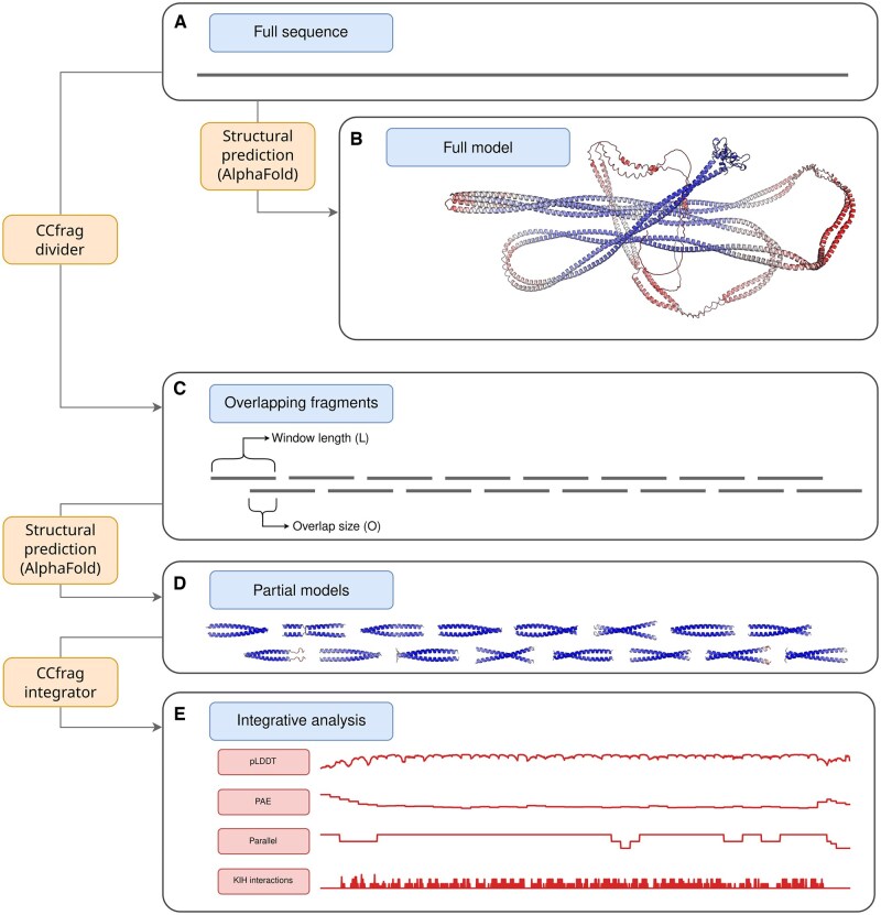
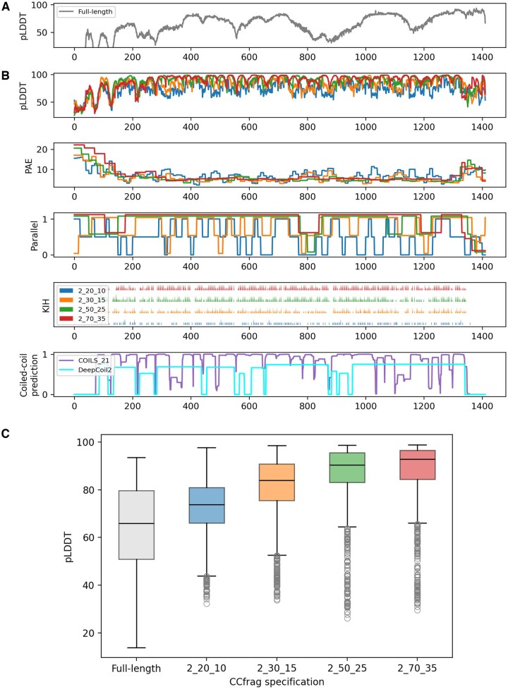
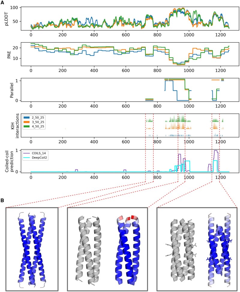
Structured abstract: Motivation: Coiled coils are a widespread structural motif consisting of multiple α-helices that wind around a central axis to bury their hydrophobic core. Although their backbone can be uniquely described by the Crick parametric equations, these have little practical application in structural prediction, given that most coiled coils in nature feature non-canonical repeats that locally distort their geometry. While AlphaFold has emerged as an effective coiled-coil modeling tool, capable of accurately predicting changes in periodicity and core geometry along coiled-coil stalks, it is not without limitations. These include the generation of spuriously bent models and the inability to effectively model globally non-canonical coiled coils. In an effort to overcome these limitations, we investigated whether dividing full-length sequences into fragments would result in better models. Results: We developed CCfrag to leverage AlphaFold for the piece-wise modeling of coiled coils. The user can create a specification, defined by window size, length of overlap, and oligomerization state, and the program produces the files necessary to run structural predictions with AlphaFold. Then, the structural models and their scores are integrated into a rich per-residue representation defined by sequence-or structure-based features, which can be visualized or employed for further analysis. Our results suggest that removing coiled-coil sequences from their native context can in some case improve the prediction confidence and avoids bent models with spurious contacts. In this paper, we present various use cases of CCfrag, and propose that fragment-based prediction is useful for understanding the properties of long, fibrous coiled coils, by showing local features not seen in full-length models. Availability and Implementation: The program is implemented as a Python module. The code and its documentation are available at https://github.com/Mikel-MG/CCfrag . Contact: mikel.martinez@tuebingen.mpg.de
Overview
The claims and statistics of this paper haven’t been checked yet.
Run the check — free, no account needed. It reads the open-access text and checks the sources it cites.
Integrity signals
Registry
Cites
Statistics
Figures
Verification
Claims the paper makes
The claims and statistics of this paper haven’t been checked yet.
Run the check — free, no account needed. It reads the open-access text and checks the sources it cites.
Self-check
Percentages, sums and reported differences re-computed from the paper’s own numbers.
The self-check needs the paper’s full text.
Citations
Cites
Highly accurate protein structure prediction with AlphaFold
Jumper, J., Evans, R., Pritzel, A., Green, T., Figurnov, M., Ronneberger, O., Tunyasuvunakool, K., Bates, R., Žídek, A., Potapenko, A., et al. (2021) · Nature · doi:10.1038/s41586-021-03819-2 · Cited by 43,764
Predicting multiple conformations via sequence clustering and AlphaFold2
Wayment-Steele, H. K., Ojoawo, A., Otten, R., Apitz, J. M., Pitsawong, W., Hömberger, M., Ovchinnikov, S., Colwell, L., & Kern, D. (2024) · Nature · doi:10.1038/s41586-023-06832-9 · Cited by 545
Research works printed but not linked: 17
- 10.1107/S0365110X53001964 · DOI · Paper page
- 10.1107/S0365110X53001952 · DOI · Paper page
- 2006, Structures and Polymorphic Interactions of Two Heptad-Repeat Regions of the SARS Virus S2 Protein, Structure (London, England: 1993), 14, 889, 10.1016/j.str.2006.03.007 · DOI
- Evans R , O’Neill M , Pritzel A et al. Protein Complex Prediction with AlphaFold-Multimer. Bioinformatics, 2021. · DOI · Paper page
- 10.1126/science.8248779 · DOI · Paper page
- 10.1093/protein/gzm071 · DOI
- 2021, Socket2: a program for locating, visualizing and analyzing coiled-coil interfaces in protein structures, Bioinformatics, 37, 4575, 10.1093/bioinformatics/btab631 · DOI · Paper page
- Lin Z , Akin H , Rao R et al. Evolutionary-Scale Prediction of Atomic Level Protein Structure with a Language Model. Synthetic Biology, 2022. · DOI · Paper page
- 10.1093/bioinformatics/bty1062 · DOI
- 10.1126/science.252.5009.1162 · DOI · Paper page
- 10.1016/j.tibs.2016.10.007 · DOI
- 2017, The Structure and Topology of α-Helical Coiled Coils, Fibrous Proteins: Structures and Mechanisms, 82, 95
- Madaj R , Martinez-Goikoetxea M , Kaminski K et al. Applicability of AlphaFold2 in the modelling of coiled-coil domains. 2024:2024.03.07.583852. · DOI
- 2023, New protein families with hendecad coiled coils in the proteome of life, Journal of Structural Biology, 215, 108007, 10.1016/j.jsb.2023.108007 · DOI
- 10.1038/nature19326 · DOI
- 10.1016/J.CELL.2024.01.022 · DOI · Paper page
- 10.1002/pro.4846 · DOI
Metadata
- Authors
- Martinez-Goikoetxea, M.
- Venue
- Bioinformatics Advances
- Publisher
- openRxiv
- Published
- 2024-05-30
- Volume
- 5
- Issue
- 1
- Pages
- vbae195-vbae195
- ISSN
- 2635-0041 (ISSN-L)
- PMC
- PMC11676326
- OpenAlex
- W4405223295
- Cited by
- 3
- References
- 2
- Citations by year
- 2026: 1 · 2025: 2
- FWCI
- 0.41
- Type
- journal article
- Language
- en
- Open access
- gold
- doi.org
- Licence
- cc-by
- Locations
- Bioinformatics Advances (publishedVersion, cc-by)
- bioRxiv (Cold Spring Harbor Laboratory) (acceptedVersion, cc-by-nc)
- PubMed Central (publishedVersion, cc-by)
- PubMed (publishedVersion)
- Funders
- Max-Planck-Gesellschaft
- Topics
- Life Sciences › Biochemistry, Genetics and Molecular Biology › Molecular Biology › Protein Structure and Dynamics
- Life Sciences › Biochemistry, Genetics and Molecular Biology › Molecular Biology › Machine Learning in Bioinformatics
- Life Sciences › Biochemistry, Genetics and Molecular Biology › Structural Biology › Enzyme Structure and Function
- Keywords
- coiled coil, AlphaFold, oligomeric state, fibrous proteins, protein structure prediction
About these checks
Registry, retraction, Cites and statistics results are computed, not judged, with one exception: a printed reference that no index lists is read once by a model before it counts as a citation error. Claims are extracted by a model and kept only when their quote appears verbatim in the text; each cited source is then checked, with a model’s help, for whether it says what the claim relies on. Those verdicts can be wrong — read the claim’s quote and the cited source before relying on one.
Statistics and arithmetic are recomputed, not judged.
Confidence bands are shown only for fields calibrated on our annotated test set. None is calibrated yet, so no band is shown.
What counts as a claim ›How we measure confidence ›How we check this ›
Are you an author, or do you see a problem with this page? Request a review
