PreprintCited by 5Chromosomal and Genetic VariationsRNA Interference and Gene DeliveryPlant Molecular Biology Research

The DOI matches its Crossref record; 1 of the 1 references it cites were found in a registry or an index.

Molecular insight into the Drosophila piRNA pathway network through a combination of systematic protein interaction screening and structural prediction

Open access

Preprint — published as 10.1093/nar/gkaf993

Abstract

SUMMARY: piRNA-bound PIWI proteins mediate the silencing of transposons at both the transcriptional and post-transcriptional levels, processes that are critical for genome integrity and fertility in animals. While numerous additional proteins are known to be essential for piRNA biogenesis and function in Drosophila and other animals, their molecular and mechanistic functions have remained largely unknown. To improve our molecular understanding of the Drosophila piRNA pathway, we used a cell culture-based protein-protein interaction assay called ReLo to perform a systematic pairwise interaction screen involving 22 factors operating in the cytoplasm, including PIWI proteins, Tudor domain-containing proteins (TDRDs), RNA helicases, and mitochondrial surface proteins. Through additional ReLo interaction testing and structural modeling using AlphaFold-Multimer, we have characterized six protein complexes at the molecular and structural levels. We believe that the results of this screen and our methodological approach are likely to guide future research into the molecular mechanisms underlying piRNA biogenesis and function.

Overview

The claims and statistics of this paper haven’t been checked yet.

Run the check — free, no account needed. It reads the open-access text and checks the sources it cites.

A few free checks a day · papers already verified open instantly · results are public

Integrity signals

Registry

Crossref · 10.1101/2024.05.31.596839

Cites

1 of 1 references verified0 citation errors · 0 not checkable

Statistics

Figures

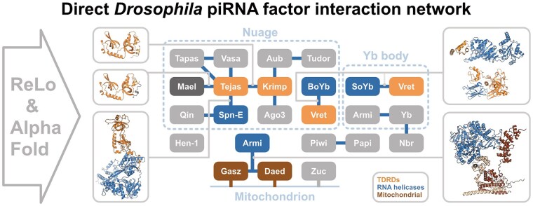
Graphical Abstract
Graphical Abstract CC BY-NC · PubMed Central
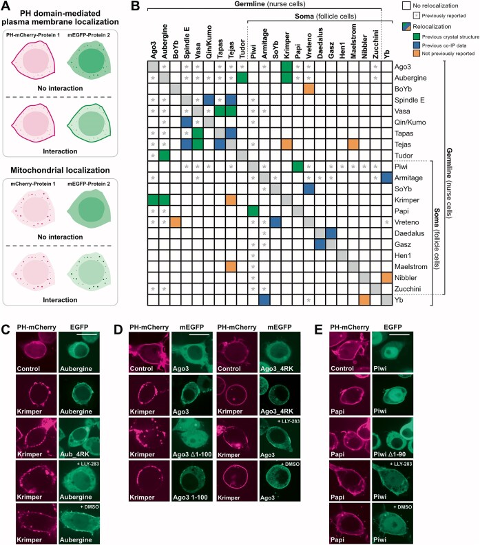
Figure 1. ReLo assay-based PPI screen of cytoplasmic and mitochondrial surface piRNA pathway factors. (A) Principle of the ReLo assay. Two constructs, as indicated, were coexpressed in S2R+ cells, and an interaction between them was inferred if unanchored protein 2 relocalized to the site of membrane-anchored protein 1. Adapted from Fig. 1 of Salgania et al. [53]. (B) Summary of the PPI screen. Colored rectangles indicate PPIs identified by ReLo assays and categorized as previously characterized by crystal structures (green), previously observed in co-IPs (blue), or previously unreported (orange). Homodimer formation was not tested (gray). Boxes marked with an asterisk (*) indicate previously reported interactions based on co-IP, Y2H, CL-IP, but not crystal structures (see Supplementary Table S1) that could not be detected using the ReLo assay. Although redundant, a square layout was chosen over a triangular one to facilitate quick access to interaction partners of any given protein. (C) The interaction between Krimper and Aubergine was sDMA-dependent in ReLo assays. The interactions between Krimper and Ago3 (D) and between Papi and Piwi (E) were sDMA-independent. LLY-283 and DMSO concentrations were 0.5 μM and 0.005%, respectively, in panels (C) and (E) and 1 μM and 0.01%, respectively, in panel (D). The 4RK mutations in Aub and Ago3 were R11K/R13K/R15K/R17K and R4K/R68K/R70K/R72K, respectively. Scale bars are 10 μm. Arginine methylation of Aubergine in DMSO control samples and its absence in LLY-283-treated samples was verified by mass spectrometry (Supplementary Fig. S4).
Figure 1. ReLo assay-based PPI screen of cytoplasmic and mitochondrial surface piRNA pathway factors. (A) Principle of the ReLo assay. Two constructs, as indicated, were coexpressed in S2R+ cells, and an interaction between them was inferred if unanchored protein 2 relocalized to the site of membrane-anchored protein 1. Adapted from Fig. 1 of Salgania et al. [53]. (B) Summary of the PPI screen. Colored rectangles indicate PPIs identified by ReLo assays and categorized as previously characterized by crystal structures (green), previously observed in co-IPs (blue), or previously unreported (orange). Homodimer formation was not tested (gray). Boxes marked with an asterisk (*) indicate previously reported interactions based on co-IP, Y2H, CL-IP, but not crystal structures (see Supplementary Table S1) that could not be detected using the ReLo assay. Although redundant, a square layout was chosen over a triangular one to facilitate quick access to interaction partners of any given protein. (C) The interaction between Krimper and Aubergine was sDMA-dependent in ReLo assays. The interactions between Krimper and Ago3 (D) and between Papi and Piwi (E) were sDMA-independent. LLY-283 and DMSO concentrations were 0.5 μM and 0.005%, respectively, in panels (C) and (E) and 1 μM and 0.01%, respectively, in panel (D). The 4RK mutations in Aub and Ago3 were R11K/R13K/R15K/R17K and R4K/R68K/R70K/R72K, respectively. Scale bars are 10 μm. Arginine methylation of Aubergine in DMSO control samples and its absence in LLY-283-treated samples was verified by mass spectrometry (Supplementary Fig. S4).CC BY-NC · PubMed Central
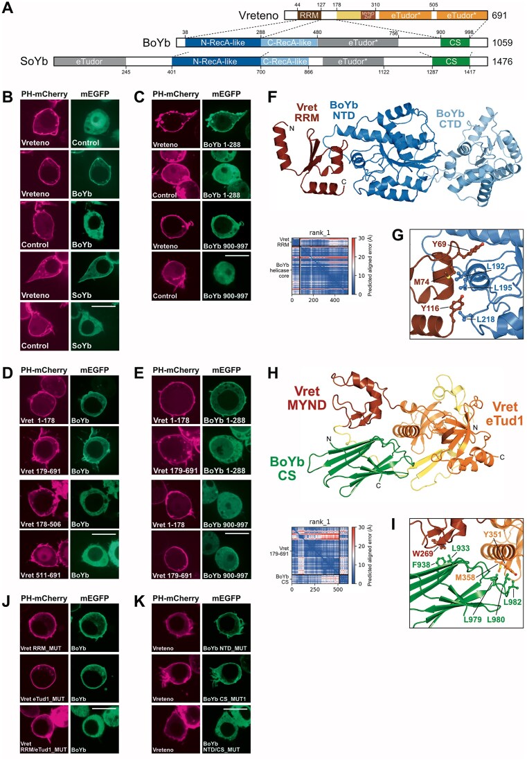
Figure 2. Interaction between Vreteno and BoYb. (A) Domain organization of Vreteno, BoYb, and SoYb. RRM, RNA recognition motif; ZnF, zinc finger; N-RecA-like, NTD; C-RecA-like, CTD; CS, CHORD and Sgt1 domain. The asterisk indicates that the eTudor domain lacks an intact aromatic cage. (B) Vreteno binds to BoYb and SoYb in ReLo assays. (C) The NTD and the CS domains of BoYb each contribute to Vret binding. (D) The N- and C-terminal portions of Vret each contribute to BoYb binding. (E) The N-terminal part of Vret binds the NTD of BoYb and the C-terminal part of Vret binds the CS domain of BoYb. (F) AlphaFold-Multimer-generated structural model of the complex consisting of the Vret–RRM (aa 43–117) and the helicase core of BoYb (aa 38–476). The PAE plot indicates the high confidence of the prediction (see also Supplementary Fig. S6). (G) Detail of the interface formed by the Vret-RRM and the BoYb core. Residues mutated for subsequent studies are highlighted in ball-and-stick representation. (H) AlphaFold-Multimer-generated structural model of the complex consisting of the C-terminal part of Vret (aa 179–691) with the MYND domain and the eTudor domain bound to the CS domain of BoYb (aa 901–998). The PAE plot shows the high confidence of the model. (I) Detail of the interface formed by the C-terminal part of Vret and the BoYb CS domain. Residues mutated for subsequent studies are highlighted in ball-and-stick representation. (J) Combined mutations in the RRM (RRM_MUT: Y69E/M74E/Y116E) and eTudor (eTud1_MUT: Y351E/M358E) surfaces of Vret are required to abolish the BoYb interaction. (K) Mutations in both the NTD (NTD_MUT: L192E/L195E/L218E) and the CS domain (CS_MUT1: L979E/L980E/L982E) of BoYb are required to abolish the Vret interaction. Scale bars are 10 μm.
Figure 2. Interaction between Vreteno and BoYb. (A) Domain organization of Vreteno, BoYb, and SoYb. RRM, RNA recognition motif; ZnF, zinc finger; N-RecA-like, NTD; C-RecA-like, CTD; CS, CHORD and Sgt1 domain. The asterisk indicates that the eTudor domain lacks an intact aromatic cage. (B) Vreteno binds to BoYb and SoYb in ReLo assays. (C) The NTD and the CS domains of BoYb each contribute to Vret binding. (D) The N- and C-terminal portions of Vret each contribute to BoYb binding. (E) The N-terminal part of Vret binds the NTD of BoYb and the C-terminal part of Vret binds the CS domain of BoYb. (F) AlphaFold-Multimer-generated structural model of the complex consisting of the Vret–RRM (aa 43–117) and the helicase core of BoYb (aa 38–476). The PAE plot indicates the high confidence of the prediction (see also Supplementary Fig. S6). (G) Detail of the interface formed by the Vret-RRM and the BoYb core. Residues mutated for subsequent studies are highlighted in ball-and-stick representation. (H) AlphaFold-Multimer-generated structural model of the complex consisting of the C-terminal part of Vret (aa 179–691) with the MYND domain and the eTudor domain bound to the CS domain of BoYb (aa 901–998). The PAE plot shows the high confidence of the model. (I) Detail of the interface formed by the C-terminal part of Vret and the BoYb CS domain. Residues mutated for subsequent studies are highlighted in ball-and-stick representation. (J) Combined mutations in the RRM (RRM_MUT: Y69E/M74E/Y116E) and eTudor (eTud1_MUT: Y351E/M358E) surfaces of Vret are required to abolish the BoYb interaction. (K) Mutations in both the NTD (NTD_MUT: L192E/L195E/L218E) and the CS domain (CS_MUT1: L979E/L980E/L982E) of BoYb are required to abolish the Vret interaction. Scale bars are 10 μm.CC BY-NC · PubMed Central
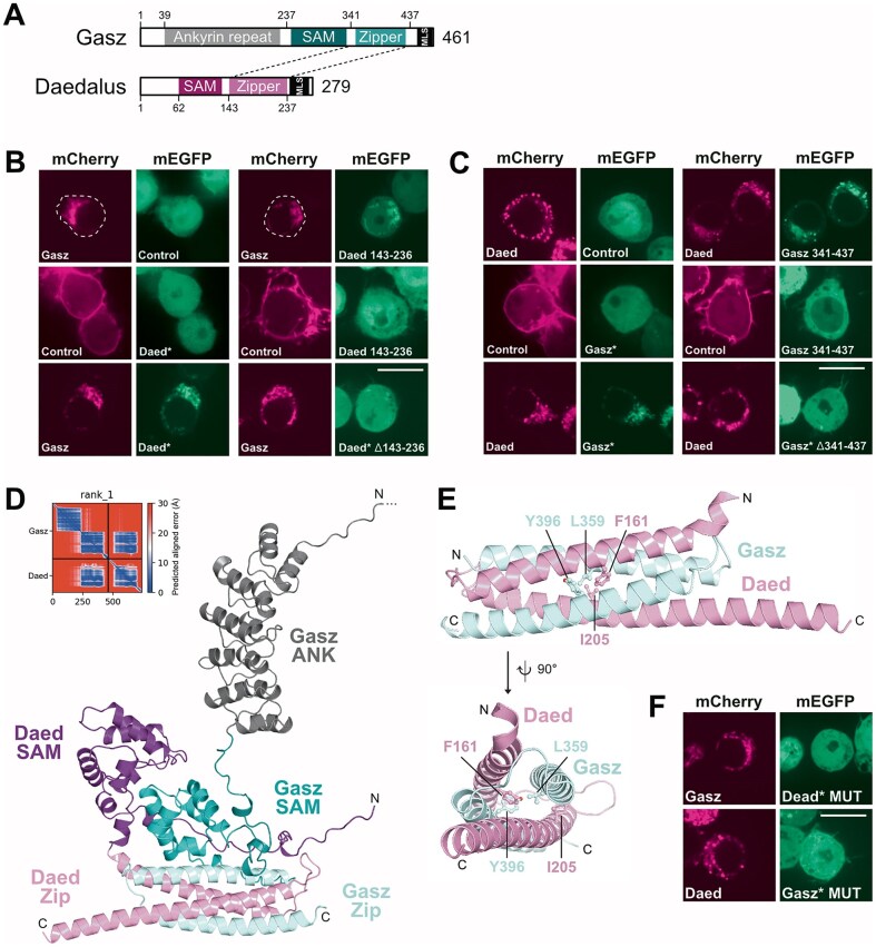
Figure 3. Interaction between Gasz and Daed. (A) Domain organization of Gasz and Daed. SAM, sterile α motif; MLS, mitochondrial localization signal. (BandC) Daed bound to Gasz in ReLo assays. The interaction required the Zipper domain of Daed (B) and Gasz (C). An asterisk indicates that the protein (additionally) lacks the C-terminal MLS, corresponding to aa 438–461 in Gasz and aa 237–279 in Daed. The mCherry-tagged controls also contained a PH domain. (D) AlphaFold-Multimer-generated structural model of the Daed-Gasz complex (missing the MLSs). The PAE plot indicates the high confidence of the prediction (see also Supplementary Fig. S9E). (E) Detail of the interface formed by the Zipper domains of Gasz and Daed. Residues mutated for subsequent studies are highlighted in ball-and-stick representation. (F) Mutations in the Zipper interface prevented the formation of the Gasz–Daed complex. Scale bars are 10 μm.
Figure 3. Interaction between Gasz and Daed. (A) Domain organization of Gasz and Daed. SAM, sterile α motif; MLS, mitochondrial localization signal. (BandC) Daed bound to Gasz in ReLo assays. The interaction required the Zipper domain of Daed (B) and Gasz (C). An asterisk indicates that the protein (additionally) lacks the C-terminal MLS, corresponding to aa 438–461 in Gasz and aa 237–279 in Daed. The mCherry-tagged controls also contained a PH domain. (D) AlphaFold-Multimer-generated structural model of the Daed-Gasz complex (missing the MLSs). The PAE plot indicates the high confidence of the prediction (see also Supplementary Fig. S9E). (E) Detail of the interface formed by the Zipper domains of Gasz and Daed. Residues mutated for subsequent studies are highlighted in ball-and-stick representation. (F) Mutations in the Zipper interface prevented the formation of the Gasz–Daed complex. Scale bars are 10 μm.CC BY-NC · PubMed Central
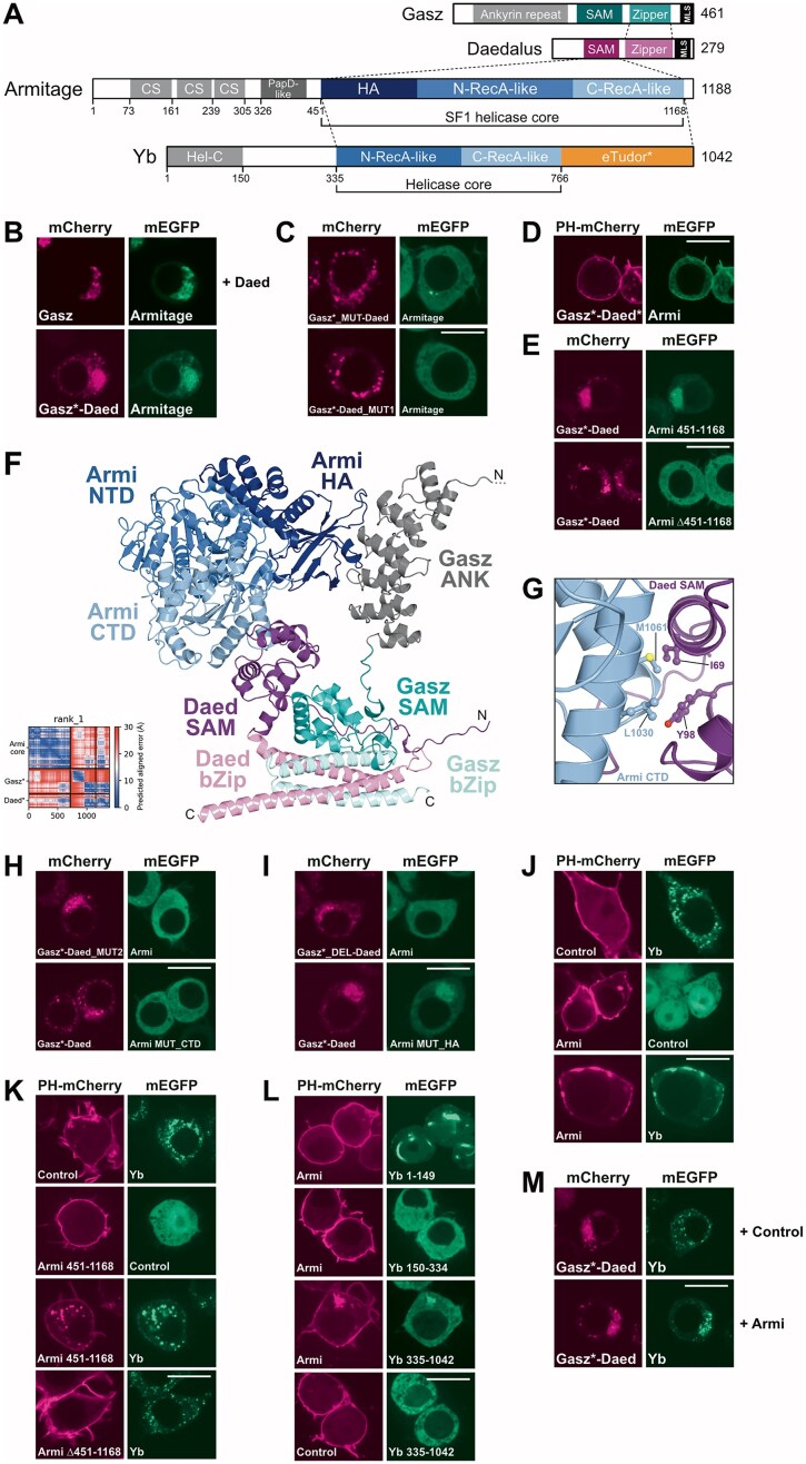
Figure 4. Interaction between Armi and the Gasz–Daed complex. (A) Domain organization of Armi, Gasz, Daed, and Yb. N-RecA-like, NTD; C-RecA-like, CTD; CS, cold-shock domain; HA, helicase-associated domain, SAM, sterile α motif; MLS, mitochondrial localization signal. The asterisk indicates that the eTudor domain lacks an intact aromatic cage. (B) Armi binds to Gasz in the presence of Daed. A Gasz*–Daed fusion lacking the Gasz–MLS recruits Armi to mitochondria. (C) Armi did not bind to the Gasz-Daed fusion carrying the L359/Y396E mutation in the Gasz part (Gasz*_MUT-Daed) or the F161/I205 mutation in the Daed part (Gasz*-Daed_MUT1). (D) A Gasz–Daed fusion carrying the PH domain at the N-terminus and lacking both MLS (Gasz*–Daed*) recruited Armi to the plasma membrane. (E) The Gasz*–Daed fusion bound to the helicase core of Armi (aa 451–1168). (F) AlphaFold-Multimer-generated structural model of the Daed–Gasz complex (lacking the MLSs) bound to the helicase core (aa 451–1168) of Armi. The PAE plot showing the confidence of the prediction is depicted on the right (see also Supplementary Fig. 10C). (G) Detail of the interface formed by the CTD of Armi and the SAM domain of Daed. Residues mutated for subsequent studies are highlighted in ball-and-stick representation. (H) Mutations in the CTD–SAM interface prevented the formation of the Gasz–Daed–Armi complex. The Gasz*–Daed MUT2 is I69E/Y98E located in the Daed part and the Armi MUT_CTD is L1030E/M1061E. (I) Deletion of the Gasz Ankyrin repeat (aa 1–236) from the Gasz*–Daed fusion (Gasz*_DEL-Daed) abolished Armi binding. Mutations at the HA surface of Armi (MUT_HA, W592E/D631A) did not prevent binding to the Gasz–Daed fusion. (J) Yb interacts with Armi in ReLo assays. (K) The Armi helicase core (aa 451–1168) is necessary and sufficient for Yb binding. (L) Armi binds to the C-terminal portion (aa 335–1042) of Yb, which includes the helicase core and the eTudor domain. (M) Yb binds to the Gasz–Daed fusion in the presence of Armi. The asterisk (*) in Gasz indicates that the MLS was deleted from the protein. Scale bars are 10 μm.
Figure 4. Interaction between Armi and the Gasz–Daed complex. (A) Domain organization of Armi, Gasz, Daed, and Yb. N-RecA-like, NTD; C-RecA-like, CTD; CS, cold-shock domain; HA, helicase-associated domain, SAM, sterile α motif; MLS, mitochondrial localization signal. The asterisk indicates that the eTudor domain lacks an intact aromatic cage. (B) Armi binds to Gasz in the presence of Daed. A Gasz*–Daed fusion lacking the Gasz–MLS recruits Armi to mitochondria. (C) Armi did not bind to the Gasz-Daed fusion carrying the L359/Y396E mutation in the Gasz part (Gasz*_MUT-Daed) or the F161/I205 mutation in the Daed part (Gasz*-Daed_MUT1). (D) A Gasz–Daed fusion carrying the PH domain at the N-terminus and lacking both MLS (Gasz*–Daed*) recruited Armi to the plasma membrane. (E) The Gasz*–Daed fusion bound to the helicase core of Armi (aa 451–1168). (F) AlphaFold-Multimer-generated structural model of the Daed–Gasz complex (lacking the MLSs) bound to the helicase core (aa 451–1168) of Armi. The PAE plot showing the confidence of the prediction is depicted on the right (see also Supplementary Fig. 10C). (G) Detail of the interface formed by the CTD of Armi and the SAM domain of Daed. Residues mutated for subsequent studies are highlighted in ball-and-stick representation. (H) Mutations in the CTD–SAM interface prevented the formation of the Gasz–Daed–Armi complex. The Gasz*–Daed MUT2 is I69E/Y98E located in the Daed part and the Armi MUT_CTD is L1030E/M1061E. (I) Deletion of the Gasz Ankyrin repeat (aa 1–236) from the Gasz*–Daed fusion (Gasz*_DEL-Daed) abolished Armi binding. Mutations at the HA surface of Armi (MUT_HA, W592E/D631A) did not prevent binding to the Gasz–Daed fusion. (J) Yb interacts with Armi in ReLo assays. (K) The Armi helicase core (aa 451–1168) is necessary and sufficient for Yb binding. (L) Armi binds to the C-terminal portion (aa 335–1042) of Yb, which includes the helicase core and the eTudor domain. (M) Yb binds to the Gasz–Daed fusion in the presence of Armi. The asterisk (*) in Gasz indicates that the MLS was deleted from the protein. Scale bars are 10 μm.CC BY-NC · PubMed Central
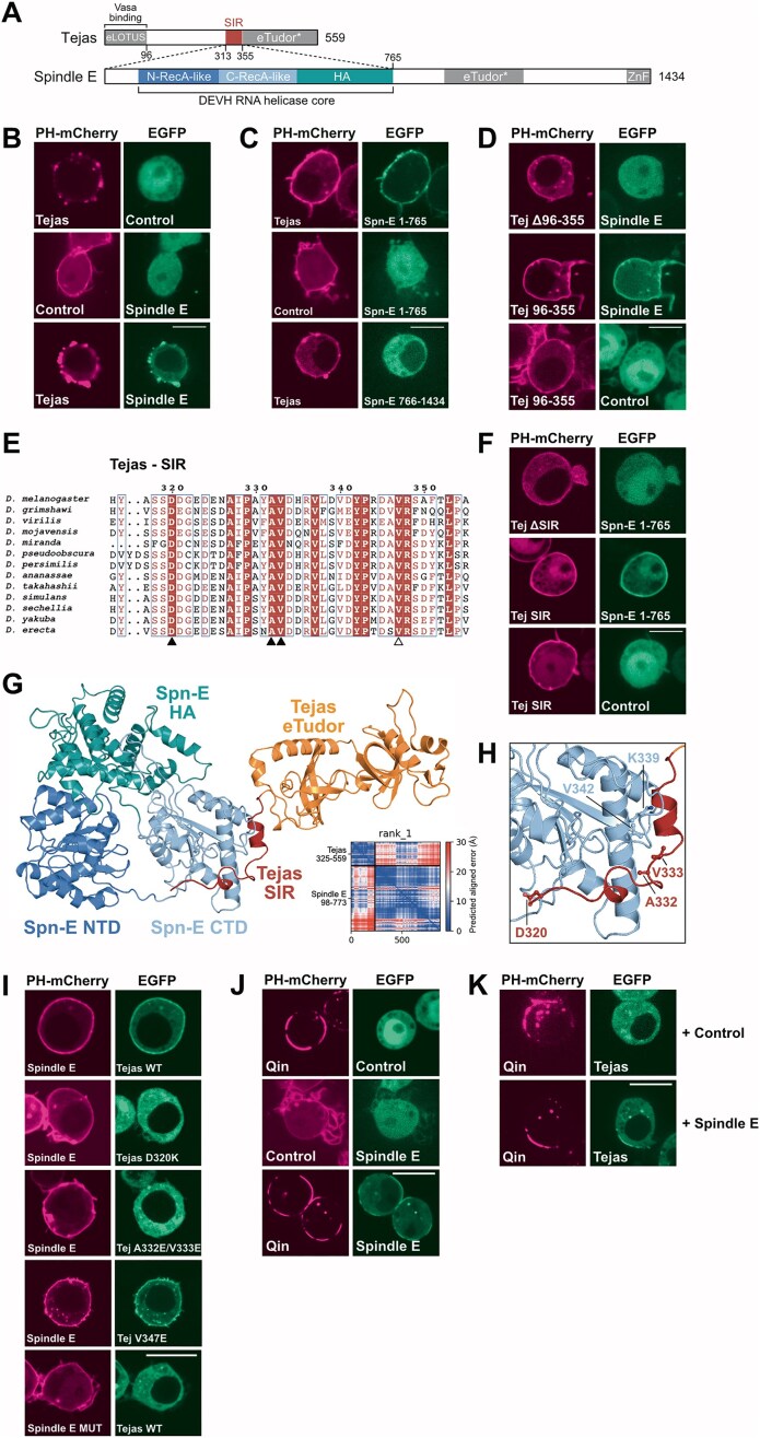
Figure 5. The interaction between Tejas and Spindle E. (legend on next page). (A) Domain organization of Tejas and Spindle E. SIR, Spindle E-interacting region; N-RecA-like, NTD; C-RecA-like, CTD; HA, helicase-associated domain; ZnF, zinc finger. The asterisk indicates that the eTudor domain lacks an intact aromatic cage. (B) Tejas interacts with Spindle E in ReLo assays. (CandD) The interaction is mediated by the N-terminal portion of Spindle E (C) and the predominantly unstructured region of Tejas (D). (E) Multiple sequence alignment showing the SIR, which is the most conserved region of the predominantly unstructured region of Tejas and extends N-terminally of the eTudor domain. (F) The SIR of Tejas is necessary and sufficient for binding to Spindle E. (G) AlphaFold-Multimer-generated structural model of the complex formed by the helicase core of Spindle E (aa 98–773) and the C-terminal part of Tejas comprising the SIR and the eTudor domain (aa 325–559). The PAE plot indicates the confidence of the prediction (see also Supplementary Fig. S12D). (H) Detail of the interface formed by the Tejas–Spindle E complex. Residues mutated for subsequent studies are highlighted in ball-and-stick representation. (I) Mutations in the predicted interface, as indicated, prevented formation of the Tejas–Spindle E complex. Spindle E MUT is K339/V342E. (J) Spindle E bound to Qin in ReLo assays. (K) Qin bound to Tejas in the presence of Spindle E. Scale bars are 10 μm.
Figure 5. The interaction between Tejas and Spindle E. (legend on next page). (A) Domain organization of Tejas and Spindle E. SIR, Spindle E-interacting region; N-RecA-like, NTD; C-RecA-like, CTD; HA, helicase-associated domain; ZnF, zinc finger. The asterisk indicates that the eTudor domain lacks an intact aromatic cage. (B) Tejas interacts with Spindle E in ReLo assays. (CandD) The interaction is mediated by the N-terminal portion of Spindle E (C) and the predominantly unstructured region of Tejas (D). (E) Multiple sequence alignment showing the SIR, which is the most conserved region of the predominantly unstructured region of Tejas and extends N-terminally of the eTudor domain. (F) The SIR of Tejas is necessary and sufficient for binding to Spindle E. (G) AlphaFold-Multimer-generated structural model of the complex formed by the helicase core of Spindle E (aa 98–773) and the C-terminal part of Tejas comprising the SIR and the eTudor domain (aa 325–559). The PAE plot indicates the confidence of the prediction (see also Supplementary Fig. S12D). (H) Detail of the interface formed by the Tejas–Spindle E complex. Residues mutated for subsequent studies are highlighted in ball-and-stick representation. (I) Mutations in the predicted interface, as indicated, prevented formation of the Tejas–Spindle E complex. Spindle E MUT is K339/V342E. (J) Spindle E bound to Qin in ReLo assays. (K) Qin bound to Tejas in the presence of Spindle E. Scale bars are 10 μm.CC BY-NC · PubMed Central
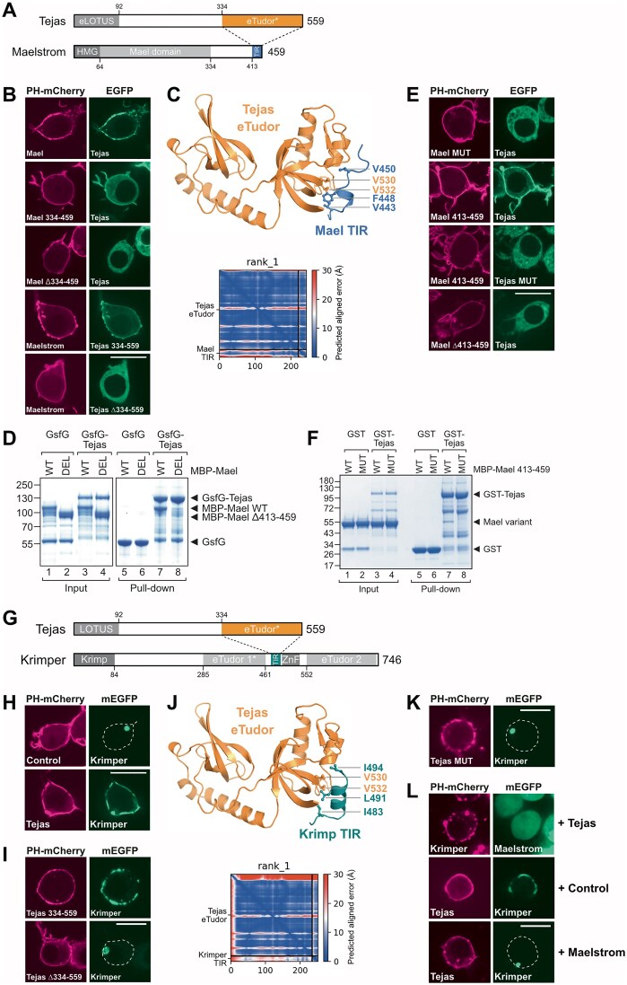
Figure 6. The interaction between Tejas and Maelstrom or Krimper. (A) Domain organization of Tejas and Maelstrom. HMG, high mobility group box. The asterisk indicates that the eTudor domain lacks an intact aromatic cage. (B) Tejas interacts with Maelstrom in ReLo assays and the interaction is mediated by the C-terminal region of Maelstrom (aa 334–459) and the eTudor domain of Tejas (aa 334–559). (C) AlphaFold-Multimer-generated structural model of the complex formed by the eTudor domain of Tejas and the TIR of Maelstrom. Residues mutated for subsequent studies are highlighted in ball-and-stick representation. The PAE plot indicates the confidence of the prediction (see also Supplementary Fig. 13C). (D) GST pull-down assay showing binding of GST–superfolder–GFP (GsfG)–Tejas to full-length MBP–Maelstrom but not to Maelstrom lacking aa 413–459. (E) Mutations in the predicted interface prevent Tejas–Maelstrom complex formation. (F) GST pull-down assay showing binding of GST-Tejas to MBP-fused Mael C-terminus (aa 413–459) but not to a mutated version of it. (G) Domain organization of Tejas and Krimper. The asterisk indicates that the eTudor domain lacks an intact aromatic cage. (H) Tejas interacts with Krimper in ReLo assays. (I) The Krimper interaction is mediated by the eTudor domain of Tejas (aa 334–559). (J) AlphaFold-Multimer-generated structural model of the complex formed by the eTudor domain of Tejas and the TIR (aa 481–495) of Krimper. Residues mutated for subsequent studies are highlighted in ball-and-stick representation. The PAE plot indicates the confidence of the prediction (see also Supplementary Fig. 15E). (K) Mutations in the predicted Tejas interface prevented Tejas–Krimper complex formation. (L) In ReLo assays, Tejas did not bridge Krimper and Maelstrom (top panels). The Tejas–Krimper interaction is abolished by coexpression of Maelstrom (middle and lower panels). Scale bars are 10 μm.
Figure 6. The interaction between Tejas and Maelstrom or Krimper. (A) Domain organization of Tejas and Maelstrom. HMG, high mobility group box. The asterisk indicates that the eTudor domain lacks an intact aromatic cage. (B) Tejas interacts with Maelstrom in ReLo assays and the interaction is mediated by the C-terminal region of Maelstrom (aa 334–459) and the eTudor domain of Tejas (aa 334–559). (C) AlphaFold-Multimer-generated structural model of the complex formed by the eTudor domain of Tejas and the TIR of Maelstrom. Residues mutated for subsequent studies are highlighted in ball-and-stick representation. The PAE plot indicates the confidence of the prediction (see also Supplementary Fig. 13C). (D) GST pull-down assay showing binding of GST–superfolder–GFP (GsfG)–Tejas to full-length MBP–Maelstrom but not to Maelstrom lacking aa 413–459. (E) Mutations in the predicted interface prevent Tejas–Maelstrom complex formation. (F) GST pull-down assay showing binding of GST-Tejas to MBP-fused Mael C-terminus (aa 413–459) but not to a mutated version of it. (G) Domain organization of Tejas and Krimper. The asterisk indicates that the eTudor domain lacks an intact aromatic cage. (H) Tejas interacts with Krimper in ReLo assays. (I) The Krimper interaction is mediated by the eTudor domain of Tejas (aa 334–559). (J) AlphaFold-Multimer-generated structural model of the complex formed by the eTudor domain of Tejas and the TIR (aa 481–495) of Krimper. Residues mutated for subsequent studies are highlighted in ball-and-stick representation. The PAE plot indicates the confidence of the prediction (see also Supplementary Fig. 15E). (K) Mutations in the predicted Tejas interface prevented Tejas–Krimper complex formation. (L) In ReLo assays, Tejas did not bridge Krimper and Maelstrom (top panels). The Tejas–Krimper interaction is abolished by coexpression of Maelstrom (middle and lower panels). Scale bars are 10 μm.CC BY-NC · PubMed Central
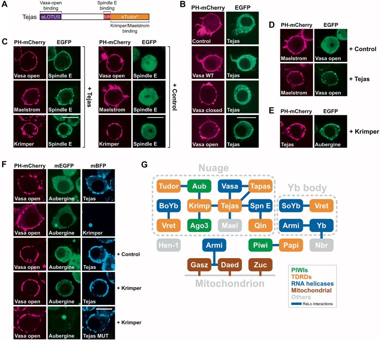
Figure 7. The Tejas interaction network. (A) Tejas and its binding partners. (B) Tejas relocalized to Vasa only when Vasa carried an ATP-binding mutation that kept it in an open conformation (Vasa-open). (C) Spindle interacted with Vasa-open, Maelstrom, or Krimper when Tejas was coexpressed. (D) Maelstrom interacted with Vasa-open when Tejas was coexpressed. (E) Tejas interacted with Aubergine when Krimper was coexpressed. (F) Vasa-open interacted with Aubergine when both Tejas and Krimper were coexpressed. Coexpression of a Tejas mutant that abolishes Krimper binding (Tejas MUT, V530E/V532E) did not mediate the Vasa-open–Aub interaction. mBFP, monomeric blue fluorescent protein. Scale bars are 10 μm. (G) Summary of interactions observed in the ReLo PPI screen. Interactions are indicated by a blue connector. Proteins are grouped according to subcellular localization and colored according to protein features: PIWI proteins (green), TDRDs (orange), RNA helicases (blue), mitochondrially anchored proteins (brown), and others (gray). Note that BoYb, SoYb, and Spindle E contain both eTudor domains and RNA helicase cores.
Figure 7. The Tejas interaction network. (A) Tejas and its binding partners. (B) Tejas relocalized to Vasa only when Vasa carried an ATP-binding mutation that kept it in an open conformation (Vasa-open). (C) Spindle interacted with Vasa-open, Maelstrom, or Krimper when Tejas was coexpressed. (D) Maelstrom interacted with Vasa-open when Tejas was coexpressed. (E) Tejas interacted with Aubergine when Krimper was coexpressed. (F) Vasa-open interacted with Aubergine when both Tejas and Krimper were coexpressed. Coexpression of a Tejas mutant that abolishes Krimper binding (Tejas MUT, V530E/V532E) did not mediate the Vasa-open–Aub interaction. mBFP, monomeric blue fluorescent protein. Scale bars are 10 μm. (G) Summary of interactions observed in the ReLo PPI screen. Interactions are indicated by a blue connector. Proteins are grouped according to subcellular localization and colored according to protein features: PIWI proteins (green), TDRDs (orange), RNA helicases (blue), mitochondrially anchored proteins (brown), and others (gray). Note that BoYb, SoYb, and Spindle E contain both eTudor domains and RNA helicase cores.CC BY-NC · PubMed Central

Verification

Claims the paper makes

The claims and statistics of this paper haven’t been checked yet.

Run the check — free, no account needed. It reads the open-access text and checks the sources it cites.

A few free checks a day · papers already verified open instantly · results are public

Self-check

Percentages, sums and reported differences re-computed from the paper’s own numbers.

The self-check needs the paper’s full text.

Citations

Cites

Research works printed but not linked: 121

  1. 2024, Accurate structure prediction of biomolecular interactions with AlphaFold 3, Nature, 630, 493, 10.1038/s41586-024-07487-w · DOI · Paper page
  2. 10.1038/nature04716 · DOI · Paper page
  3. 10.1038/emboj.2011.449 · DOI
  4. 10.1186/1745-6150-5-1 · DOI
  5. 10.1016/j.jmb.2013.09.027 · DOI
  6. 10.1038/s41586-021-03856-x · DOI
  7. 10.1101/gad.1424106 · DOI
  8. 10.1021/acsmedchemlett.8b00014 · DOI
  9. 10.1016/j.cell.2007.01.043 · DOI · Paper page
  10. Bronkhorst AW , Lee CY , Möckel MM , Ruegenberg S , de Jesus Domingues AM , Sadouki S , Piccinno R , Sumiyoshi T , Siomi MC , Stelzl L , et al. 2023. An extended Tudor domain within Vreteno interconnects Gtsf1L and Ago3 for piRNA biogenesis in Bombyx mori. EMBO J. https://pubmed.ncbi.nlm.nih.gov/37984437/ (Accessed November 27, 2023). · DOI
  11. Buchwald G , Schüssler S , Basquin C , Le Hir H , Conti E. 2013. Crystal structure of the human eIF4AIII-CWC22 complex shows how a DEAD-box protein is inhibited by a MIF4G domain. Proc Natl Acad Sci U S A 110. · DOI
  12. 10.1093/bioinformatics/btq122 · DOI
  13. 10.1016/j.molcel.2018.10.038 · DOI
  14. 10.1261/rna.049437.114 · DOI
  15. 10.1016/j.ymeth.2019.07.002 · DOI
  16. 10.1016/j.tibs.2015.12.008 · DOI
  17. 10.1016/j.molcel.2013.04.007 · DOI · Paper page
  18. 10.1073/pnas.1213283110 · DOI
  19. 10.1074/jbc.272.34.21213 · DOI
  20. Evans R , O’Neill M , Pritzel A , Antropova N , Senior A , Green T , Žídek A , Bates R , Blackwell S , Yim J , et al. 2021. Protein complex prediction with AlphaFold-Multimer. bioRxiv 2021.10.04.463034. https://www.biorxiv.org/content/10.1101/2021.10.04.463034v1 (Accessed March 2, 2022). · DOI · Paper page
  21. 10.1242/dev.00310 · DOI
  22. 10.1080/10409238.2019.1603199 · DOI
  23. 10.1016/S0014-5793(02)03321-5 · DOI
  24. 10.1371/journal.pone.0131080 · DOI
  25. 10.1101/gad.9.20.2495 · DOI
  26. 1988, A conserved NTP-motif in putative helicases, Nature, 333, 22, 10.1038/333022a0 · DOI
  27. 10.1126/science.1140494 · DOI
  28. 10.1155/2016/6809105 · DOI
  29. 10.1101/gad.1968110 · DOI
  30. 10.1126/science.aaa1264 · DOI
  31. 10.1016/j.molcel.2013.04.031 · DOI
  32. 10.1038/emboj.2011.308 · DOI
  33. 10.1093/nar/gkq1127 · DOI
  34. 10.15252/embr.201947732 · DOI
  35. 10.1016/j.bbagrm.2015.09.002 · DOI
  36. 2010, Dali server: Conservation mapping in 3D, Nucleic Acids Res, 38, 545, 10.1093/nar/gkq366 · DOI · Paper page
  37. 10.1016/j.celrep.2015.06.030 · DOI
  38. 10.1083/jcb.201401002 · DOI
  39. 10.1016/j.tig.2017.09.002 · DOI
  40. 10.1038/s41467-020-20241-w · DOI · Paper page
  41. 10.1016/j.molcel.2010.05.014 · DOI
  42. 10.1038/nature11502 · DOI
  43. 10.1016/j.celrep.2015.06.035 · DOI
  44. 10.1016/j.celrep.2019.04.029 · DOI
  45. 10.1016/j.molcel.2010.05.015 · DOI
  46. 10.1038/nature14254 · DOI
  47. 10.1101/gad.297051.117 · DOI
  48. 2021, Mutations in Hsp90 Cochaperones Result in a Wide Variety of Human Disorders, Front Mol Biosci, 8, 787260, 10.3389/fmolb.2021.787260 · DOI
  49. 10.1038/ncb1872 · DOI
  50. 10.1261/rna.1869710 · DOI
  51. 10.1073/pnas.1106676108 · DOI
  52. 10.1074/jbc.273.49.32973 · DOI
  53. 2020, LOTUS-domain proteins - Developmental effectors from a molecular perspective, Biol Chem, 402, 7, 10.1515/hsz-2020-0270 · DOI
  54. 10.1073/pnas.2304385120 · DOI · Paper page
  55. 10.1016/j.celrep.2014.08.013 · DOI
  56. 10.1101/gad.245514.114 · DOI
  57. 10.1073/pnas.0701920104 · DOI
  58. Lin Y , Suyama R , Kawaguchi S , Iki T , Kai T . 2023. Tejas functions as a core component in nuage assembly and precursor processing in Drosophila piRNA biogenesis. J Cell Biol 222. http://www.ncbi.nlm.nih.gov/pubmed/37555815 (Accessed August 14, 2023). · DOI
  59. 10.1101/gad.1956010 · DOI
  60. 10.1073/pnas.1013106107 · DOI
  61. 10.1242/dev.059287 · DOI
  62. 10.1016/j.cell.2009.03.040 · DOI
  63. 10.1261/rna.034181.112 · DOI
  64. 10.1016/j.cell.2016.09.002 · DOI
  65. 10.1016/j.celrep.2015.03.030 · DOI
  66. 2022, ColabFold: making protein folding accessible to all, Nature Methods, 19, 679, 10.1038/s41592-022-01488-1 · DOI · Paper page
  67. 2010, A direct role for Hsp90 in pre-RISC formation in Drosophila, Nature Structural & Molecular Biology, 17, 1024, 10.1038/nsmb.1875 · DOI
  68. 10.1126/science.aaa1039 · DOI
  69. 10.1101/gad.325662.119 · DOI
  70. 10.1016/j.celrep.2014.05.043 · DOI
  71. 10.1016/j.isci.2022.103914 · DOI
  72. 10.1261/rna.063891.117 · DOI
  73. 10.1016/j.celrep.2014.12.013 · DOI
  74. 10.1038/emboj.2009.365 · DOI · Paper page
  75. 10.1038/nature11509 · DOI
  76. 10.1101/gad.1335305 · DOI
  77. 10.1016/j.molcel.2012.07.021 · DOI
  78. 10.1038/emboj.2010.212 · DOI
  79. Onishi R , Sato K , Murano K , Negishi L , Siomi H , Siomi MC . 2020. Piwi suppresses transcription of Brahma-dependent transposons via Maelstrom in ovarian somatic cells . Sci Adv 6. https://www.science.org/doi/10.1126/sciadv.aaz7420 (Accessed September 15, 2023). · DOI
  80. 10.1038/s41576-018-0073-3 · DOI
  81. 10.1371/journal.pgen.1006563 · DOI
  82. 10.1186/1741-7007-12-1 · DOI
  83. 10.1016/j.cub.2010.02.046 · DOI
  84. 10.1016/j.cub.2010.11.051 · DOI
  85. 10.1038/s41586-023-06588-2 · DOI · Paper page
  86. 10.1261/rna.034405.112 · DOI
  87. 10.1074/jbc.M110.193888 · DOI
  88. 10.1038/cr.2014.63 · DOI
  89. 2024, Insights into Hsp90 mechanism and in vivo functions learned from studies in the yeast, Saccharomyces cerevisiae, Front Mol Biosci, 11, 1325590, 10.3389/fmolb.2024.1325590 · DOI · Paper page
  90. 10.1101/gad.303214.117 · DOI
  91. 10.1261/rna.1820009 · DOI
  92. 10.1038/nature08501 · DOI
  93. 10.1101/gad.1989510 · DOI
  94. 10.1101/gad.1989510 · DOI
  95. 2024, ReLo is a simple and rapid colocalization assay to identify and characterize direct protein–protein interactions, Nature Communications, 15, 1
  96. 10.1093/nar/gkx136 · DOI
  97. 10.1016/j.molcel.2015.06.024 · DOI
  98. 10.1073/pnas.0800418105 · DOI
  99. 10.1016/j.cell.2006.01.054 · DOI
  100. 10.1016/j.cell.2012.10.040 · DOI
  101. Specchia V , Piacentini L , Tritto P , Fanti L , Dalessandro R , Palumbo G , Pimpinelli S , Bozzetti MP . 2010. Hsp90 prevents phenotypic variation by suppressing the mutagenic activity of transposons. Nature 2010 463:7281 463: 662–665. https://www.nature.com/articles/nature08739 (Accessed January 26, 2024). · DOI
  102. 10.1074/jbc.272.12.8007 · DOI
  103. 10.1083/jcb.200903034 · DOI
  104. 10.1126/science.1129333 · DOI
  105. 10.1261/rna.034967.112 · DOI
  106. 10.1002/j.1460-2075.1982.tb01276.x · DOI · Paper page
  107. 10.1261/rna.5906 · DOI
  108. 10.1016/j.molcel.2015.07.017 · DOI
  109. 10.1073/pnas.2023989118 · DOI
  110. 10.1016/j.molcel.2012.07.019 · DOI
  111. 10.1016/j.cell.2014.05.018 · DOI
  112. 10.1038/s41467-019-13889-6 · DOI · Paper page
  113. 2014, PiRNA clusters and open chromatin structure, Mob DNA, 5, 1
  114. Yamashiro H , Negishi M , Kinoshita T , Ishizu H , Ohtani H , Siomi MC . 2020. Armitage determines Piwi−piRISC processing from precursor formation and quality control to inter-organelle translocation. EMBO Rep 21. · DOI
  115. 10.1021/acs.chemrev.7b00393 · DOI
  116. 10.1210/me.16.6.1168 · DOI
  117. 10.1242/dev.069187 · DOI
  118. 10.1073/pnas.1711486114 · DOI
  119. 10.15252/embr.201540846 · DOI
  120. 10.1038/emboj.2008.190 · DOI
  121. 10.1073/pnas.1717116115 · DOI

Metadata

Authors
Salgania, H. K., Metz, J., Lingren, E., Bleischwitz, C., Hauser, D., Oliveras Máté, K., Bollack, D., Lahr, F., Garbelyanski, A., & Jeske, M.
Affiliations
Heidelberg University (DE)
Corresponding author
Jeske, Mandy
Venue
Nucleic Acids Research
Publisher
openRxiv
Published
2024-06-01
Volume
53
Issue
19
ISSN
0305-1048 (ISSN-L)
OpenAlex
W4415197847
Cited by
5
References
1
Citations by year
2026: 2 · 2025: 2 · 2024: 1
FWCI
2.16
Type
journal article
Language
en
Open access
Locations
  • Nucleic Acids Research (publishedVersion, cc-by-nc)
  • bioRxiv (Cold Spring Harbor Laboratory) (acceptedVersion, cc-by-nc-nd)
  • Europe PMC (PubMed Central) (publishedVersion, cc-by-nc)
  • PubMed (publishedVersion)
Funders
  • California Department of Fish and Game (INST 35/1503-1 FUGG, INST 35/1314-1 FUGG)
  • Deutsche Forschungsgemeinschaft (INST 35/1314-1 FUGG., INST35/1503‐1FUGG, JE827/1-1, JE-827/1-2)
  • Universität Heidelberg
  • Ministerium für Wissenschaft, Forschung und Kunst Baden-Württemberg
Topics
  • Life Sciences › Biochemistry, Genetics and Molecular Biology › Genetics › Chromosomal and Genetic Variations
  • Life Sciences › Biochemistry, Genetics and Molecular Biology › Molecular Biology › RNA Interference and Gene Delivery
  • Life Sciences › Agricultural and Biological Sciences › Plant Science › Plant Molecular Biology Research
Keywords
piRNA pathway, PIWI proteins, piRNA biogenesis, transposon silencing, protein-protein interaction, protein complex structure, RNA helicase, Drosophila
MeSH terms
Piwi-Interacting RNA, Animals, Drosophila melanogaster, Models, Molecular, Protein Binding, RNA Helicases, Protein Interaction Mapping, Drosophila Proteins, RNA, Small Interfering, Argonaute Proteins

About these checks

Registry, retraction, Cites and statistics results are computed, not judged, with one exception: a printed reference that no index lists is read once by a model before it counts as a citation error. Claims are extracted by a model and kept only when their quote appears verbatim in the text; each cited source is then checked, with a model’s help, for whether it says what the claim relies on. Those verdicts can be wrong — read the claim’s quote and the cited source before relying on one.

Statistics and arithmetic are recomputed, not judged.

Confidence bands are shown only for fields calibrated on our annotated test set. None is calibrated yet, so no band is shown.

What counts as a claim ›How we measure confidence ›How we check this ›

Are you an author, or do you see a problem with this page? Request a review