Cited by 18RNA Research and SplicingDevelopmental Biology and Gene RegulationProtein Structure and Dynamics

The DOI matches its Crossref record; 2 of the 2 references it cites were found in a registry or an index.

Structural basis for binding of Drosophila Smaug to the GPCR Smoothened and to the germline inducer Oskar

Open access
Kubíková, Jana, Ubartaitė, Gabrielė, Metz, Jutta, Jeske, Mandy·2023·Proceedings of the National Academy of Sciences

Published version of a preprint

Abstract

Drosophila Smaug and its orthologs comprise a family of mRNA repressor proteins that exhibit various functions during animal development. Smaug proteins contain a characteristic RNA-binding sterile-α motif (SAM) domain and a conserved but uncharacterized N-terminal domain (NTD). Here, we resolved the crystal structure of the NTD of the human SAM domain-containing protein 4A (SAMD4A, a.k.a. Smaug1) to 1.6 Å resolution, which revealed its composition of a homodimerization D subdomain and a subdomain with similarity to a pseudo-HEAT-repeat analogous topology (PHAT) domain. Furthermore, we show that Drosophila Smaug directly interacts with the Drosophila germline inducer Oskar and with the Hedgehog signaling transducer Smoothened through its NTD. We determined the crystal structure of the NTD of Smaug in complex with a Smoothened α-helical peptide to 2.0 Å resolution. The peptide binds within a groove that is formed by both the D and PHAT subdomains. Structural modeling supported by experimental data suggested that an α-helix within the disordered region of Oskar binds to the NTD of Smaug in a mode similar to Smoothened. Together, our data uncover the NTD of Smaug as a peptide-binding domain.

Overview

The claims and statistics of this paper haven’t been checked yet.

Run the check — free, no account needed. It reads the open-access text and checks the sources it cites.

A few free checks a day · papers already verified open instantly · results are public

Integrity signals

Registry

Crossref · 10.1073/pnas.2304385120

Cites

2 of 2 references verified0 citation errors · 0 not checkable

Statistics

Figures

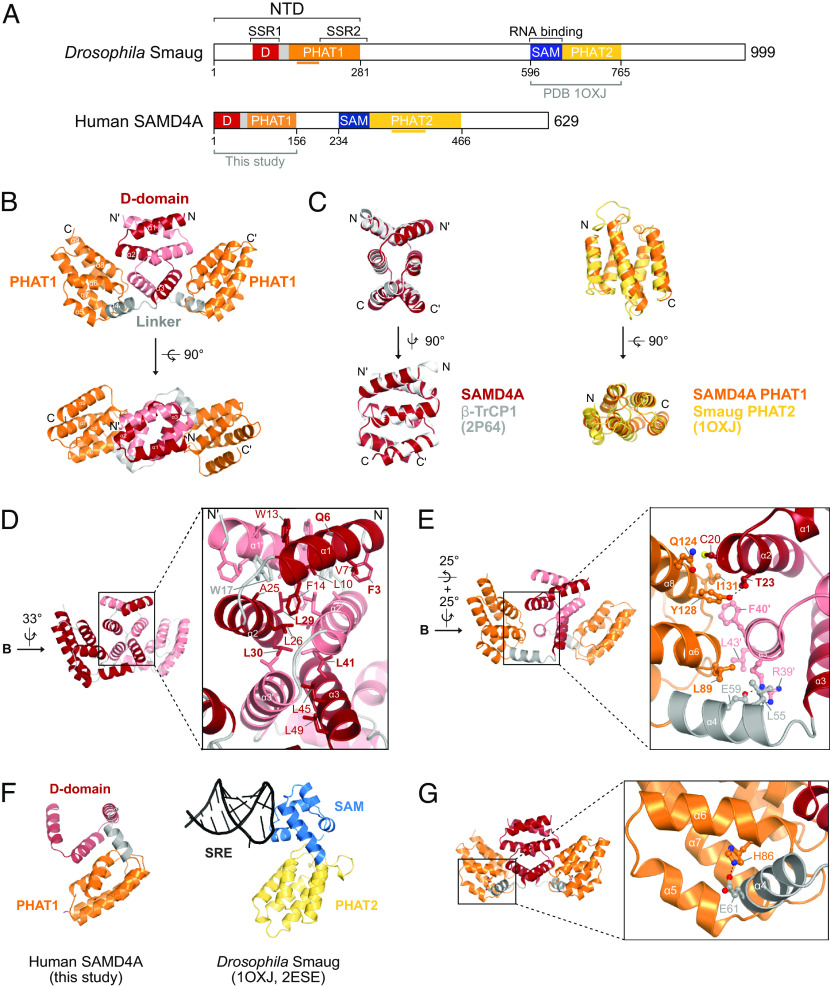
Fig. 1. Crystal structure of the NTD of human SAMD4A. (A) Domain organization of Drosophila Smaug and human SAMD4A proteins. SSR, Smaug similarity region; NTD, N-terminal domain. The horizontal orange or yellow lines below the protein schemes indicate insertions into the respective PHAT domains. (B) Crystal structure of the NTD dimer of the human SAMD4A. The D subdomain, the PHAT1 subdomain, and the linker α-helix are indicated. (C) Structural superimposition of the D subdomains of SAMD4A onto the D domain of β-TrCP1 (Left) and of the PHAT1 domain of SAMD4A onto the PHAT2 domain of Smaug (Right). PDB codes are indicated in parentheses. (D) Details of the D subdomain dimer interface of SAMD4A. Amino acid residues are labeled for one protein chain only. Residues in bold are 100% conserved across animals. The orientation of the structure is relative to the one depicted in the Upper panel of Fig. 1B. (E) Details of the interface formed by the D and the PHAT1 subdomains. Residues in bold are 100% conserved across animals. The orientation of the structure is relative to the one depicted in the Upper panel of Fig. 1B. (F) Structural comparison between the D-PHAT1 domain of SAMD4A and the SAM-PHAT2 domain of Smaug. SRE, Smaug recognition elements. The model of the SAM-PHAT2 domain–SRE complex was prepared by superimposition of the two crystal structures indicated by the PDB codes in parenthesis. (G) Structural detail indicating the position of the H86 residue, which is mutated in spmd mice.
Fig. 1. Crystal structure of the NTD of human SAMD4A. (A) Domain organization of Drosophila Smaug and human SAMD4A proteins. SSR, Smaug similarity region; NTD, N-terminal domain. The horizontal orange or yellow lines below the protein schemes indicate insertions into the respective PHAT domains. (B) Crystal structure of the NTD dimer of the human SAMD4A. The D subdomain, the PHAT1 subdomain, and the linker α-helix are indicated. (C) Structural superimposition of the D subdomains of SAMD4A onto the D domain of β-TrCP1 (Left) and of the PHAT1 domain of SAMD4A onto the PHAT2 domain of Smaug (Right). PDB codes are indicated in parentheses. (D) Details of the D subdomain dimer interface of SAMD4A. Amino acid residues are labeled for one protein chain only. Residues in bold are 100% conserved across animals. The orientation of the structure is relative to the one depicted in the Upper panel of Fig. 1B. (E) Details of the interface formed by the D and the PHAT1 subdomains. Residues in bold are 100% conserved across animals. The orientation of the structure is relative to the one depicted in the Upper panel of Fig. 1B. (F) Structural comparison between the D-PHAT1 domain of SAMD4A and the SAM-PHAT2 domain of Smaug. SRE, Smaug recognition elements. The model of the SAM-PHAT2 domain–SRE complex was prepared by superimposition of the two crystal structures indicated by the PDB codes in parenthesis. (G) Structural detail indicating the position of the H86 residue, which is mutated in spmd mice.CC BY-NC-ND · PubMed Central
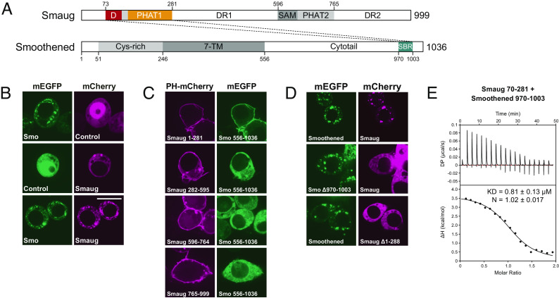
Fig. 2. The Smaug–Smoothened interaction. (A) Domain organization of Drosophila Smaug and Smoothened proteins. DR, disordered region; TM, transmembrane; SBR, Smaug-binding region. (B) ReLo assays showed a colocalization between Smaug and Smoothened. Controls are plasmids without an insert. (The scale bar is 10 µm.) (C) Of the four Smaug fragments indicated, only the NTD (Smaug 1-281) interacted with the Smoothened cytotail (Smo 556-1036) in the ReLo assay. (The scale bar is 10 µm.) (D) The Smoothened deletion and the Smaug deletion were unable to interact with Smaug and Smoothened, respectively, in the ReLo assay. (The scale bar is 10 µm.) (E) ITC using Smaug NTD (aa 70-281, His-tag removed) and a Smoothened peptide (Smo 970-1003).
Fig. 2. The Smaug–Smoothened interaction. (A) Domain organization of Drosophila Smaug and Smoothened proteins. DR, disordered region; TM, transmembrane; SBR, Smaug-binding region. (B) ReLo assays showed a colocalization between Smaug and Smoothened. Controls are plasmids without an insert. (The scale bar is 10 µm.) (C) Of the four Smaug fragments indicated, only the NTD (Smaug 1-281) interacted with the Smoothened cytotail (Smo 556-1036) in the ReLo assay. (The scale bar is 10 µm.) (D) The Smoothened deletion and the Smaug deletion were unable to interact with Smaug and Smoothened, respectively, in the ReLo assay. (The scale bar is 10 µm.) (E) ITC using Smaug NTD (aa 70-281, His-tag removed) and a Smoothened peptide (Smo 970-1003).CC BY-NC-ND · PubMed Central
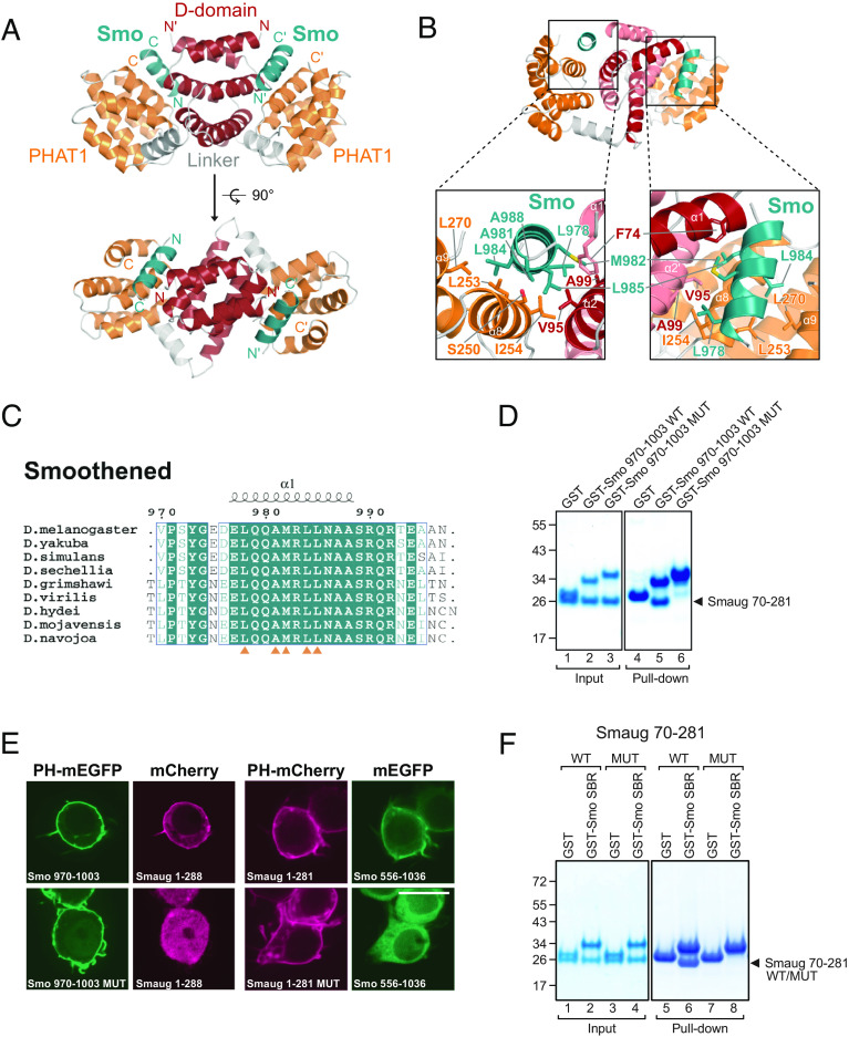
Fig. 3. Crystal structure of Smaug-NTD in complex with Smoothened. (A) Crystal structure of the NTD of Drosophila Smaug in complex with a Smoothened peptide (colored deep teal). (B) Structural detail of the Smaug–Smoothened interface. (C) Sequence alignment of the Smaug-binding region of Smoothened. Orange triangles indicate central residues involved in the Smaug interaction. (D) GST pull-down assay using 1 nmol GST-Smoothened 970-1003 (SBR) wild-type (WT) or L978E/L984E/L985E mutant (MUT) proteins and 2 nmol Smaug 70-281 (His-tag removed). Protein markers in kDa are indicated on the left. Input and pull-down samples originated from one experiment; the gel was split as it contained also the data shown in SI Appendix, Fig. S6D. (E) Mutant Smoothened SBR (MUT; L978E/L984E/L985E) did not interact with the NTD of Smaug, and mutant Smaug NTD (MUT; S250E/L253E) did not interact with the Smoothened cytotail in the ReLo assay. (The scale bar is 10 µm.) (F) GST pull-down assay using 1 nmol GST or GST-Smoothened SBR (970-1003) and 1 nmol Smaug NTD (70-281) wild-type or S250E/L253E mutant proteins (His-tag removed). Protein markers in kDa are indicated on the left. Input and pull-down samples originated from one experiment but were loaded onto two gels.
Fig. 3. Crystal structure of Smaug-NTD in complex with Smoothened. (A) Crystal structure of the NTD of Drosophila Smaug in complex with a Smoothened peptide (colored deep teal). (B) Structural detail of the Smaug–Smoothened interface. (C) Sequence alignment of the Smaug-binding region of Smoothened. Orange triangles indicate central residues involved in the Smaug interaction. (D) GST pull-down assay using 1 nmol GST-Smoothened 970-1003 (SBR) wild-type (WT) or L978E/L984E/L985E mutant (MUT) proteins and 2 nmol Smaug 70-281 (His-tag removed). Protein markers in kDa are indicated on the left. Input and pull-down samples originated from one experiment; the gel was split as it contained also the data shown in SI Appendix, Fig. S6D. (E) Mutant Smoothened SBR (MUT; L978E/L984E/L985E) did not interact with the NTD of Smaug, and mutant Smaug NTD (MUT; S250E/L253E) did not interact with the Smoothened cytotail in the ReLo assay. (The scale bar is 10 µm.) (F) GST pull-down assay using 1 nmol GST or GST-Smoothened SBR (970-1003) and 1 nmol Smaug NTD (70-281) wild-type or S250E/L253E mutant proteins (His-tag removed). Protein markers in kDa are indicated on the left. Input and pull-down samples originated from one experiment but were loaded onto two gels.CC BY-NC-ND · PubMed Central
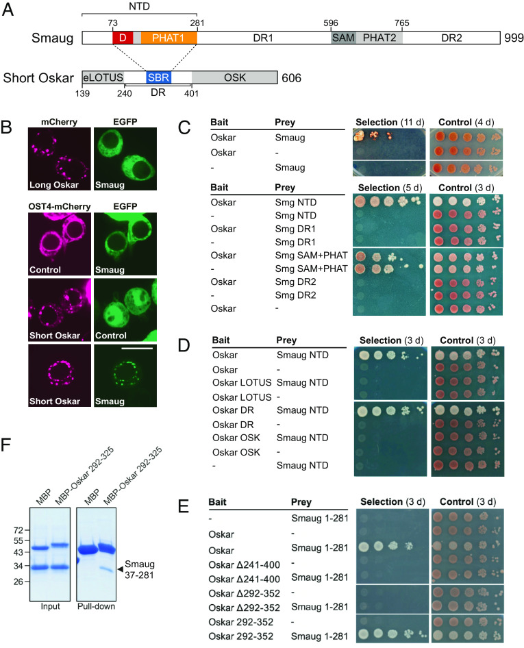
Fig. 4. The Smaug–Short Oskar interaction. (A) Domain organization of Drosophila Smaug and Oskar. The Short Oskar isoform is generated by alternate translation from M139 onward. NTD, N-terminal domain; DR, disordered region; SBR, Smaug-binding region. (B) Smaug interacted with Short Oskar but not Long Oskar in the ReLo assay. Plasmids lacking an insert served as controls. (The scale bar is 10 µm.) (C) Oskar (only the short isoform was tested) and Smaug interacted in split-ubiquitin yeast two-hybrid (Y2H) assays (Upper). Short Oskar binding mapped to the NTD of Smaug (Lower). Ten-fold dilutions are shown. Selection plates lacked adenine and histidine as compared to control plates. (D) The NTD of Smaug bound to the DR of Short Oskar in Y2H assays. Ten-fold dilutions are shown. Selection plates lacked adenine and histidine as compared to control plates. (E) Oskar 292-352 (SBR) was necessary and sufficient for Smaug-NTD binding in Y2H assays. Ten-fold dilutions are shown. Selection plates lacked adenine and histidine as compared to control plates. (F) MBP pull-down assay using 1 nmol His-MBP-Oskar 292-325 and 5 nmol Smaug NTD (37 to 281; His-tag removed). Input and pull-down samples originated from one experiment; the gel was split into two panels as it contained additional data irrelevant to the study.
Fig. 4. The Smaug–Short Oskar interaction. (A) Domain organization of Drosophila Smaug and Oskar. The Short Oskar isoform is generated by alternate translation from M139 onward. NTD, N-terminal domain; DR, disordered region; SBR, Smaug-binding region. (B) Smaug interacted with Short Oskar but not Long Oskar in the ReLo assay. Plasmids lacking an insert served as controls. (The scale bar is 10 µm.) (C) Oskar (only the short isoform was tested) and Smaug interacted in split-ubiquitin yeast two-hybrid (Y2H) assays (Upper). Short Oskar binding mapped to the NTD of Smaug (Lower). Ten-fold dilutions are shown. Selection plates lacked adenine and histidine as compared to control plates. (D) The NTD of Smaug bound to the DR of Short Oskar in Y2H assays. Ten-fold dilutions are shown. Selection plates lacked adenine and histidine as compared to control plates. (E) Oskar 292-352 (SBR) was necessary and sufficient for Smaug-NTD binding in Y2H assays. Ten-fold dilutions are shown. Selection plates lacked adenine and histidine as compared to control plates. (F) MBP pull-down assay using 1 nmol His-MBP-Oskar 292-325 and 5 nmol Smaug NTD (37 to 281; His-tag removed). Input and pull-down samples originated from one experiment; the gel was split into two panels as it contained additional data irrelevant to the study.CC BY-NC-ND · PubMed Central
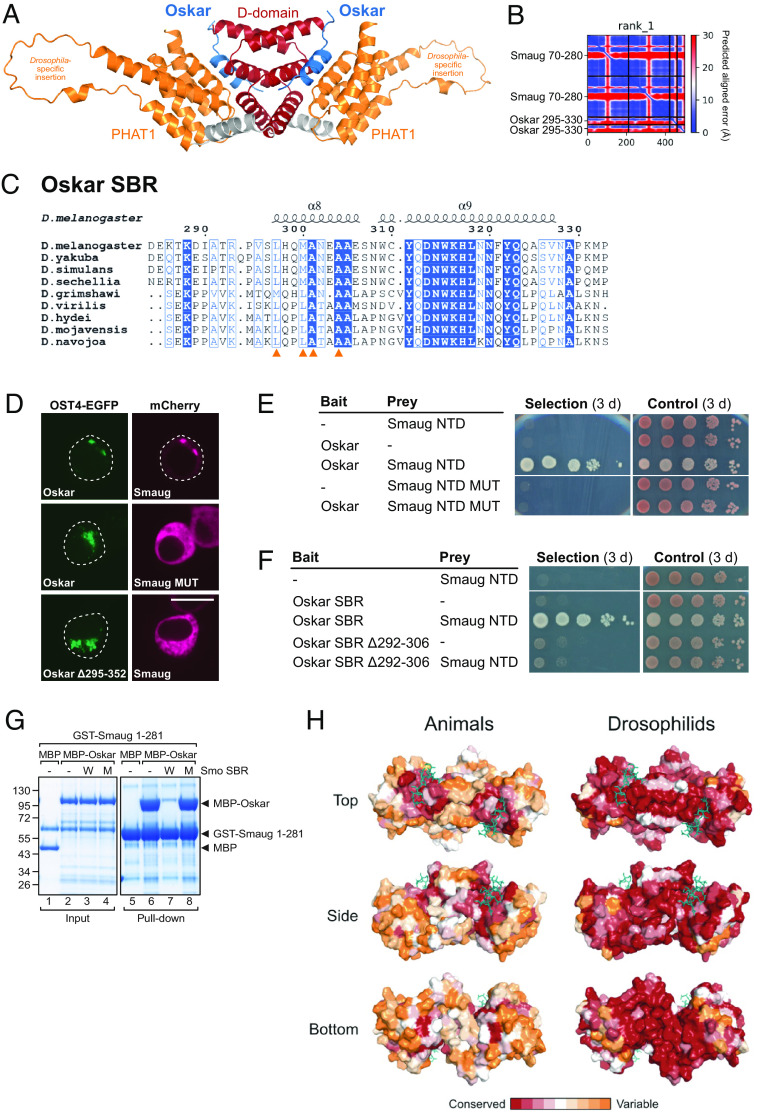
Fig. 5. Predicted structure of Smaug-NTD in complex with the Oskar SBR. (A) Structural model of the Drosophila Smaug-NTD in complex with the Oskar-SBR (blue) generated using AlphaFold2-multimer-version3 (58–60). The model contains the Drosophila-specific insertion (aa 156-196) of the Smaug-NTD. (B) Predicted aligned error plot generated from the prediction run, showing a low positional error of the N-terminal part of the Oskar-SBR relative to the Smaug-NTD. (C) Sequence alignment of Oskar covering the SBR. Orange triangles indicate Oskar residues that are critical for Smaug binding based on the predicted structural model. (D) OST4-EGFP and mCherry fusions to the proteins indicated were coexpressed in S2R+ cells, and their localization was analyzed by microscopy. The Smaug MUT (S250E, L253E) and the deletion of the SBR of Oskar (295-352) prevented the Smaug–Oskar interaction. (Scale bar is 10 µm.) (E and F) Yeast two-hybrid assays showing that the Smaug NTD MUT (S250E, L253E) or the deletion of aa 292-306 from the SBR (292-352) of Oskar prevented the Smaug–Oskar interaction. Selection medium lacked adenine and histidine. (G) GST pull-down assay using 1 nmol GST-Smaug 1-281 and 2 nmol His-MBP or His-MBP-Oskar. In addition, 45 nmol of a Smo 907-1003 synthetic peptide with wild-type (W) sequence or carrying the L978E/L984E/L985E point mutations (M) were present during the experiment as indicated above the gel image. Protein markers in kDa are indicated on the left. Input and pull-down samples originated from one experiment but were loaded onto two gels. (H) Surface representation of the Smaug NTD colored according to residue conservation (61) considering sequences of Smaug orthologs from various animals (Left) or Drosophilids only (Right).
Fig. 5. Predicted structure of Smaug-NTD in complex with the Oskar SBR. (A) Structural model of the Drosophila Smaug-NTD in complex with the Oskar-SBR (blue) generated using AlphaFold2-multimer-version3 (58–60). The model contains the Drosophila-specific insertion (aa 156-196) of the Smaug-NTD. (B) Predicted aligned error plot generated from the prediction run, showing a low positional error of the N-terminal part of the Oskar-SBR relative to the Smaug-NTD. (C) Sequence alignment of Oskar covering the SBR. Orange triangles indicate Oskar residues that are critical for Smaug binding based on the predicted structural model. (D) OST4-EGFP and mCherry fusions to the proteins indicated were coexpressed in S2R+ cells, and their localization was analyzed by microscopy. The Smaug MUT (S250E, L253E) and the deletion of the SBR of Oskar (295-352) prevented the Smaug–Oskar interaction. (Scale bar is 10 µm.) (E and F) Yeast two-hybrid assays showing that the Smaug NTD MUT (S250E, L253E) or the deletion of aa 292-306 from the SBR (292-352) of Oskar prevented the Smaug–Oskar interaction. Selection medium lacked adenine and histidine. (G) GST pull-down assay using 1 nmol GST-Smaug 1-281 and 2 nmol His-MBP or His-MBP-Oskar. In addition, 45 nmol of a Smo 907-1003 synthetic peptide with wild-type (W) sequence or carrying the L978E/L984E/L985E point mutations (M) were present during the experiment as indicated above the gel image. Protein markers in kDa are indicated on the left. Input and pull-down samples originated from one experiment but were loaded onto two gels. (H) Surface representation of the Smaug NTD colored according to residue conservation (61) considering sequences of Smaug orthologs from various animals (Left) or Drosophilids only (Right).CC BY-NC-ND · PubMed Central

Verification

Claims the paper makes

The claims and statistics of this paper haven’t been checked yet.

Run the check — free, no account needed. It reads the open-access text and checks the sources it cites.

A few free checks a day · papers already verified open instantly · results are public

Self-check

Percentages, sums and reported differences re-computed from the paper’s own numbers.

The self-check needs the paper’s full text.

Citations

Cites

Show all 2 references

Research works printed but not linked: 79

  1. 10.1002/wrna.70 · DOI
  2. 10.1080/15476286.2017.1338231 · DOI
  3. 10.1038/nsmb.2296 · DOI
  4. 10.1016/j.tig.2019.12.007 · DOI
  5. 10.1126/science.283.5403.833 · DOI
  6. 10.1002/pro.5560060128 · DOI
  7. 10.1126/stke.2862005re7 · DOI
  8. 10.1523/JNEUROSCI.2172-15.2015 · DOI
  9. 10.1038/nsb956 · DOI
  10. 10.1074/jbc.M508374200 · DOI
  11. 10.1016/S1097-2765(03)00178-3 · DOI
  12. 10.1038/celldisc.2016.50 · DOI
  13. 10.1038/nsmb1039 · DOI
  14. 10.1038/nsmb1053 · DOI
  15. 10.1038/nsmb1038 · DOI
  16. 10.1016/j.cell.2007.04.042 · DOI · Paper page
  17. 10.1073/pnas.1406511111 · DOI
  18. 10.1101/gad.10.20.2610 · DOI
  19. 10.1016/S1097-2765(00)80368-8 · DOI
  20. 10.1101/gad.10.20.2600 · DOI
  21. 10.1261/rna.062208.117 · DOI
  22. 10.1038/emboj.2010.283 · DOI
  23. 10.1038/sj.emboj.7600026 · DOI
  24. 10.1016/j.cub.2005.01.048 · DOI
  25. 10.1242/dev.02649 · DOI
  26. F. Pekovic RNA binding proteins Smaug and Cup induce CCR4-NOT-dependent deadenylation of the nanos mRNA in a reconstituted system. Nucleic Acids Res. 51 3950–3970 (2023). · DOI
  27. 10.1038/embor.2012.192 · DOI
  28. 10.1016/0092-8674(91)90110-K · DOI
  29. 10.1002/aja.1001990204 · DOI
  30. 10.1016/0092-8674(92)90358-J · DOI
  31. 10.1038/369315a0 · DOI
  32. 10.1242/dev.031815 · DOI
  33. 10.1186/gb-2014-15-1-r4 · DOI
  34. 10.1186/gb-2012-13-2-r11 · DOI
  35. 10.1016/j.devcel.2006.10.005 · DOI · Paper page
  36. 10.1016/j.celrep.2020.107783 · DOI
  37. 10.1017/S1355838299991392 · DOI
  38. 10.1093/genetics/iyab177 · DOI
  39. 10.15252/embr.201948425 · DOI
  40. 10.1101/gad.938601 · DOI
  41. 10.1093/nar/gkq366 · DOI · Paper page
  42. 10.1016/j.jmb.2007.05.022 · DOI
  43. 10.1073/pnas.1411798111 · DOI
  44. H. K. Salgania J. Metz M. Jeske ReLo is a simple and quick colocalization assay to identify and characterize direct protein-protein interactions. bioRxiv [Preprint] (2022). https://www.biorxiv.org/content/10.1101/2022.03.04.482790v2 (Accessed 17 July 2023). · DOI · Paper page
  45. 10.1016/j.celrep.2015.07.030 · DOI
  46. 10.1038/nature09465 · DOI
  47. 10.1242/dev.121.11.3723 · DOI
  48. 10.1016/j.devcel.2007.03.002 · DOI
  49. 10.1101/gad.297051.117 · DOI
  50. 10.1016/j.celrep.2015.06.055 · DOI
  51. 10.1371/journal.pone.0014362 · DOI
  52. C. Ottone, S. Gigliotti, A. Giangrande, F. Graziani, A. V. di Pianella, The translational repressor cup is required for germ cell development in Drosophila. J. Cell Sci. 125, 3114–3123 (2012).
  53. 10.1242/dev.027698 · DOI
  54. 10.1101/gad.10.17.2179 · DOI
  55. 10.1093/nar/gkaa652 · DOI
  56. 10.1073/pnas.1515568112 · DOI
  57. R. Evans Protein complex prediction with AlphaFold-Multimer. bioRxiv [Preprint] (2021). https://www.biorxiv.org/content/10.1101/2021.10.04.463034v2 (Accessed 17 July 2023). · DOI
  58. 10.1038/s41592-022-01488-1 · DOI · Paper page
  59. 10.1093/nar/gkw408 · DOI · Paper page
  60. 10.1016/S0168-9525(02)02792-0 · DOI
  61. 10.1093/genetics/iyac035 · DOI
  62. 10.1101/gr.213694.116 · DOI
  63. M. van den Heuvel, P. W. Ingham, Required for hedgehog signalling. Lett. Nat. 382, 413–417 (1996).
  64. 10.1016/j.devcel.2013.06.011 · DOI
  65. 10.1098/rsob.200319 · DOI
  66. 10.1038/nrc2396 · DOI
  67. 10.1016/j.devcel.2005.10.006 · DOI
  68. J. Fehilly Condensate formation of the human RNA-binding protein SMAUG1 is controlled by its intrinsically disordered regions and interactions with 14-3-3 proteins. bioRxiv [Preprint] (2023). https://doi.org/10.1101/2023.02.09.527857 (Accessed 17 July 2023). · DOI
  69. 10.3389/fmolb.2022.1016071 · DOI
  70. 10.1021/pr700696a · DOI
  71. 10.1016/j.ydbio.2006.10.042 · DOI
  72. 10.1101/gad.1424106 · DOI
  73. 10.1016/j.pep.2005.01.016 · DOI
  74. 10.1038/nmeth.2019 · DOI · Paper page
  75. 10.1107/S0907444909052925 · DOI · Paper page
  76. 10.1107/S0907444912001308 · DOI · Paper page
  77. 10.1107/S0907444904019158 · DOI · Paper page
  78. J. Kubikova, M. Jeske, D-PHAT domain (NTD) of human SAMD4A. Protein Data Bank. https://doi.org/10.2210/pdb8oik/pdb. Accessed 17 July 2023b. · DOI
  79. G. Ubartaite, J. Kubikova, M. Jeske, Drosophila Smaug-Smoothened complex. Protein Data Bank. https://doi.org/10.2210/pdb8oij/pdb. Accessed 17 July 2023a. · DOI

Metadata

Authors
Kubíková, J., Ubartaitė, G., Metz, J., & Jeske, M.
Affiliations
Heidelberg University (DE)
Corresponding author
Jeske, Mandy
Venue
Proceedings of the National Academy of Sciences
Published
2023-07-31
Volume
120
Issue
32
Pages
e2304385120-e2304385120
ISSN
  • 0027-8424 (ISSN-L)
  • 1091-6490
OpenAlex
W4385409666
Cited by
18
References
2
Citations by year
2026: 1 · 2025: 7 · 2024: 9 · 2023: 1
FWCI
1.60
Type
journal article
Language
en
Open access
Locations
  • Proceedings of the National Academy of Sciences (publishedVersion, cc-by-nc-nd)
  • bioRxiv (Cold Spring Harbor Laboratory) (acceptedVersion, cc-by-nc-nd)
  • PubMed Central (publishedVersion, cc-by-nc-nd)
  • PubMed (publishedVersion)
  • Desy Publications Database (Deutsches Elektronen-Synchrotron DESY) (submittedVersion, other-oa)
  • DESY Publication Database (PUBDB) (Deutsches Elektronen-Synchrotron)
Funders
Deutsche Forschungsgemeinschaft (JE827/1-1)
Topics
  • Life Sciences › Biochemistry, Genetics and Molecular Biology › Molecular Biology › RNA Research and Splicing
  • Life Sciences › Biochemistry, Genetics and Molecular Biology › Molecular Biology › Developmental Biology and Gene Regulation
  • Life Sciences › Biochemistry, Genetics and Molecular Biology › Molecular Biology › Protein Structure and Dynamics
Keywords
Smaug, sterile alpha motif domain, N-terminal domain, Smoothened, Oskar, Hedgehog signaling, peptide binding, protein-protein interaction, Drosophila, crystal structure
MeSH terms
Smoothened Receptor, Animals, Drosophila, Germ Cells, Humans, Repressor Proteins, RNA, Messenger, RNA-Binding Proteins, Drosophila Proteins, Receptors, G-Protein-Coupled, Hedgehog Proteins

About these checks

Registry, retraction, Cites and statistics results are computed, not judged, with one exception: a printed reference that no index lists is read once by a model before it counts as a citation error. Claims are extracted by a model and kept only when their quote appears verbatim in the text; each cited source is then checked, with a model’s help, for whether it says what the claim relies on. Those verdicts can be wrong — read the claim’s quote and the cited source before relying on one.

Statistics and arithmetic are recomputed, not judged.

Confidence bands are shown only for fields calibrated on our annotated test set. None is calibrated yet, so no band is shown.

What counts as a claim ›How we measure confidence ›How we check this ›

Are you an author, or do you see a problem with this page? Request a review

How to cite

Cite CoChat for the checks on this page, and the paper for its own findings.

This page
CoChat. (2026). Citation check: Structural basis for binding of Drosophila Smaug to the GPCR Smoothened and to the germline inducer Oskar. https://app.cochat.ai/papers/10.1073/pnas.2304385120
The paper
Kubíková, J., Ubartaitė, G., Metz, J., & Jeske, M. (2023). Structural basis for binding of Drosophila Smaug to the GPCR Smoothened and to the germline inducer Oskar. Proceedings of the National Academy of Sciences. https://doi.org/10.1073/pnas.2304385120