

The DOI matches its Crossref record; 1 of the 1 references it cites were found in a registry or an index.
Microbial Hauberks: Composition and Function of Surface Layer Proteins in Gammaproteobacterial Methanotrophs
Open accessPreprint — published as 10.1128/aem.01364-24 · Paper page
Abstract
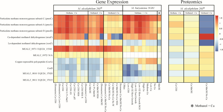
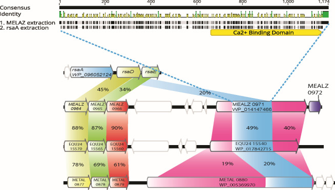
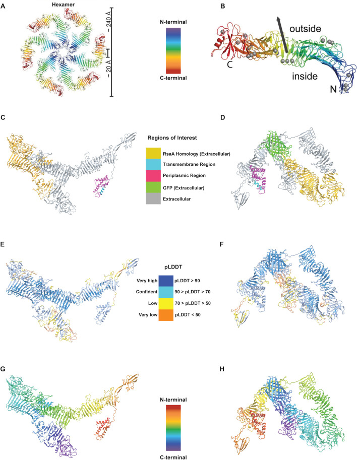
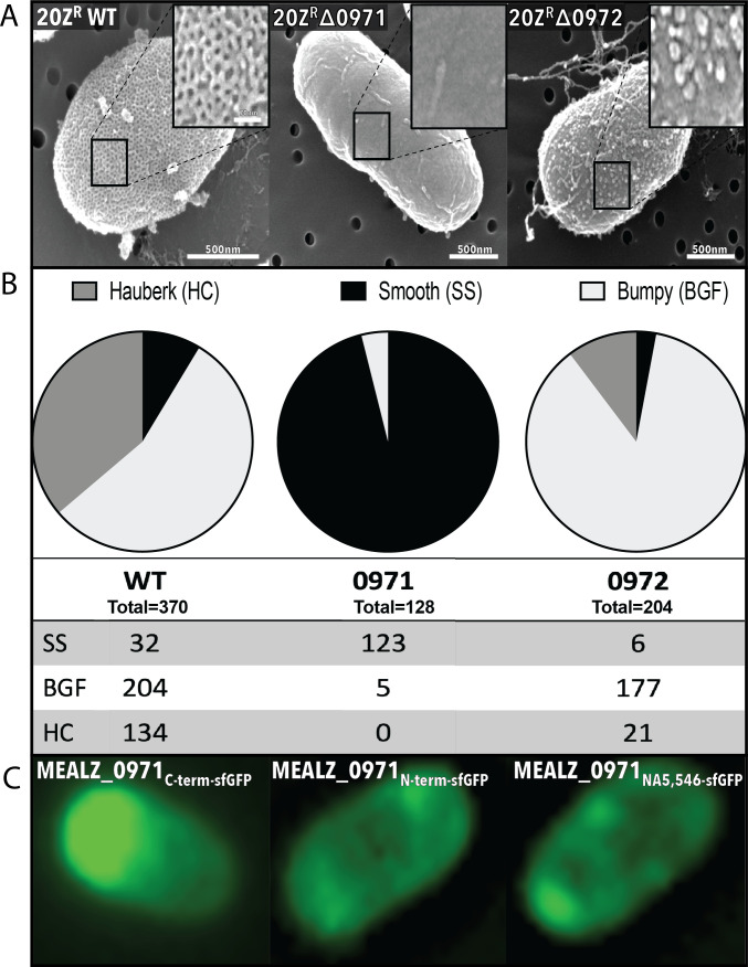
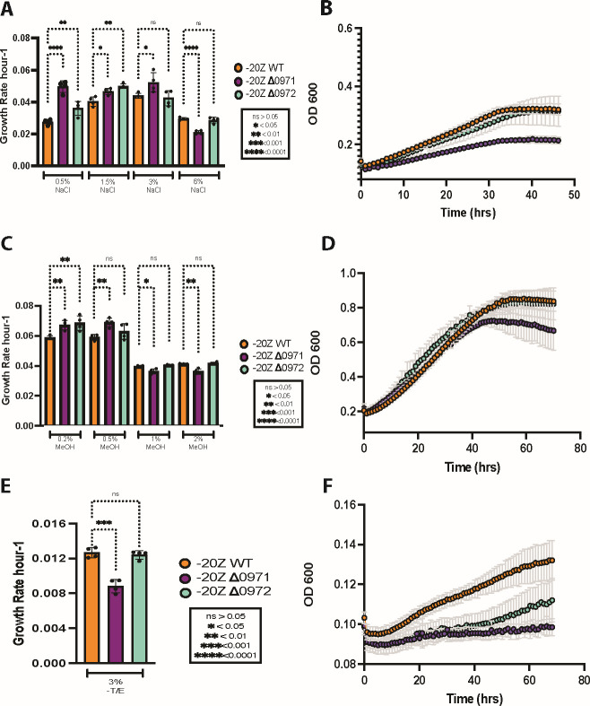
Many species of proteobacterial methane-consuming bacteria (methanotrophs) form a hauberk-like envelope represented by a surface (S-) layer protein matrix. While several proteins were predicted to be associated with the cell surface, the composition and function of the hauberk matrix remained elusive. Here we report the identification of the genes encoding the hauberk-forming protein in two gamma-proteobacterial (Type I) methanotrophs, Methylotuvimicrobium buryatense 5GB1 (EQU24_15540) and Methylotuvimicrobium alcaliphilum 20Z R (MEALZ_0971 and MEALZ_0972). The proteins share 40% AA identity with each other and are distantly related to the RsaA proteins from Caulobacter crescentus (20% AA identity). Deletion of these genes resulted in loss of the characteristic hauberk pattern on the cell surface. A set of transcriptional fusions between the MEALZ_0971 and a superfolder green fluorescent protein (sfGFP) further confirmed its surface localization. The functional roles of the hauberk and cell-surface associated proteins, including MEALZ_0971, MEALZ_0972, EQU24_15540, and a copper-induced CorA protein, were further investigated via gene expression studies and phenotypic tests. The hauberk core protein shows constitutive expression across 18 growth conditions. The M. alcaliphilum 20Z R Δ0971 showed reduced growth at high salinity, high methanol and metal-limited conditions, suggesting a role in cell-envelope stability and metal scavenging. Overall, understanding the genetics, composition and cellular functions of the S-layers contributes to our knowledge of methanotroph adaptation to environmental perturbations and opens a promising prospect for (nano)biotechnology applications.
Overview
The claims and statistics of this paper haven’t been checked yet.
Run the check — free, no account needed. It reads the open-access text and checks the sources it cites.
Integrity signals
Registry
Cites
Statistics
Figures
Verification
Claims the paper makes
The claims and statistics of this paper haven’t been checked yet.
Run the check — free, no account needed. It reads the open-access text and checks the sources it cites.
Self-check
Percentages, sums and reported differences re-computed from the paper’s own numbers.
The self-check needs the paper’s full text.
Citations
Cites
Highly accurate protein structure prediction with AlphaFold
Jumper, J., Evans, R., Pritzel, A., Green, T., Figurnov, M., Ronneberger, O., Tunyasuvunakool, K., Bates, R., Žídek, A., Potapenko, A., et al. (2021) · Nature · doi:10.1038/s41586-021-03819-2 · Cited by 43,764
Research works printed but not linked: 47
- 1997, V. Functions of S-layers. FEMS microbiology reviews, 20, 99
- 2000, S-layer proteins. Journal of bacteriology, 182, 859
- Sleytr, U.B. , et al., Crystalline bacterial cell surface proteins. 1996: Elsevier.
- 2023, Surface-layer protein is a public-good matrix exopolymer for microbial community organisation in environmental anammox biofilms, The ISME journal, 17, 803, 10.1038/s41396-023-01388-y · DOI
- 10.1128/JB.180.12.3062-3069.1998 · DOI
- 2017, Structure of the hexagonal surface layer on Caulobacter crescentus cells, Nature microbiology, 2, 1
- 10.1016/j.cub.2020.07.033 · DOI
- 10.1139/W10-036 · DOI
- 1978, Electron microscopy of the cell wall complex of Methylomonas albus, Archives of Microbiology, 119, 227, 10.1007/BF00964278 · DOI
- 2010, Structural and functional features of methanotrophs from hypersaline and alkaline lakes, Microbiology, 79, 472, 10.1134/S0026261710040090 · DOI
- 10.1007/s002030050786 · DOI
- 10.1078/0723-2020-00028 · DOI
- 10.1134/S0026261713050068 · DOI
- 1985, Some features of the ultrastructure of Methylococcus capsulatus grown under different conditions, Prikl. Biokhim. Mikrobiol, 21, 770
- 10.1128/genomea · DOI
- 2011, Primary characterization of dominant cell surface proteins of halotolerant methanotroph Methylomicrobium alcaliphilum 20Z, Microbiology, 80, 608, 10.1134/S0026261711050122 · DOI
- Berson, O. and M.E. Lidstrom , Cloning and characterization of corA, a gene encoding a copper-repressible polypeptide in the type I methanotroph, Methylomicrobium albus BG8. FEMS microbiology letters, 1997. 148(2): p. 169-174. · DOI
- 10.1099/mic.0.037119-0 · DOI
- 10.1371/journal.pone.0087750 · DOI
- 2023, Identification and characterisation of a major outer membrane protein from Methylacidiphilum fumariolicum SolV, Antonie van Leeuwenhoek, 116, 1227, 10.1007/s10482-023-01879-0 · DOI
- 10.1186/s12934-015-0267-8 · DOI
- 10.1046/j.1365-2958.1997.4801846.x · DOI
- Akberdin, I.R. , et al., Rare earth elements alter redox balance in Methylomicrobium alcaliphilum 20ZR . Frontiers in microbiology, 2018: p. 2735. · DOI
- 10.1038/s41598-018-35504-2 · DOI
- 10.1128/JB.180.24.6450-6458.1998 · DOI
- 10.1038/nrmicro3213 · DOI
- 10.1073/pnas.1909798116 · DOI
- 10.1016/S0378-1119(96)00829-3 · DOI
- 10.1128/AEM.03724-15 · DOI
- 2017, Outer membrane vesicles (OMVs) of gram-negative bacteria: a perspective update, Frontiers in microbiology, 8, 254572
- 10.1038/nrmicro3525 · DOI · Paper page
- 2018, Isolation, characterization, and metal response of novel, acid-tolerant Penicillium spp. from extremely metal-rich waters at a mining site in Transbaikal (Siberia, Russia), . Microbial ecology, 76, 911, 10.1007/s00248-018-1186-0 · DOI
- 10.1128/AEM.03795-14 · DOI
- 10.1093/bib/bbx108 · DOI · Paper page
- 10.1093/nar/gkt389 · DOI · Paper page
- 10.1093/bioinformatics/bts199 · DOI · Paper page
- 2022, ColabFold: making protein folding accessible to all, Nature methods, 19, 679, 10.1038/s41592-022-01488-1 · DOI · Paper page
- Hallgren, J. , et al., DeepTMHMM predicts alpha and beta transmembrane proteins using deep neural networks. BioRxiv, 2022: p. 2022.04. 08.487609. · DOI · Paper page
- 10.1093/nar/gkab314 · DOI · Paper page
- 10.1093/bioinformatics/bty560 · DOI · Paper page
- 2017, Salmon provides fast and bias-aware quantification of transcript expression, Nature methods, 14, 417, 10.1038/nmeth.4197 · DOI · Paper page
- 10.1186/gb-2014-15-1-r1 · DOI · Paper page
- 2008, Development of a broad-host-range sacB-based vector for unmarked allelic exchange, BMC research notes, 1, 1, 10.1186/1756-0500-1-1 · DOI
- 10.1128/jb · DOI
- Hemmann, J.L. , et al., Lanpepsy is a novel lanthanide-binding protein involved in the lanthanide response of the obligate methylotroph Methylobacillus flagellatus . Journal of Biological Chemistry, 2023. 299(3). · DOI
- 10.1101/2022.01.19.476857 · DOI
- 2022, Siderophore for lanthanide and iron uptake for methylotrophy and plant growth promotion in Methylobacterium aquaticum strain 22A, Frontiers in Microbiology, 13, 921635, 10.3389/fmicb.2022.921635 · DOI
Metadata
- Authors
- Hamilton, R., Gebbie, W., Bowman, C., Mantanona, A., & Kalyuzhnaya, M. G.
- Affiliations
- San Diego State University (US)
- Corresponding author
- Kalyuzhnaya, Marina G.
- Venue
- Applied and Environmental Microbiology
- Publisher
- openRxiv
- Published
- 2024-07-09
- Volume
- 91
- Issue
- 1
- Pages
- e0136424-e0136424
- ISSN
- 0099-2240 (ISSN-L)
- PMC
- PMC11784148
- OpenAlex
- W4405944986
- Cited by
- 1
- References
- 1
- Citations by year
- 2026: 1
- FWCI
- 0.14
- Type
- journal article
- Language
- en
- Open access
- hybrid
- doi.org
- Licence
- cc-by
- Locations
- Applied and Environmental Microbiology (publishedVersion, cc-by)
- bioRxiv (Cold Spring Harbor Laboratory) (acceptedVersion)
- PubMed Central (publishedVersion, cc-by)
- PubMed (publishedVersion)
- DOAJ (DOAJ: Directory of Open Access Journals) (submittedVersion, cc-by-sa)
- Funders
- U.S. Department of Defense
- Defense Advanced Research Projects Agency
- Advanced Research Projects Agency (DE-AR000350)
- Topics
- Life Sciences › Biochemistry, Genetics and Molecular Biology › Molecular Biology › Microbial metabolism and enzyme function
- Life Sciences › Biochemistry, Genetics and Molecular Biology › Genetics › Bacterial Genetics and Biotechnology
- Physical Sciences › Environmental Science › Environmental Chemistry › Methane Hydrates and Related Phenomena
- Keywords
- surface layer protein, methanotrophic bacteria, Gammaproteobacteria, cell envelope, gene deletion, gene expression, metal scavenging, salt tolerance, methanol tolerance
- MeSH terms
- Bacterial Proteins, Membrane Glycoproteins, Methane, Methylococcaceae, Gammaproteobacteria
About these checks
Registry, retraction, Cites and statistics results are computed, not judged, with one exception: a printed reference that no index lists is read once by a model before it counts as a citation error. Claims are extracted by a model and kept only when their quote appears verbatim in the text; each cited source is then checked, with a model’s help, for whether it says what the claim relies on. Those verdicts can be wrong — read the claim’s quote and the cited source before relying on one.
Statistics and arithmetic are recomputed, not judged.
Confidence bands are shown only for fields calibrated on our annotated test set. None is calibrated yet, so no band is shown.
What counts as a claim ›How we measure confidence ›How we check this ›
Are you an author, or do you see a problem with this page? Request a review
How to cite
Cite CoChat for the checks on this page, and the paper for its own findings.
- This page
- CoChat. (2026). Citation check: Microbial Hauberks: Composition and Function of Surface Layer Proteins in Gammaproteobacterial Methanotrophs. https://app.cochat.ai/papers/10.1101/2024.07.09.602694
- The paper
- Hamilton, R., Gebbie, W., Bowman, C., Mantanona, A., & Kalyuzhnaya, M. G. (2024). Microbial Hauberks: Composition and Function of Surface Layer Proteins in Gammaproteobacterial Methanotrophs. Applied and Environmental Microbiology. https://doi.org/10.1101/2024.07.09.602694
Data sources
Bibliographic data: Semantic Scholar, OpenAlex, Crossref.
Contains information from the Semantic Scholar Academic Graph Datasets, made available under the ODC Attribution License (ODC-BY 1.0).
Semantic Scholar’s data is described in: Kinney, R., et al. (2023). The Semantic Scholar Open Data Platform. arXiv:2301.10140.
