

The DOI matches its Crossref record; 1 of the 1 references it cites were found in a registry or an index.
Large serine integrases utilise scavenged phage proteins as directionality cofactors
Open accessPreprint — published as 10.1093/nar/gkaf050
Abstract
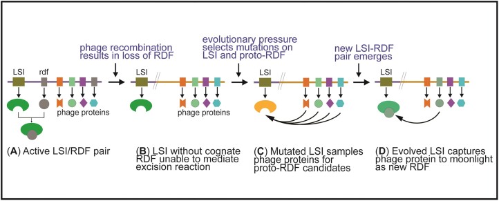
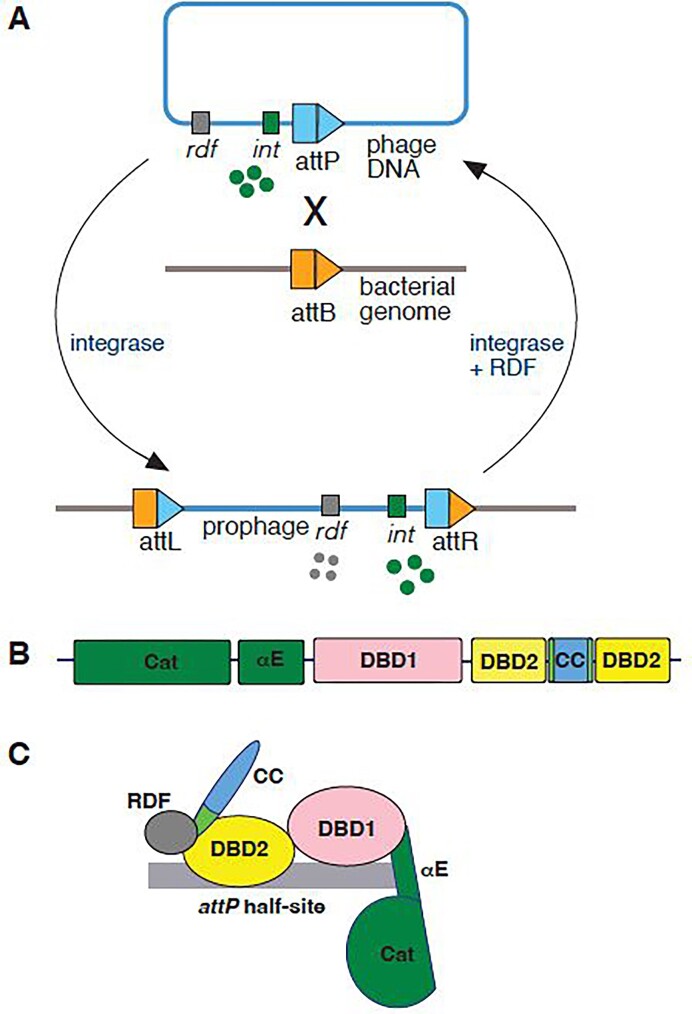
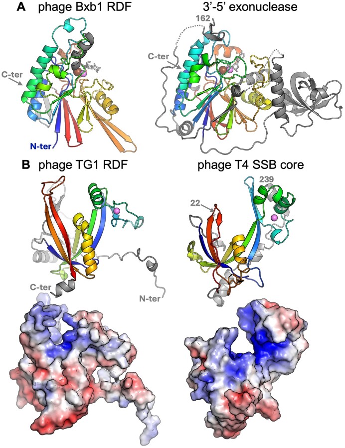
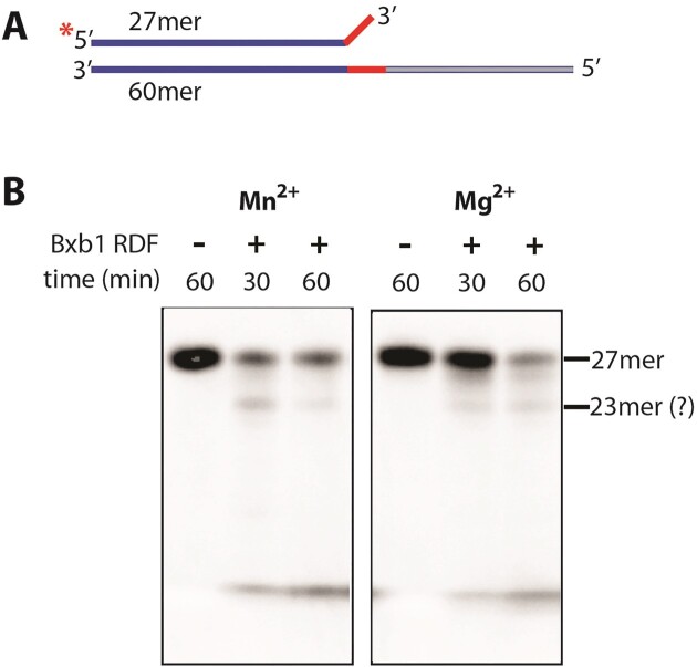
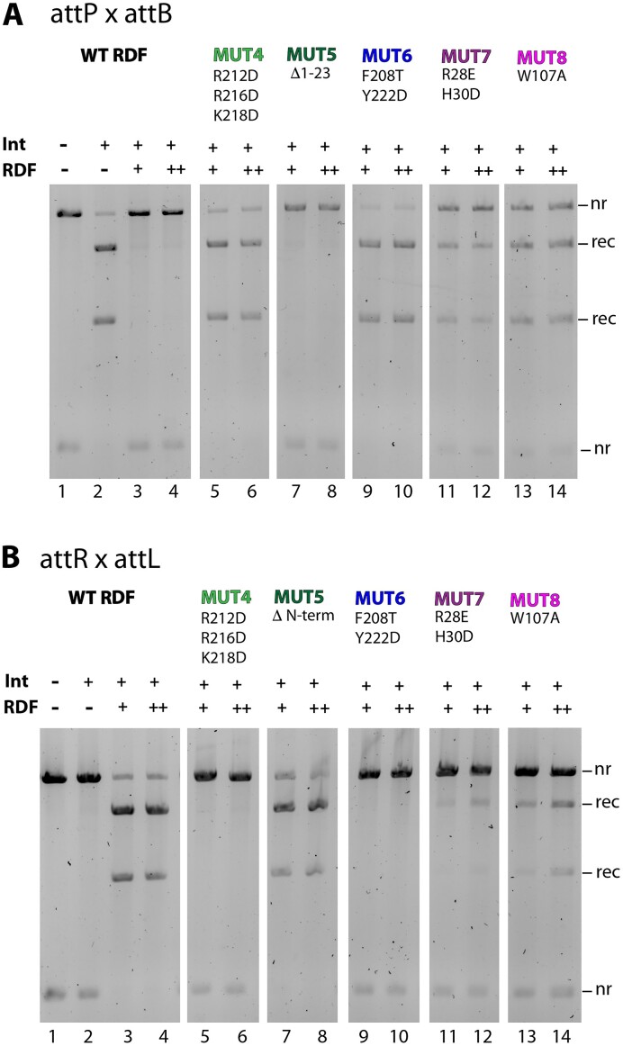
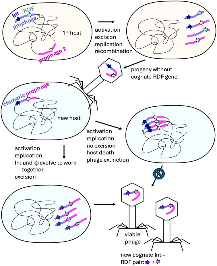
Recombination directionality factors (RDFs) for large serine integrases (LSIs) are cofactor proteins that control the directionality of recombination to favor excision over insertion. Although RDFs are predicted to bind their cognate LSIs in similar ways, there is no overall common structural theme across LSI RDFs, leading to the suggestion that some of them may be moonlighting proteins with other primary functions. To test this hypothesis, we searched for characterized proteins with structures similar to the predicted structures of known RDFs. Our search shows that the RDFs for two LSIs, TG1 integrase and Bxb1 integrase, show high similarities to a single stranded DNA binding (SSB) protein and an editing exonuclease, respectively. We present experimental data to show that TG1 RDF is a functional SSB protein. We used mutational analysis to validate the integrase-RDF interface predicted by AlphaFold2 multimer for TG1 integrase and its RDF, and establish that control of recombination directionality is mediated via protein-protein interaction at the junction of recombinase’s second DNA binding domain and the base of the coiled coil domain.
Overview
The claims and statistics of this paper haven’t been checked yet.
Run the check — free, no account needed. It reads the open-access text and checks the sources it cites.
Integrity signals
Registry
Cites
Statistics
Figures
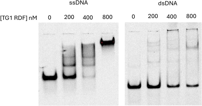
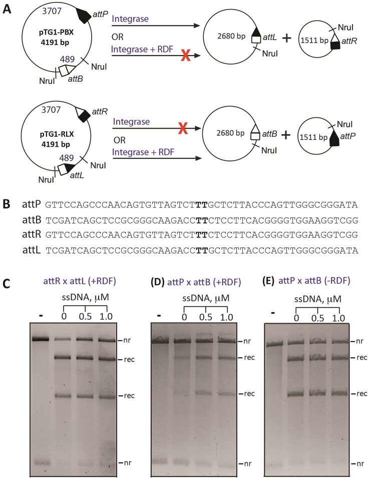
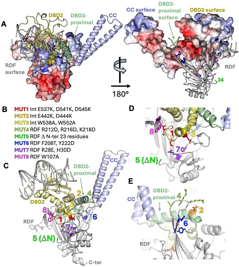
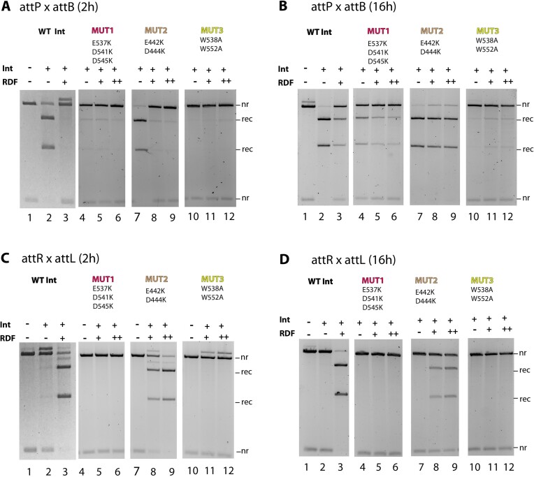
Verification
Claims the paper makes
The claims and statistics of this paper haven’t been checked yet.
Run the check — free, no account needed. It reads the open-access text and checks the sources it cites.
Self-check
Percentages, sums and reported differences re-computed from the paper’s own numbers.
The self-check needs the paper’s full text.
Citations
Cites
Highly accurate protein structure prediction with AlphaFold
Jumper, J., Evans, R., Pritzel, A., Green, T., Figurnov, M., Ronneberger, O., Tunyasuvunakool, K., Bates, R., Žídek, A., Potapenko, A., et al. (2021) · Nature · doi:10.1038/s41586-021-03819-2 · Cited by 43,764
Research works printed but not linked: 52
- 2015, An Overview of Tyrosine Site-specific Recombination: From an Flp Perspective, Microbiol. Spectr, 3, 3.4.12, 10.1128/microbiolspec.MDNA3-0021-2014 · DOI
- 2015, Phage-encoded Serine Integrases and Other Large Serine Recombinases, Microbiol. Spectr, 3, 3.4.06, 10.1128/microbiolspec.MDNA3-0059-2014 · DOI
- 10.1042/BJ20151112 · DOI
- 10.1146/annurev.biochem.73.011303.073908 · DOI
- 10.1101/2021.12.07.471667 · DOI
- 10.1111/mmi.14579 · DOI
- 10.1016/j.molcel.2008.02.023 · DOI
- 2016, Structure of a Holliday junction complex reveals mechanisms governing a highly regulated DNA transaction, eLife, 5, e14313, 10.7554/eLife.14313 · DOI
- Johnson, R.C. (2015) Site-specific DNA Inversion by Serine Recombinases. Microbiol. Spectr., 3, MDNA3-0047–2014. · DOI
- 10.1111/j.1365-2958.2005.04517.x · DOI
- 10.1371/journal.pbio.0040186 · DOI
- 10.1111/j.1365-2958.2011.07696.x · DOI
- 10.3109/10409238.2013.831807 · DOI
- 10.1146/annurev.biophys.30.1.87 · DOI
- 10.1038/nature03657 · DOI
- Landy, A. (2015) The λ Integrase Site-specific Recombination Pathway. Microbiol. Spectr., 3, MDNA3-0051–2014. · DOI
- 2024, Bacteriophage lambda site-specific recombination, Mol. Microbiol, 121, 895, 10.1111/mmi.15241 · DOI
- 10.1093/nar/29.11.2205 · DOI
- 10.1101/2024.06.11.598349 · DOI · Paper page
- 10.1101/2021.10.04.463034 · DOI · Paper page
- 10.1038/S41586-024-07487-W · DOI · Paper page
- 10.1016/j.gene.2011.12.003 · DOI
- 10.1128/jb.175.24.7848-7855.1993 · DOI
- 10.1111/j.1365-2958.1994.tb00462.x · DOI
- 1987, The cox protein of bacteriophage P2 inhibits the formation of the repressor protein and autoregulates the early operon, EMBO J, 6, 3191, 10.1002/j.1460-2075.1987.tb02631.x · DOI
- 10.1006/viro.1997.8646 · DOI
- 10.1038/nature09065 · DOI
- 10.1107/S0907444913021136 · DOI
- 2017, On the domains of T4 phage sliding clamp gp45: An intermolecular crosstalk governs structural stability and biological activity, Biochim. Biophys. Acta BBA - Gen. Subj, 1861, 3300, 10.1016/j.bbagen.2016.08.012 · DOI
- 10.1101/2024.04.03.587898 · DOI
- 10.1101/2021.08.15.456425 · DOI · Paper page
- 10.1002/pro.3280 · DOI
- 10.1002/pro.4519 · DOI
- 10.1038/ncomms12227 · DOI
- 2022, OB-fold Families of Genome Guardians: A Universal Theme Constructed From the Small β-barrel Building Block, Front. Mol. Biosci, 9, 784451, 10.3389/fmolb.2022.784451 · DOI
- 10.1038/376362a0 · DOI
- 2023, High fidelity one-pot DNA assembly using orthogonal serine integrases, Biotechnol. J, 18, e2200411, 10.1002/biot.202200411 · DOI
- 2017, Control of serine integrase recombination directionality by fusion with the directionality factor, Nucleic Acids Res, 45, 8635, 10.1093/nar/gkx567 · DOI
- 2019, Control of ϕC31 integrase-mediated site-specific recombination by protein trans-splicing, Nucleic Acids Res, 47, 11452, 10.1093/nar/gkz936 · DOI
- 2021, Extreme C-terminal element of SprA serine integrase is a potential component of the ‘molecular toggle switch’ which controls the recombination and its directionality, Mol. Microbiol, 115, 1110, 10.1111/mmi.14654 · DOI
- 2017, Control of Recombination Directionality by the Listeria Phage A118 Protein Gp44 and the Coiled-Coil Motif of Its Serine Integrase, J. Bacteriol, 199, e00019
- 10.1046/j.1365-2958.2002.03130.x · DOI
- 2015, The Phage-Inducible Chromosomal Islands: A Family of Highly Evolved Molecular Parasites, Annu. Rev. Virol, 2, 181, 10.1146/annurev-virology-031413-085446 · DOI
- 2017, Pirating conserved phage mechanisms promotes promiscuous staphylococcal pathogenicity island transfer, eLife, 6, e26487, 10.7554/eLife.26487 · DOI
- 10.1093/nar/gkx1233 · DOI
- 10.1093/nar/gkn269 · DOI
- 10.1093/nar/gkt580 · DOI
- 10.1371/journal.pgen.1004181 · DOI
- 2021, Pervasive prophage recombination occurs during evolution of spore-forming Bacilli, ISME J, 15, 1344, 10.1038/s41396-020-00854-1 · DOI
- 10.1126/science.aat5867 · DOI
- 10.1038/s41467-018-04786-5 · DOI
- 10.1111/mmi.12253 · DOI
Metadata
- Authors
- Alsaleh, A., Pena Reyes, T., Baksh, A., Taiwo-Aiyerin, O. T., Holland, A., Pigli, Y., Rice, P. A., & Olorunniji, F. J.
- Affiliations
- Liverpool John Moores University (GB)
- University of Chicago (US)
- Corresponding author
- Rice, Phoebe A.
- Olorunniji, Femi J.
- ORCID
- Venue
- Nucleic Acids Research
- Publisher
- openRxiv
- Published
- 2024-08-21
- Volume
- 53
- Issue
- 3
- ISSN
- 0305-1048 (ISSN-L)
- PMC
- PMC11795197
- OpenAlex
- W4407177236
- Cited by
- 6
- References
- 1
- Citations by year
- 2026: 4 · 2025: 2
- FWCI
- 1.80
- Type
- journal article
- Language
- en
- Open access
- gold
- doi.org
- Licence
- Locations
- Nucleic Acids Research (publishedVersion, cc-by-nc)
- bioRxiv (Cold Spring Harbor Laboratory) (acceptedVersion, cc-by-nc-nd)
- PubMed Central (publishedVersion, cc-by-nc)
- PubMed (publishedVersion)
- Liverpool John Moores University (acceptedVersion, cc-by)
- Funders
- National Science Foundation (2223480)
- Directorate for Biological Sciences
- Biotechnology and Biological Sciences Research Council (BB/X012085/1, BBSRC BB/X012085/1)
- Topics
- Life Sciences › Biochemistry, Genetics and Molecular Biology › Molecular Biology › CRISPR and Genetic Engineering
- Life Sciences › Biochemistry, Genetics and Molecular Biology › Genetics › Bacterial Genetics and Biotechnology
- Life Sciences › Biochemistry, Genetics and Molecular Biology › Molecular Biology › Biochemical and Structural Characterization
- Keywords
- Bxb1 integrase, single-stranded DNA binding protein, exonuclease, moonlighting proteins, protein-protein interaction, coiled-coil domain, site-specific recombination
- MeSH terms
- Amino Acid Sequence, Bacteriophages, Coenzymes, DNA-Binding Proteins, Exonucleases, Models, Molecular, Protein Binding, Recombination, Genetic, Serine, Viral Proteins, Integrases
About these checks
Registry, retraction, Cites and statistics results are computed, not judged, with one exception: a printed reference that no index lists is read once by a model before it counts as a citation error. Claims are extracted by a model and kept only when their quote appears verbatim in the text; each cited source is then checked, with a model’s help, for whether it says what the claim relies on. Those verdicts can be wrong — read the claim’s quote and the cited source before relying on one.
Statistics and arithmetic are recomputed, not judged.
Confidence bands are shown only for fields calibrated on our annotated test set. None is calibrated yet, so no band is shown.
What counts as a claim ›How we measure confidence ›How we check this ›
Are you an author, or do you see a problem with this page? Request a review
