PreprintCited by 9Vibrio bacteria research studiesBacterial biofilms and quorum sensingPolysaccharides Composition and Applications

The DOI matches its Crossref record; 1 of the 1 references it cites were found in a registry or an index.

Vibrio cholerae RbmB is an α−1,4-polysaccharide lyase with biofilm-dispersal activity against Vibrio Polysaccharide (VPS)

Open access

Preprint — published as 10.1371/journal.ppat.1012750 · Paper page

Abstract

Many pathogenic bacteria form biofilms as a protective measure against environmental and host hazards. The underlying structure of the biofilm matrix consists of secreted macromolecules, usually including exopolysaccharides. To escape the biofilm, bacteria may produce a number of matrix-degrading enzymes, including glycosidic enzymes that digest exopolysaccharide scaffolds. The human pathogen Vibrio cholerae assembles and secretes an exopolysaccharide called VPS ( V ibrio p oly s accharide) which is essential in most cases for the formation of biofilms and consists of a repeating tetrasaccharide unit. Previous studies have indicated that a secreted glycosidase called RbmB is involved in Vibrio cholerae biofilm dispersal, although the mechanism by which this occurs is not understood. To approach the question of RbmB function, we recombinantly expressed and purified RbmB and tested its activity against purified VPS. Using a fluorescence-based biochemical assay, we show that RbmB specifically cleaves VPS in vitro under physiological conditions. Analysis of the cleavage products using mass spectrometry, solid-state NMR, and solution NMR indicates that RbmB cleaves VPS at a specific site (at the α−1,4 linkage between D-galactose and a modified L-guluronic acid) into a mixture of tetramers and octamers. We demonstrate that the product of the cleavage contains a double bond in the modified-guluronic acid ring, strongly suggesting that RbmB is cleaving using a glycoside lyase mechanism. Finally, we show that recombinant RbmB from Vibrio cholerae and the related aquatic species Vibrio coralliilyticus are both able to disperse living Vibrio cholerae biofilms. Our results support the role of RbmB as a polysaccharide lyase involved in biofilm dispersal as well as an additional glycolytic enzyme to add to the toolbox of potential therapeutic antibacterial enzymes. Author summary: Biofilms are protective sheaths produced by many bacteria that play important roles in survival in the environment and in hosts during infection. Understanding how biofilms form and disperse is essential in the fight against harmful bacterial pathogens. Vibrio cholerae is a historically significant human pathogen that forms biofilms made of a polysaccharide called VPS and secreted accessory proteins. Within the V. cholerae biofilm gene cluster is a gene encoding a protein called RbmB, which is known to play a role in biofilm dispersal. Using a combination of biochemical and analytical tools, we show that RbmB indeed cleaves VPS into 4- and 8-monosaccharide fragments at a unique site and demonstrate that it falls into the family of polysaccharide lyase enzymes (as opposed to the more common hydrolases). Furthermore, recombinant RbmB disperses living V. cholerae biofilms further indicating that it may play a role in VPS processing or degradation.

Overview

The claims and statistics of this paper haven’t been checked yet.

Run the check — free, no account needed. It reads the open-access text and checks the sources it cites.

A few free checks a day · papers already verified open instantly · results are public

Integrity signals

Registry

Crossref · 10.1101/2024.08.27.609776

Cites

1 of 1 references verified0 citation errors · 0 not checkable

Statistics

Figures

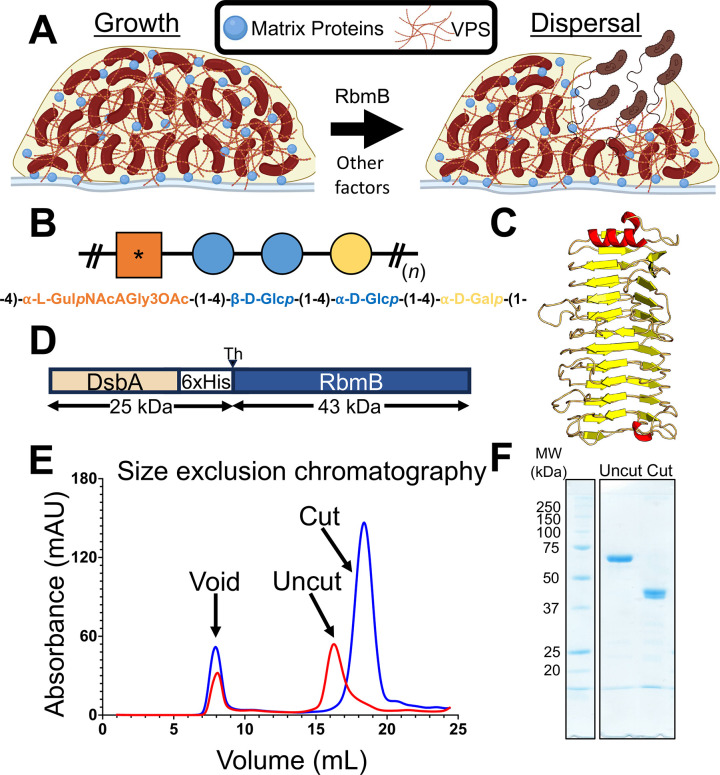
Fig 1 RbmB plays a role in V. cholerae biofilm dispersal. (A) Schematic of a working model suggesting cellular escape is helped by RbmB-mediated cleavage of VPS. Created in BioRender. Olson, R. (2022) BioRender.com/m42l648 and Olson, R. (2022) BioRender.com/n45c416. (B) Schematic version of the tetrasaccharide structure of VPS based on [21]. The asterisk represents the modified gulose moiety. (C) AlphaFold2 [24] model for RbmB structure. (D) RbmB was expressed as a thrombin-cleavable fusion protein with DsbA and a periplasmic secretion signal peptide at the N-terminus. (E) Size exclusion chromatography trace showing purified RbmB fusion before and after cleavage of the DsbA fusion partner. (F) SDS-PAGE gel showing purified DsbA-RbmB fusion and cleaved and purified RbmB. The expected molecular weight for cleaved RbmB is ~42.7 kDa.
Fig 1 RbmB plays a role in V. cholerae biofilm dispersal. (A) Schematic of a working model suggesting cellular escape is helped by RbmB-mediated cleavage of VPS. Created in BioRender. Olson, R. (2022) BioRender.com/m42l648 and Olson, R. (2022) BioRender.com/n45c416. (B) Schematic version of the tetrasaccharide structure of VPS based on [21]. The asterisk represents the modified gulose moiety. (C) AlphaFold2 [24] model for RbmB structure. (D) RbmB was expressed as a thrombin-cleavable fusion protein with DsbA and a periplasmic secretion signal peptide at the N-terminus. (E) Size exclusion chromatography trace showing purified RbmB fusion before and after cleavage of the DsbA fusion partner. (F) SDS-PAGE gel showing purified DsbA-RbmB fusion and cleaved and purified RbmB. The expected molecular weight for cleaved RbmB is ~42.7 kDa.CC BY · PubMed Central
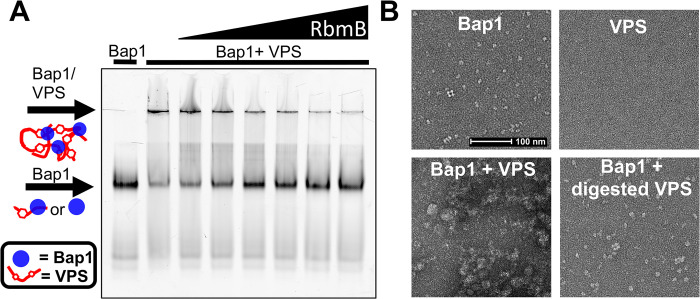
Fig 2 RbmB digests VPS in vitro. (A) Fluorescent-imaged native-PAGE gel using Bap1-GFPUV to label VPS polymer. Addition of Bap1 to uncut VPS shows a shift to a large complex that is unable to enter the gel. Increasing amounts of RbmB show a decrease in the aggregates and increase in free Bap1, suggesting cleavage of VPS into small fragments. Lanes left to right: Bap1 alone, Bap1+VPS, Bap1+VPS pre-treated with 0.05 μg RbmB, Bap1+VPS pre-treated with 1 μg RbmB, Bap1+VPS pre-treated with 2.5 μg RbmB, Bap1+VPS pre-treated with 5 μg RbmB, Bap1+VPS pre-treated with 10 μg RbmB, and Bap1+VPS pre-treated with 15 μg RbmB. (B) Negative-stained EM images of Bap1/VPS complexes. Bap1 particles form dense aggregates of size >0.5 μm when added to VPS. Digestion with RbmB disrupts aggregates back to smaller Bap1/VPS particles of size <0.1 μm.
Fig 2 RbmB digests VPS in vitro. (A) Fluorescent-imaged native-PAGE gel using Bap1-GFPUV to label VPS polymer. Addition of Bap1 to uncut VPS shows a shift to a large complex that is unable to enter the gel. Increasing amounts of RbmB show a decrease in the aggregates and increase in free Bap1, suggesting cleavage of VPS into small fragments. Lanes left to right: Bap1 alone, Bap1+VPS, Bap1+VPS pre-treated with 0.05 μg RbmB, Bap1+VPS pre-treated with 1 μg RbmB, Bap1+VPS pre-treated with 2.5 μg RbmB, Bap1+VPS pre-treated with 5 μg RbmB, Bap1+VPS pre-treated with 10 μg RbmB, and Bap1+VPS pre-treated with 15 μg RbmB. (B) Negative-stained EM images of Bap1/VPS complexes. Bap1 particles form dense aggregates of size >0.5 μm when added to VPS. Digestion with RbmB disrupts aggregates back to smaller Bap1/VPS particles of size <0.1 μm.CC BY · PubMed Central
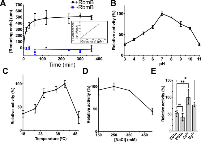
Fig 3 VPS cleavage activity of purified RbmB. (A) Quantification of reducing ends as a function of time formed during RbmB-mediated cleavage of VPS. A standard curve using galactose as a substrate is shown in the inset and S2 Fig. The effects of pH (B), temperature (C), salt concentration (D), and divalent cations (E) on the activity of RbmB against VPS are also shown. All data are represented as the mean ± SD (n = 3). 100% activity in each graph is calculated based on the condition with the highest activity. Statistical significance was determined using an unpaired, two-tailed t-test with Welch’s correction. ns = not significant, *p<0.05, **p<0.01. Solid lines indicate which samples were compared in the statistical tests. All experiments were conducted with 25 μg of VPS and 20 μg of RbmB.
Fig 3 VPS cleavage activity of purified RbmB. (A) Quantification of reducing ends as a function of time formed during RbmB-mediated cleavage of VPS. A standard curve using galactose as a substrate is shown in the inset and S2 Fig. The effects of pH (B), temperature (C), salt concentration (D), and divalent cations (E) on the activity of RbmB against VPS are also shown. All data are represented as the mean ± SD (n = 3). 100% activity in each graph is calculated based on the condition with the highest activity. Statistical significance was determined using an unpaired, two-tailed t-test with Welch’s correction. ns = not significant, *p<0.05, **p<0.01. Solid lines indicate which samples were compared in the statistical tests. All experiments were conducted with 25 μg of VPS and 20 μg of RbmB.CC BY · PubMed Central
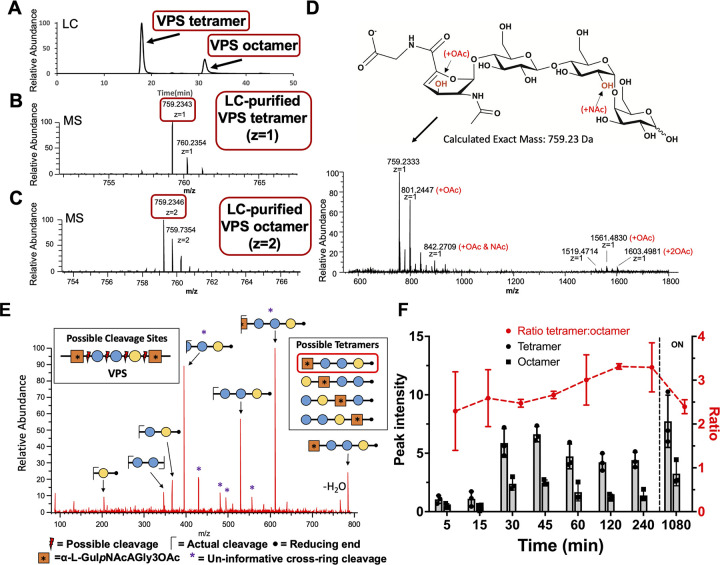
Fig 4 Mass spectrometry analysis of VPS fragments produced by RbmB cleavage. (A) Liquid chromatographic analysis of an overnight digestion of VPS by RbmB indicates a separable mixture of tetra- and octameric oligosaccharides of VPS. (B,C) Mass spectrometry analysis of liquid chromatography peaks indicates a primary species consistent with the published tetrameric form of VPS, including formation of a C4-C5 double bond in the gulose moiety due to a lyase mechanism. (D) Full negative mode mass spectrometry spectrum showing modified species and the chemical structure of the primary species. The spectra also indicate species consistent with O-acetylation and N-acetylation of the tetrasaccharide, presumably on the gulose and glucose saccharides as indicated in red. Annotation of major peaks in the spectrum is shown in S1 Table. (E) MS2 analysis is consistent with cleavage between the galactose and gulose moieties. Four possible cleavage sites would produce four different tetrameric species (shown in inset boxes). To identify the exact cleavage site, VPS was borate-reduced following RbmB cleavage and subjected to MS2 mass spectrometry analysis. The fragmentation pattern is consistent with a single cleavage site between the galactose and gulose, designating RbmB as an α-1,4 lyase (red box indicates resulting tetrasaccharide). (F) Integration of peak intensity for the tetra- and octasaccharide over time (ON = overnight). The tetramer:octamer peak intensity ratio (red) remains relatively constant throughout the digestion. All data are represented as the mean ± SD (n = 3).
Fig 4 Mass spectrometry analysis of VPS fragments produced by RbmB cleavage. (A) Liquid chromatographic analysis of an overnight digestion of VPS by RbmB indicates a separable mixture of tetra- and octameric oligosaccharides of VPS. (B,C) Mass spectrometry analysis of liquid chromatography peaks indicates a primary species consistent with the published tetrameric form of VPS, including formation of a C4-C5 double bond in the gulose moiety due to a lyase mechanism. (D) Full negative mode mass spectrometry spectrum showing modified species and the chemical structure of the primary species. The spectra also indicate species consistent with O-acetylation and N-acetylation of the tetrasaccharide, presumably on the gulose and glucose saccharides as indicated in red. Annotation of major peaks in the spectrum is shown in S1 Table. (E) MS2 analysis is consistent with cleavage between the galactose and gulose moieties. Four possible cleavage sites would produce four different tetrameric species (shown in inset boxes). To identify the exact cleavage site, VPS was borate-reduced following RbmB cleavage and subjected to MS2 mass spectrometry analysis. The fragmentation pattern is consistent with a single cleavage site between the galactose and gulose, designating RbmB as an α-1,4 lyase (red box indicates resulting tetrasaccharide). (F) Integration of peak intensity for the tetra- and octasaccharide over time (ON = overnight). The tetramer:octamer peak intensity ratio (red) remains relatively constant throughout the digestion. All data are represented as the mean ± SD (n = 3).CC BY · PubMed Central
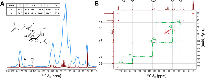
Fig 5 NMR analysis of uncleaved and cleaved VPS. (A) Superposition of 1D 13C NMR spectra collected on uncleaved VPS (using CPMAS solid-state NMR spinning at 10,000 Hz, blue) and RbmB-cleaved VPS (solution NMR, red) shows the presence of new peaks in the cleaved sample (carbons numbered as shown on proposed modified gulose structure). The chemical shifts of these two new peaks are consistent with formation of alkene carbons predicted to result from lyase activity on the gulose moiety. The inset table shows chemical shift values (in ppm) for uncleaved VPS as reported in [21] and cleaved VPS from the solution NMR data. (B) Solution 2D [13C, 13C] COSY experiment run on RbmB-cleaved VPS shows assignments for carbons around the gulose ring. Green dotted lines illustrate the connectivity of carbon atoms around the gulose ring as labeled in (A).
Fig 5 NMR analysis of uncleaved and cleaved VPS. (A) Superposition of 1D 13C NMR spectra collected on uncleaved VPS (using CPMAS solid-state NMR spinning at 10,000 Hz, blue) and RbmB-cleaved VPS (solution NMR, red) shows the presence of new peaks in the cleaved sample (carbons numbered as shown on proposed modified gulose structure). The chemical shifts of these two new peaks are consistent with formation of alkene carbons predicted to result from lyase activity on the gulose moiety. The inset table shows chemical shift values (in ppm) for uncleaved VPS as reported in [21] and cleaved VPS from the solution NMR data. (B) Solution 2D [13C, 13C] COSY experiment run on RbmB-cleaved VPS shows assignments for carbons around the gulose ring. Green dotted lines illustrate the connectivity of carbon atoms around the gulose ring as labeled in (A).CC BY · PubMed Central
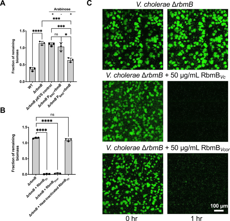
Fig 6 RbmB disrupts V. cholerae biofilms. (A) After biofilm growth in M9 medium with 0.5% glucose and 0.5% casamino acids as nutrient source, we induced nutrient limitation by removing the carbon sources in the medium. Biomasses before and after dispersal for 5 hours at 30°C were quantified using a confocal microscope. The ΔrbmB mutant retains more surface biofilm mass following incubation, suggesting a weaker dispersal phenotype. Induction of rbmB from a plasmid under arabinose control shows partial recovery of the dispersal phenotype. Statistical analyses were performed using an unpaired, two-tailed t-test with Welch’s correction. ns = not significant, *p < 0.05, ***p < 0.001, ****p < 0.0001. (B) Recombinantly purified RbmB (50 μg/mL) from V. cholerae (Vc) or V. coralliilyticus (Vcor) is sufficient to nearly completely disrupt V. cholerae biofilms within 1 hour, but heat-inactivated RbmB has no effect. All data are depicted as mean ± SD. Statistical analyses were performed using an unpaired, two-tailed t-test with Welch’s correction. ns = not significant, ****p < 0.0001. (C) Cross-sectional images of V. cholerae biofilms at the bottom cell layer, before and after 1 hour of nutrient limitation. 50 μg/mL RbmB from V. cholerae (Vc) or V. coralliilyticus (Vcor) were present during the nutrient limitation step, in the middle and bottom rows, respectively. All data in this figure are depicted as mean ± SD (n = 3 biologically independent samples).
Fig 6 RbmB disrupts V. cholerae biofilms. (A) After biofilm growth in M9 medium with 0.5% glucose and 0.5% casamino acids as nutrient source, we induced nutrient limitation by removing the carbon sources in the medium. Biomasses before and after dispersal for 5 hours at 30°C were quantified using a confocal microscope. The ΔrbmB mutant retains more surface biofilm mass following incubation, suggesting a weaker dispersal phenotype. Induction of rbmB from a plasmid under arabinose control shows partial recovery of the dispersal phenotype. Statistical analyses were performed using an unpaired, two-tailed t-test with Welch’s correction. ns = not significant, *p < 0.05, ***p < 0.001, ****p < 0.0001. (B) Recombinantly purified RbmB (50 μg/mL) from V. cholerae (Vc) or V. coralliilyticus (Vcor) is sufficient to nearly completely disrupt V. cholerae biofilms within 1 hour, but heat-inactivated RbmB has no effect. All data are depicted as mean ± SD. Statistical analyses were performed using an unpaired, two-tailed t-test with Welch’s correction. ns = not significant, ****p < 0.0001. (C) Cross-sectional images of V. cholerae biofilms at the bottom cell layer, before and after 1 hour of nutrient limitation. 50 μg/mL RbmB from V. cholerae (Vc) or V. coralliilyticus (Vcor) were present during the nutrient limitation step, in the middle and bottom rows, respectively. All data in this figure are depicted as mean ± SD (n = 3 biologically independent samples).CC BY · PubMed Central

Verification

Claims the paper makes

The claims and statistics of this paper haven’t been checked yet.

Run the check — free, no account needed. It reads the open-access text and checks the sources it cites.

A few free checks a day · papers already verified open instantly · results are public

Self-check

Percentages, sums and reported differences re-computed from the paper’s own numbers.

The self-check needs the paper’s full text.

Citations

Cites

Show all 1 references

Research works printed but not linked: 45

  1. 10.3389/fmicb.2021.686793 · DOI
  2. 10.1038/s41579-022-00791-0 · DOI
  3. 10.1111/mmi.13698 · DOI
  4. 10.1038/s41579-020-0385-0 · DOI
  5. 10.1016/j.jmb.2005.03.082 · DOI
  6. 10.1128/JB.185.16.4693-4698.2003 · DOI · Paper page
  7. 10.1038/nrmicro3433 · DOI
  8. 10.1146/annurev-micro-111021-053553 · DOI
  9. 10.1128/IAI.00048-10 · DOI
  10. 10.1073/pnas.1916571117 · DOI
  11. 10.1073/pnas.0237386100 · DOI
  12. 10.1073/pnas.96.7.4028 · DOI
  13. 10.1099/mic.0.040196-0 · DOI
  14. 10.1128/JB.188.3.1049-1059.2006 · DOI
  15. 10.1128/JB.01569-06 · DOI
  16. 10.1126/science.1222981 · DOI
  17. 10.1371/journal.ppat.1002210 · DOI
  18. 10.1038/s41467-023-37660-0 · DOI
  19. 10.1016/j.bpj.2014.10.015 · DOI
  20. 10.1073/pnas.2021166117 · DOI
  21. 10.1371/journal.pone.0086751 · DOI
  22. 10.1038/s41579-022-00712-1 · DOI · Paper page
  23. 10.1093/nar/gkab1112 · DOI · Paper page
  24. 10.1126/science.8502994 · DOI
  25. 10.1093/nar/gkac993 · DOI
  26. 10.1093/nar/gkaa1079 · DOI · Paper page
  27. 10.1093/nar/gkaa937 · DOI
  28. 10.1021/ja00420a036 · DOI
  29. 10.1038/ismej.2017.33 · DOI
  30. 10.1016/j.ijbiomac.2020.07.199 · DOI
  31. 10.1074/jbc.M116.766030 · DOI
  32. 10.1371/journal.ppat.1008745 · DOI
  33. 10.1038/nature05267 · DOI
  34. 10.1146/annurev.biochem.75.103004.142545 · DOI
  35. 10.3389/fcimb.2020.00379 · DOI
  36. 10.1016/j.bioflm.2021.100061 · DOI
  37. 10.1021/acs.chemrev.0c01338 · DOI
  38. 10.1002/pro.102 · DOI
  39. 10.1074/jbc.RA119.008335 · DOI
  40. 10.1016/0003-2697(80)90143-8 · DOI
  41. 10.1016/0003-2697(89)90004-3 · DOI
  42. 10.1021/pr7008252 · DOI
  43. 10.1063/1.470372 · DOI
  44. 10.1016/S1090-7807(03)00082-X · DOI
  45. 10.1007/BF01049915 · DOI

Metadata

Authors
Weerasekera, R., Moreau, A., Huang, X., Nam, K.-M., Hinbest, A., Huynh, Y., Liu, X., Ashwood, C., Pepi, L. E., Paulson, E., Cegelski, L., Yan, J., & Olson, R.
Affiliations
  • Wesleyan University (US)
  • Yale University (US)
  • Stanford University (US)
  • Beth Israel Deaconess Medical Center (US)
Corresponding author
Olson, Rich
Venue
PLoS Pathogens
Publisher
openRxiv
Published
2024-08-27
Volume
20
Issue
12
Pages
e1012750-e1012750
ISSN
1553-7366 (ISSN-L)
OpenAlex
W4404914127
Cited by
9
References
1
Citations by year
2026: 5 · 2025: 3 · 2024: 1
FWCI
2.67
Type
journal article
Language
en
Open access
Locations
  • PLoS Pathogens (publishedVersion, cc-by)
  • bioRxiv (Cold Spring Harbor Laboratory) (acceptedVersion)
  • PubMed Central (publishedVersion, cc-by)
  • PubMed (publishedVersion)
  • DOAJ (DOAJ: Directory of Open Access Journals) (submittedVersion)
Funders
  • National Science Foundation (2205006)
  • Alfred P. Sloan Foundation (FG-2023-20857)
  • Yale University
  • National Institutes of Health
  • National Institute of General Medical Sciences (DP2GM146253, R15GM152959, R01GM117278)
  • Division of Materials Research
Topics
  • Life Sciences › Immunology and Microbiology › Microbiology › Vibrio bacteria research studies
  • Life Sciences › Biochemistry, Genetics and Molecular Biology › Molecular Biology › Bacterial biofilms and quorum sensing
  • Life Sciences › Agricultural and Biological Sciences › Food Science › Polysaccharides Composition and Applications
Keywords
Vibrio cholerae, biofilm dispersal, biofilm matrix degradation, Vibrio coralliilyticus, antibacterial enzymes, glycolytic enzymes
MeSH terms
Bacterial Proteins, Cholera, Humans, Polysaccharide-Lyases, Polysaccharides, Bacterial, Vibrio cholerae, Biofilms

About these checks

Registry, retraction, Cites and statistics results are computed, not judged, with one exception: a printed reference that no index lists is read once by a model before it counts as a citation error. Claims are extracted by a model and kept only when their quote appears verbatim in the text; each cited source is then checked, with a model’s help, for whether it says what the claim relies on. Those verdicts can be wrong — read the claim’s quote and the cited source before relying on one.

Statistics and arithmetic are recomputed, not judged.

Confidence bands are shown only for fields calibrated on our annotated test set. None is calibrated yet, so no band is shown.

What counts as a claim ›How we measure confidence ›How we check this ›

Are you an author, or do you see a problem with this page? Request a review

How to cite

Cite CoChat for the checks on this page, and the paper for its own findings.

This page
CoChat. (2026). Citation check: Vibrio cholerae RbmB is an α−1,4-polysaccharide lyase with biofilm-dispersal activity against Vibrio Polysaccharide (VPS). https://app.cochat.ai/papers/10.1101/2024.08.27.609776
The paper
Weerasekera, R., Moreau, A., Huang, X., Nam, K.-M., Hinbest, A., Huynh, Y., Liu, X., Ashwood, C., Pepi, L. E., Paulson, E., Cegelski, L., Yan, J., & Olson, R. (2024). Vibrio cholerae RbmB is an α−1,4-polysaccharide lyase with biofilm-dispersal activity against Vibrio Polysaccharide (VPS). PLoS Pathogens. https://doi.org/10.1101/2024.08.27.609776