

The DOI matches its Crossref record; 1 of the 1 references it cites were found in a registry or an index.
The Helicobacter pylori TlpD cytoplasmic chemoreceptor requires an intact C-terminus for polar localization and function
Open accessPreprint — published as 10.1128/jb.00394-25 · Paper page
Abstract
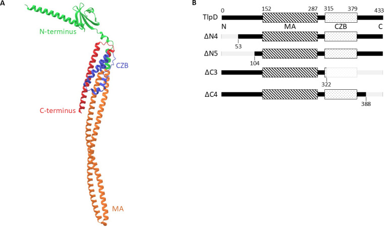
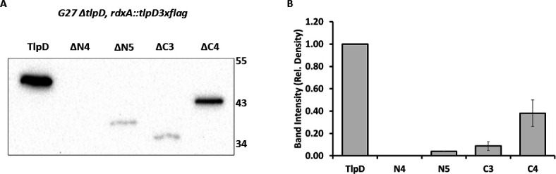
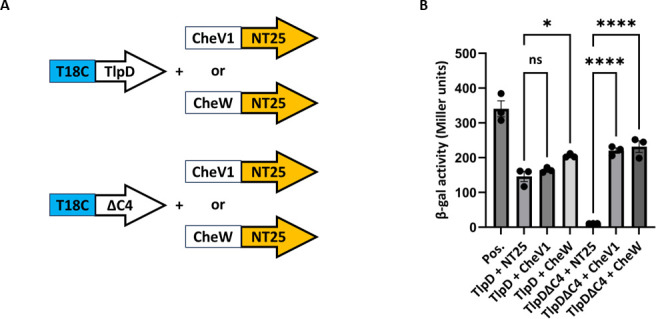
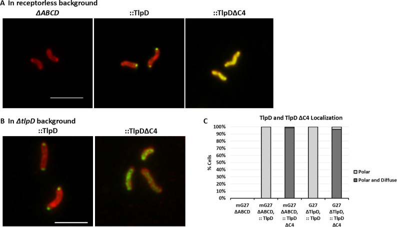
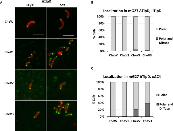
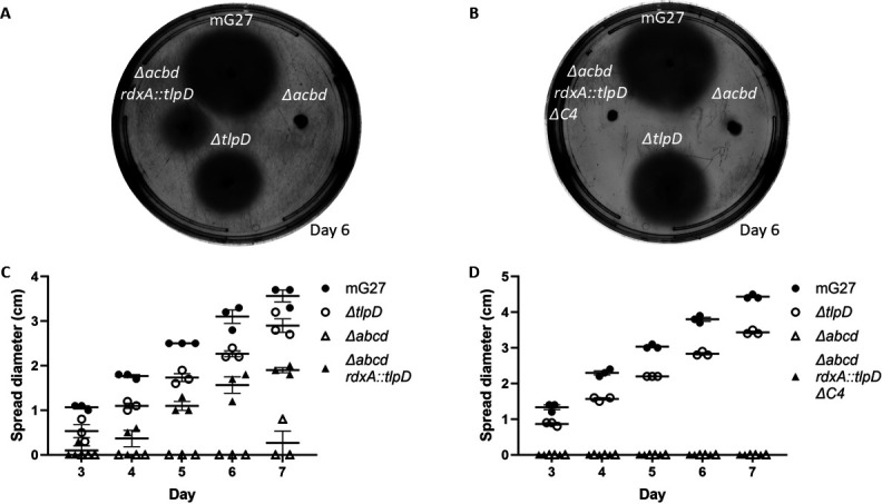
Bacteria localize proteins to distinct subcellular locations, including chemoreceptors, which frequently localize to the bacterial pole. Although some polarity-promoting mechanisms have been described, many chemoreceptors lack clear routes to becoming polar. TlpD of the bacterial pathogen Helicobacter pylori is one such protein. This cytoplasmic chemoreceptor localizes to the pole in a manner that is independent of the other chemoreceptors. In this work, we evaluated the role of TlpD domains in its function. Truncated proteins were created that lacked different amounts of the N- or C-termini and expressed in H. pylori in place of native tlpD or as the sole chemoreceptor. These TlpD variants were examined for expression, protein localization, association with chemotaxis signaling proteins, and effect on motility. TlpD that lacked any portion of the N-terminal 104 amino acids did not produce detectable protein. In contrast, TlpD retained expression with loss of the C-terminal 45 amino acids. TlpD lacking the last 45 amino acids (TlpDΔC4) preserved the ability to interact with CheW and CheV proteins based on bacterial two-hybrid analysis but were unable to localize to the pole either on their own or in the presence of other chemoreceptors. TlpDΔC4 was found diffuse in the cytoplasm, and interacted with CheV1, CheV2, and CheV3 at this location but not CheW. TlpDΔC4 did not confer chemotactic abilities in soft agar chemotaxis assays. These findings suggest the C-terminal end of TlpD plays a previously unappreciated role in promoting TlpD polar localization and function. Importance: Bacteria place their proteins in specific locations that are required for the proteins to function including the bacterial pole. How the bacterial cell identifies which proteins go to the pole is not fully understood. In this work, we dissect parts of a protein called TlpD that naturally goes to the pole. We find that mutants lacking one end of TlpD lose their polar placement, but retain other abilities. TlpD allows directed motility known as chemotaxis. This ability is critical for infection in H. pylori and numerous other pathogens. When TlpD loses its polar placement, the protein no longer functions for chemotaxis, laying the foundation for future studies that can dissect how this segment promotes function and eventually translate into therapies for H. pylori infection.
Overview
The claims and statistics of this paper haven’t been checked yet.
Run the check — free, no account needed. It reads the open-access text and checks the sources it cites.
Integrity signals
Registry
Cites
Statistics
Figures
Verification
Claims the paper makes
The claims and statistics of this paper haven’t been checked yet.
Run the check — free, no account needed. It reads the open-access text and checks the sources it cites.
Self-check
Percentages, sums and reported differences re-computed from the paper’s own numbers.
The self-check needs the paper’s full text.
Citations
Cites
Highly accurate protein structure prediction with AlphaFold
Jumper, J., Evans, R., Pritzel, A., Green, T., Figurnov, M., Ronneberger, O., Tunyasuvunakool, K., Bates, R., Žídek, A., Potapenko, A., et al. (2021) · Nature · doi:10.1038/s41586-021-03819-2 · Cited by 43,764
Research works printed but not linked: 53
- Malfertheiner P , Camargo MC , El-Omar E , Liou JM , Peek R , Schulz C , Smith SI , Suerbaum S . 2023. Helicobacter pylori infection. Nat Rev Dis Prim 9. · DOI · Paper page
- Collins KD , Hu S , Grasberger H , Kao JY , Ottemann KM . 2018. Chemotaxis allows bacteria to overcome host-generated reactive oxygen species that constrain gland colonization. Infect Immun 86:IAI.00878-17. · DOI
- 10.1093/femsre/fuw025 · DOI
- 10.1053/j.gastro.2013.07.011 · DOI
- 10.1007/s00011-010-0245-x · DOI
- 10.1016/j.mib.2017.11.019 · DOI
- 10.1016/j.mib.2017.07.004 · DOI
- 10.1016/j.tim.2015.03.003 · DOI · Paper page
- 10.1038/nrmicro2505 · DOI · Paper page
- 10.1038/nrm1524 · DOI · Paper page
- 10.1128/IAI.00407-12 · DOI
- 10.1128/IAI.00750-13 · DOI
- 10.1128/IAI.68.4.2016-2023.2000 · DOI
- 10.1128/IAI.73.2.803-811.2005 · DOI
- 10.1016/j.tim.2010.07.004 · DOI
- 10.1073/pnas.1618227114 · DOI
- 10.1128/JB.00071-16 · DOI
- 10.1099/00221287-147-9-2493 · DOI
- 10.1371/journal.pcbi.1004721 · DOI
- 10.1099/mic.0.28217-0 · DOI
- 10.1099/mic.0.021857-0 · DOI
- 10.1128/MMBR.00033-14 · DOI
- 10.1128/JB.05140-11 · DOI
- 10.1371/journal.pbio.3000202 · DOI · Paper page
- 10.1128/JB.01940-07 · DOI
- 10.1093/nar/30.1.42 · DOI · Paper page
- 10.1093/nar/gkw1129 · DOI · Paper page
- 10.1093/nar/gkp256 · DOI
- 10.1093/nar/gkg519 · DOI
- 10.1073/pnas.95.10.5752 · DOI
- 10.1073/pnas.93.25.14648 · DOI · Paper page
- 10.1128/IAI.00082-07 · DOI
- 10.1111/j.1365-2958.2007.06033.x · DOI
- 10.1099/mic.0.049353-0 · DOI
- 10.1111/mmi.13086 · DOI
- 10.1038/s41598-016-0001-8 · DOI · Paper page
- 10.1080/19490976.2017.1279380 · DOI
- 10.1038/35051615 · DOI · Paper page
- 10.1016/j.femsim.2004.11.008 · DOI
- 10.1074/mcp.O113.033571 · DOI
- 10.1128/JB.187.22.7840-7844.2005 · DOI
- 10.1128/JB.01286-08 · DOI
- 10.1128/JB.01255-10 · DOI
- 10.1073/pnas.0910055107 · DOI
- 10.1016/j.tim.2015.03.004 · DOI
- 10.1128/JB.188.6.2233-2243.2006 · DOI
- 10.1073/pnas.95.11.5857 · DOI
- 10.1128/JB.00852-06 · DOI
- 10.1111/j.1365-2958.2012.08051.x · DOI
- 10.1128/mbio.03338-22 · DOI
- 10.1128/IAI.70.4.1984-1990.2002 · DOI · Paper page
- 10.1128/IAI.73.3.1820-1827.2005 · DOI
- 10.1128/JB.00749-09 · DOI
Metadata
- Authors
- Lopez-Magaña, R., & Ottemann, K. M.
- Affiliations
- University of California, Santa Cruz (US)
- Corresponding author
- Ottemann, Karen M.
- Venue
- Journal of Bacteriology
- Publisher
- openRxiv
- Published
- 2024-11-08
- Volume
- 208
- Issue
- 3
- Pages
- e0039425-e0039425
- ISSN
- 0021-9193 (ISSN-L)
- PMC
- PMC13001251
- OpenAlex
- W7128395484
- Cited by
- 1
- References
- 1
- Citations by year
- 2026: 1
- FWCI
- 4.00
- Type
- journal article
- Language
- en
- Open access
- diamond
- doi.org
- Licence
- Locations
- Journal of Bacteriology (publishedVersion, cc-by)
- bioRxiv (Cold Spring Harbor Laboratory) (acceptedVersion, cc-by)
- PubMed Central (publishedVersion, cc-by)
- PubMed (publishedVersion)
- DOAJ (DOAJ: Directory of Open Access Journals) (submittedVersion, cc-by-sa)
- PubMed Central (submittedVersion)
- Funders
- National Institutes of Health
- National Institute of Allergy and Infectious Diseases (RO1AI116946, R01AI116946)
- Topics
- Life Sciences › Biochemistry, Genetics and Molecular Biology › Genetics › Bacterial Genetics and Biotechnology
- Health Sciences › Medicine › Gastroenterology › Helicobacter pylori-related gastroenterology studies
- Health Sciences › Medicine › Oncology › Chemokine receptors and signaling
- Keywords
- Helicobacter pylori, polar localization, C-terminal domain, chemotaxis, bacterial motility, CheW, chemoreceptor signaling, infection pathogenesis
About these checks
Registry, retraction, Cites and statistics results are computed, not judged, with one exception: a printed reference that no index lists is read once by a model before it counts as a citation error. Claims are extracted by a model and kept only when their quote appears verbatim in the text; each cited source is then checked, with a model’s help, for whether it says what the claim relies on. Those verdicts can be wrong — read the claim’s quote and the cited source before relying on one.
Statistics and arithmetic are recomputed, not judged.
Confidence bands are shown only for fields calibrated on our annotated test set. None is calibrated yet, so no band is shown.
What counts as a claim ›How we measure confidence ›How we check this ›
Are you an author, or do you see a problem with this page? Request a review
How to cite
Cite CoChat for the checks on this page, and the paper for its own findings.
- This page
- CoChat. (2026). Citation check: The Helicobacter pylori TlpD cytoplasmic chemoreceptor requires an intact C-terminus for polar localization and function. https://app.cochat.ai/papers/10.1101/2024.11.08.622596
- The paper
- Lopez-Magaña, R., & Ottemann, K. M. (2024). The Helicobacter pylori TlpD cytoplasmic chemoreceptor requires an intact C-terminus for polar localization and function. Journal of Bacteriology. https://doi.org/10.1101/2024.11.08.622596
Data sources
Bibliographic data: Semantic Scholar, OpenAlex, Crossref.
Contains information from the Semantic Scholar Academic Graph Datasets, made available under the ODC Attribution License (ODC-BY 1.0).
Semantic Scholar’s data is described in: Kinney, R., et al. (2023). The Semantic Scholar Open Data Platform. arXiv:2301.10140.
