PreprintCited by 0Protein Structure and DynamicsForce Microscopy Techniques and ApplicationsGlycosylation and Glycoproteins Research

The DOI matches its Crossref record; 1 of the 1 references it cites were found in a registry or an index.

Lubricin’s Mucin Domain Has Strong Polyproline Type-II Helical Character

Open access
Feng, Bibo ’Noah’, Marks, Ava J., Kim, Faith, Naik, Mandar, Schmidt, Tannin A., Elsaid, Khaled A., Jay, Gregory D., Rubenstein, Brenda·2025·Computational and Structural Biotechnology Journal

Preprint — published as 10.34133/csbj.0021 · Paper page

Abstract

Lubricin is a glycoprotein that is crucial for maintaining joint health by preventing joint wear by reducing joint friction in the boundary mode. Lubricin was recently observed to hinder the formation of uric acid crystals in the joint and prevent a form of gouty arthritis. However, despite lubricin’s great physiological importance, our current understanding of the molecular origins of lubricin’s beneficial properties is limited by a lack of detailed structural information regarding its central mucin domain: lubricin’s large size (227.5 kDa) and numerous glycosylations pose a substantial obstacle to conventional experimental methods for solving protein structures. In this work, we employ a combination of physics-based replica exchange molecular dynamics (REMD) simulations and circular dichroism (CD) experiments to shed light on the structure of lubricin’s central mucin domain. Using REMD, we model [KEPAPTTP] 2 , an amino acid repeat found throughout the mucin domain, and find that the mucin domain is likely to exhibit polyproline type II (PPII) helices, which are further stabilized by O-linked oligosaccharide chains. Motivated by these simulation results, we performed circular dichroism spectroscopy on fragments of the mucin domain that also show clear polyproline-II helical character, corroborating our computational findings. Altogether, this work provides strong evidence of a lubricin mucin domain with significant polyproline type II content. As polyproline helices are often also found in other glycoproteins with antifreeze properties, this work may also explain the atomistic underpinnings of their interfacial functions, including lubrication and competition with crystal formation. SIGNIFICANCE: Lubricin is a mucinous glycoprotein containing a central heavily glycosylated domain that plays a crucial role in the lubrication of joints. However, little is known about the structure of this large central mucin domain, which makes the development of related therapeutics or biomedical devices challenging. In this work, we provide strong evidence that lubricin’s mucin domain possesses polyproline type II (PPII) character that is enhanced by glycosylation upon the basis of a combination of molecular dynamics simulations and circular dichroism experiments. Lubricin’s PPII character provides a molecular basis for its lubricating properties, which may provide insights into related antifreeze glycoproteins (AFGP) and the development of new biocompatible lubricants and ryo-preservatives.

Overview

The claims and statistics of this paper haven’t been checked yet.

Run the check — free, no account needed. It reads the open-access text and checks the sources it cites.

A few free checks a day · papers already verified open instantly · results are public

Integrity signals

Registry

Crossref · 10.1101/2025.06.15.659778

Cites

1 of 1 references verified0 citation errors · 0 not checkable

Statistics

Figures

Graphical abstract
Graphical abstract CC BY · PubMed Central
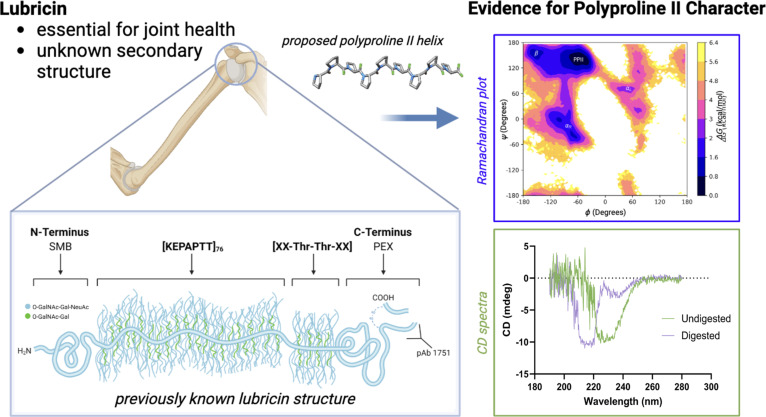
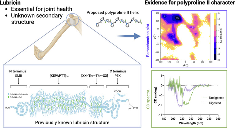
Fig. 1. (Table of contents image) In this manuscript, we demonstrate that lubricin’s mucin domain possesses polyproline type II (PPII) helical character using a combination of molecular dynamics simulations and circular dichroism (CD) spectroscopy. SMB, somatomedin B; PEX, hemopexin; NeuAc, N-acetylneuraminic acid.
Fig. 1. (Table of contents image) In this manuscript, we demonstrate that lubricin’s mucin domain possesses polyproline type II (PPII) helical character using a combination of molecular dynamics simulations and circular dichroism (CD) spectroscopy. SMB, somatomedin B; PEX, hemopexin; NeuAc, N-acetylneuraminic acid.CC BY · PubMed Central
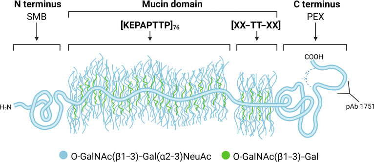
Fig. 2. A schematic of lubricin, which consists of an N-terminal somatomedin B (SMB) domain, a central mucin domain with many KEPAPTTP repeats, and a C-terminal domain referred to as a hemopexin (PEX) domain. The C terminus possesses a disulfide-bonded peptide resulting from subtilisin cleavage [48]. Common glycosidic side chains found on lubricin are also depicted.
Fig. 2. A schematic of lubricin, which consists of an N-terminal somatomedin B (SMB) domain, a central mucin domain with many KEPAPTTP repeats, and a C-terminal domain referred to as a hemopexin (PEX) domain. The C terminus possesses a disulfide-bonded peptide resulting from subtilisin cleavage [48]. Common glycosidic side chains found on lubricin are also depicted.CC BY · PubMed Central
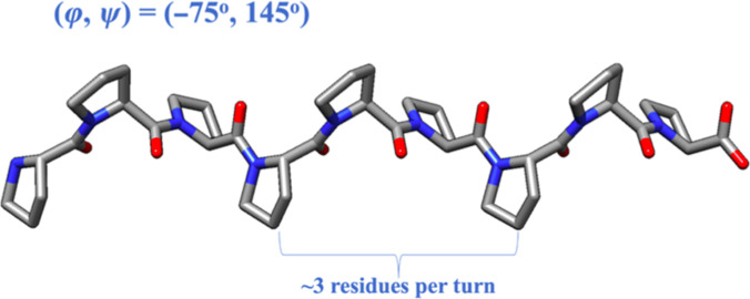
Fig. 3. A schematic of a polyproline type II (PPII) helix. PPII helices are characterized by backbone dihedral angles ϕ/ψ of −75°/145°, which result in a left-handed helix with 3 residues per turn. PPII helices also possess limited hydrogen bonding.
Fig. 3. A schematic of a polyproline type II (PPII) helix. PPII helices are characterized by backbone dihedral angles ϕ/ψ of −75°/145°, which result in a left-handed helix with 3 residues per turn. PPII helices also possess limited hydrogen bonding.CC BY · PubMed Central
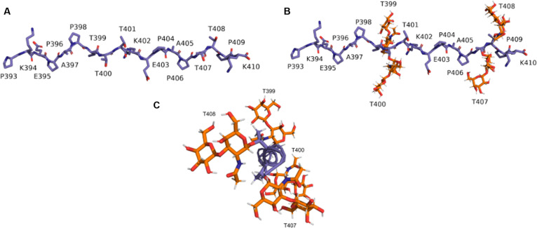
Fig. 4. (A) Nonglycosylated and (B) glycosylated subsections of the mucin domain modeled in this work. These subsections correspond to residues 393 to 410 (PKEPAPTTTKEPAPTTPK), which contain both the KEPAPTTP and KEPAPTTT repeat units. (C) End-on view of the glycosylated model peptide illustrating the positioning of the glycosylations around the peptide unit.
Fig. 4. (A) Nonglycosylated and (B) glycosylated subsections of the mucin domain modeled in this work. These subsections correspond to residues 393 to 410 (PKEPAPTTTKEPAPTTPK), which contain both the KEPAPTTP and KEPAPTTT repeat units. (C) End-on view of the glycosylated model peptide illustrating the positioning of the glycosylations around the peptide unit.CC BY · PubMed Central
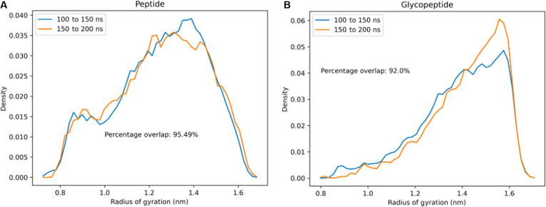
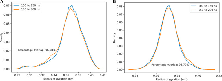
Fig. 5. Probability distributions of Rg within the time intervals 100 to 150 ns and 150 to 200 ns of replica exchange molecular dynamics (REMD) simulation at 300 K for the 2 tandem repeat model peptides: (A) without glycosylation and (B) with O-linked N-acetylgalactosamine–galactose (GalNAc–Gal) glycosylation. In both cases, there is good overlap between the Rg probability distributions within the 2 independent time intervals, indicating that the sampling has converged.
Fig. 5. Probability distributions of Rg within the time intervals 100 to 150 ns and 150 to 200 ns of replica exchange molecular dynamics (REMD) simulation at 300 K for the 2 tandem repeat model peptides: (A) without glycosylation and (B) with O-linked N-acetylgalactosamine–galactose (GalNAc–Gal) glycosylation. In both cases, there is good overlap between the Rg probability distributions within the 2 independent time intervals, indicating that the sampling has converged.CC BY · PubMed Central
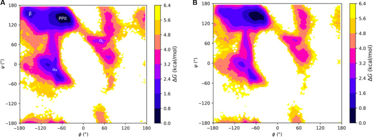
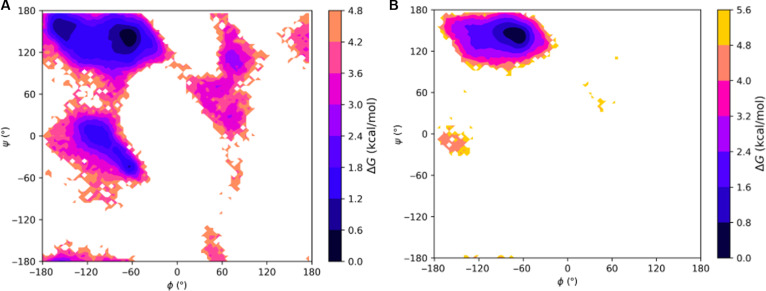
Fig. 6. Ramachandran-like free energy surfaces at 300 K of the model lubricin peptide: (A) without any glycosylations and (B) with O-linked N-acetylgalactosamine–galactose (GalNAc–Gal) glycosylations on relevant threonine residues (every threonine except the third one). The regions of the (ϕ, ψ) space corresponding to the polyproline type II (PPII) conformation, β conformation (β), right-handed α-helical conformation (αR), and left-handed α-helical conformation (αL) are labeled in (A). Free energy differences are defined relative to the minimum free energy PPII structure we observed, meaning that darker colors represent more stable structures closer in free energy to PPII helices, while lighter colors represent less stable structures further in free energy from PPII helices.
Fig. 6. Ramachandran-like free energy surfaces at 300 K of the model lubricin peptide: (A) without any glycosylations and (B) with O-linked N-acetylgalactosamine–galactose (GalNAc–Gal) glycosylations on relevant threonine residues (every threonine except the third one). The regions of the (ϕ, ψ) space corresponding to the polyproline type II (PPII) conformation, β conformation (β), right-handed α-helical conformation (αR), and left-handed α-helical conformation (αL) are labeled in (A). Free energy differences are defined relative to the minimum free energy PPII structure we observed, meaning that darker colors represent more stable structures closer in free energy to PPII helices, while lighter colors represent less stable structures further in free energy from PPII helices.CC BY · PubMed Central
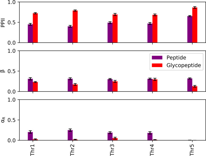
Fig. 7. Secondary structure populations of the threonine residues in the model lubricin peptide (no glycosylations) and glycopeptide at 300 K. Error bars represent 95% confidence intervals. The left-handed α-helical conformation (αL) and coil populations were negligible for both systems and are thus not included.
Fig. 7. Secondary structure populations of the threonine residues in the model lubricin peptide (no glycosylations) and glycopeptide at 300 K. Error bars represent 95% confidence intervals. The left-handed α-helical conformation (αL) and coil populations were negligible for both systems and are thus not included.CC BY · PubMed Central
Fig. 8. Probability distributions of Rg within the time intervals 100 to 150 ns and 150 to 200 ns of replica exchange molecular dynamics (REMD) simulations performed at 300 K for the 2 tandem repeat model peptides: (A) Without glycosylation and (B) with O-linked N-acetylgalactosamine–galactose (GalNAc–Gal) glycosylation. In both cases, there is good overlap between the Rg probability distributions within the 2 independent time intervals, indicating that the sampling has converged.
Fig. 8. Probability distributions of Rg within the time intervals 100 to 150 ns and 150 to 200 ns of replica exchange molecular dynamics (REMD) simulations performed at 300 K for the 2 tandem repeat model peptides: (A) Without glycosylation and (B) with O-linked N-acetylgalactosamine–galactose (GalNAc–Gal) glycosylation. In both cases, there is good overlap between the Rg probability distributions within the 2 independent time intervals, indicating that the sampling has converged.CC BY · PubMed Central
Fig. 9. Ramachandran-like free energy surfaces at 300 K of threonine tetrapeptide: (A) without glycosylations and (B) with O-linked N-acetylgalactosamine–galactose (GalNAc–Gal) glycosylations. Free energy differences are defined relative to the minimum free energy polyproline type II (PPII) structure we observed, meaning that darker colors represent more stable structures closer in free energy to PPII helices, while lighter colors represent less stable structures further in free energy from PPII helices.
Fig. 9. Ramachandran-like free energy surfaces at 300 K of threonine tetrapeptide: (A) without glycosylations and (B) with O-linked N-acetylgalactosamine–galactose (GalNAc–Gal) glycosylations. Free energy differences are defined relative to the minimum free energy polyproline type II (PPII) structure we observed, meaning that darker colors represent more stable structures closer in free energy to PPII helices, while lighter colors represent less stable structures further in free energy from PPII helices.CC BY · PubMed Central
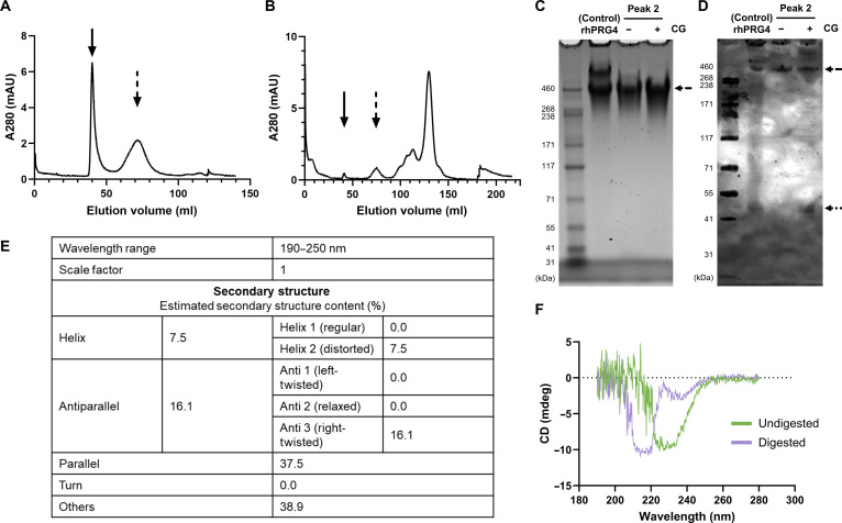
Fig. 10. Cathepsin G (CG) digestion of recombinant human PRG4 (rhPRG4) (lubricin) to partially isolate the mucin domain. (A) Fractionation of rhPRG4 over Sephacryl S-1000 in the presence of 1 M NaCl in phosphate-buffered saline (PBS) at pH 7.4. A280, absorbance at 280 nm. (B) Fractionation of the cathepsin G digestate of rhPRG4 over Sephacryl S-1000 showing digestion of the 2 principal high-molecular-weight (MW) peaks (solid and dashed arrows). (C) The partly digested second peak is detectable on sodium dodecyl sulfate polyacrylamide gel electrophoresis (SDS-PAGE) as a monomerized band with an apparent MW of 460 kDa (dashed arrow). Larger MW dimer and other larger MW forms are undetected. (D) Western blotting of the same samples reveals immunopositivity with polyclonal antibody (pAb) 1751 directed at the proximal side of the C-terminal subtilisin cleavage site observed by bands at ~46 to 48 kDa (dotted arrow). (E) BeStSel analysis of circular dichroism (CD) spectra showing the estimated secondary structure content derived from the 190- to 250-nm wavelength range. (F) Comparison of CD spectra of cathepsin G digested and undigested rhPRG4 reveals spectral signatures consistent with polyproline type II (PPII) helix in the digested state, which likely removed the C-terminal globular domain.
Fig. 10. Cathepsin G (CG) digestion of recombinant human PRG4 (rhPRG4) (lubricin) to partially isolate the mucin domain. (A) Fractionation of rhPRG4 over Sephacryl S-1000 in the presence of 1 M NaCl in phosphate-buffered saline (PBS) at pH 7.4. A280, absorbance at 280 nm. (B) Fractionation of the cathepsin G digestate of rhPRG4 over Sephacryl S-1000 showing digestion of the 2 principal high-molecular-weight (MW) peaks (solid and dashed arrows). (C) The partly digested second peak is detectable on sodium dodecyl sulfate polyacrylamide gel electrophoresis (SDS-PAGE) as a monomerized band with an apparent MW of 460 kDa (dashed arrow). Larger MW dimer and other larger MW forms are undetected. (D) Western blotting of the same samples reveals immunopositivity with polyclonal antibody (pAb) 1751 directed at the proximal side of the C-terminal subtilisin cleavage site observed by bands at ~46 to 48 kDa (dotted arrow). (E) BeStSel analysis of circular dichroism (CD) spectra showing the estimated secondary structure content derived from the 190- to 250-nm wavelength range. (F) Comparison of CD spectra of cathepsin G digested and undigested rhPRG4 reveals spectral signatures consistent with polyproline type II (PPII) helix in the digested state, which likely removed the C-terminal globular domain.CC BY · PubMed Central

Verification

Claims the paper makes

The claims and statistics of this paper haven’t been checked yet.

Run the check — free, no account needed. It reads the open-access text and checks the sources it cites.

A few free checks a day · papers already verified open instantly · results are public

Self-check

Percentages, sums and reported differences re-computed from the paper’s own numbers.

The self-check needs the paper’s full text.

Citations

Cites

Show all 1 references

Research works printed but not linked: 45

  1. 2000, Lubricin is a product of megakaryocyte stimulating factor gene expression by human synovial fibroblasts, The Journal of rheumatology, 27, 594
  2. 10.1016/S0736-0266(00)00040-1 · DOI
  3. 10.1006/bbrc.1998.0104 · DOI
  4. 10.1529/biophysj.106.088799 · DOI · Paper page
  5. 10.1016/j.biomaterials.2015.02.086 · DOI
  6. 10.1186/s13075-017-1488-5 · DOI
  7. Elsaid, K. , T. R. Merriman , L.-A. Rossitto , R. Liu-Bryan , J. Karsh , A. Phipps-Green , G. D. Jay , S. Elsayed , M. Qadri , M. Miner , et al., 2022. Amplification of inflammation by lubricin deficiency implicated in incident, erosive gout independent of hyperuricemia. Arthritis & Rheumatology. · DOI
  8. 10.1038/S41586-024-07487-W · DOI · Paper page
  9. 10.1002/art.22974 · DOI
  10. 10.1016/j.jbiomech.2012.06.036 · DOI
  11. 10.1001/jamaophthalmol.2013.2385 · DOI
  12. 10.1097/ICO.0000000000002151 · DOI
  13. 10.1021/acs.biomac.3c01348 · DOI
  14. 10.1016/S0022-2836(63)80023-6 · DOI
  15. 10.1073/pnas.93.13.6676 · DOI
  16. 10.1016/j.jmb.2013.03.018 · DOI
  17. 10.1016/S0065-3233(02)62010-8 · DOI
  18. 10.1021/bi3002924 · DOI
  19. 10.1074/jbc.M505401200 · DOI
  20. 10.1021/acs.jpcb.7b08790 · DOI
  21. 10.1021/jacs.7b13630 · DOI
  22. 10.1021/acs.biomac.2c01487 · DOI
  23. 10.1177/0363546516686965 · DOI
  24. 10.1016/j.jtos.2016.08.004 · DOI
  25. 10.1002/jcc.20084 · DOI
  26. 10.1093/glycob/cwz003 · DOI
  27. 10.1074/mcp.M114.040865 · DOI
  28. 10.1016/S0009-2614(99)01123-9 · DOI
  29. 10.1016/j.bbagen.2014.10.019 · DOI · Paper page
  30. 10.1073/pnas.0506346102 · DOI
  31. Qi, R. , G. Wei , B. Ma , and R. Nussinov , 2018. Replica exchange molecular dynamics: a practical application protocol with solutions to common problems and a peptide aggregation and self-assembly example. Peptide Self-Assembly: Methods and Protocols 101–119. · DOI
  32. 10.1002/jcc.20291 · DOI
  33. 10.1038/nmeth.4067 · DOI
  34. 10.1021/jp003020w · DOI
  35. 10.1007/s10439-015-1390-8 · DOI
  36. 10.1016/j.exer.2014.06.015 · DOI
  37. 10.1074/jbc.RA120.012882 · DOI
  38. 10.1038/s41598-020-61161-5 · DOI
  39. 10.1038/nprot.2006.202 · DOI
  40. 10.1073/pnas.1500851112 · DOI
  41. 10.1529/biophysj.108.132696 · DOI
  42. 10.1021/acs.jpcb.5b10022 · DOI
  43. 10.1073/pnas.1015356107 · DOI
  44. 10.4049/jimmunol.1002292 · DOI
  45. 10.3389/fimmu.2021.771677 · DOI

Metadata

Authors
Feng, B. ’., Marks, A. J., Kim, F., Naik, M., Schmidt, T. A., Elsaid, K. A., Jay, G. D., & Rubenstein, B.
Affiliations
  • Brown University (US)
  • UConn Health (US)
  • Chapman University (US)
Corresponding author
Feng, Bibo ’Noah’
Venue
Computational and Structural Biotechnology Journal
Publisher
openRxiv
Published
2025-06-18
Volume
35
Issue
1
Pages
0021-0021
ISSN
2001-0370 (ISSN-L)
OpenAlex
W7135214897
Cited by
0
References
1
FWCI
0.00
Type
journal article
Language
en
Open access
Locations
  • Computational and Structural Biotechnology Journal (publishedVersion, cc-by)
  • bioRxiv (Cold Spring Harbor Laboratory) (acceptedVersion, cc-by)
  • PubMed Central (publishedVersion, cc-by)
  • PubMed (publishedVersion)
  • PubMed Central (submittedVersion, cc-by)
  • DOAJ (DOAJ: Directory of Open Access Journals) (submittedVersion)
Funders
  • Brown University (UTRA/SPRINT Award)
  • Brown University Physicians Inc
Topics
  • Life Sciences › Biochemistry, Genetics and Molecular Biology › Molecular Biology › Protein Structure and Dynamics
  • Physical Sciences › Physics and Astronomy › Atomic and Molecular Physics, and Optics › Force Microscopy Techniques and Applications
  • Life Sciences › Biochemistry, Genetics and Molecular Biology › Molecular Biology › Glycosylation and Glycoproteins Research
Keywords
lubricin, mucin domain, polyproline type II helix, protein secondary structure, glycoprotein structure, O-linked oligosaccharides, joint lubrication, gout prevention, antifreeze proteins, circular dichroism, replica exchange molecular dynamics

About these checks

Registry, retraction, Cites and statistics results are computed, not judged, with one exception: a printed reference that no index lists is read once by a model before it counts as a citation error. Claims are extracted by a model and kept only when their quote appears verbatim in the text; each cited source is then checked, with a model’s help, for whether it says what the claim relies on. Those verdicts can be wrong — read the claim’s quote and the cited source before relying on one.

Statistics and arithmetic are recomputed, not judged.

Confidence bands are shown only for fields calibrated on our annotated test set. None is calibrated yet, so no band is shown.

What counts as a claim ›How we measure confidence ›How we check this ›

Are you an author, or do you see a problem with this page? Request a review

How to cite

Cite CoChat for the checks on this page, and the paper for its own findings.

This page
CoChat. (2026). Citation check: Lubricin’s Mucin Domain Has Strong Polyproline Type-II Helical Character. https://app.cochat.ai/papers/10.1101/2025.06.15.659778
The paper
Feng, B. ’., Marks, A. J., Kim, F., Naik, M., Schmidt, T. A., Elsaid, K. A., Jay, G. D., & Rubenstein, B. (2025). Lubricin’s Mucin Domain Has Strong Polyproline Type-II Helical Character. Computational and Structural Biotechnology Journal. https://doi.org/10.1101/2025.06.15.659778