Student or educator? Sign up with your school email and get 6 months of Pro free.
PreprintCited by 0Developmental Biology and Gene RegulationReproductive Biology and FertilityGenetic and Clinical Aspects of Sex Determination and Chromosomal Abnormalities

The DOI matches its Crossref record; 1 of the 1 references it cites were found in a registry or an index.

MYCBP interacts with Sakura and Otu and is essential for germline stem cell renewal and differentiation and oogenesis

Open access
Azlan, Azali, Fukunaga, Ryuya·2025·PLoS Genetics

Preprint — published as 10.1371/journal.pgen.1011792 · Paper page

Abstract

The self-renewal and differentiation of germline stem cells (GSCs) are tightly regulated during oogenesis. The Drosophila female germline provides a powerful model to study these regulatory mechanisms. We previously identified Sakura (also known as Bourbon/CG14545) as a crucial factor for maintenance and differentiation of GSCs and oogenesis, and demonstrated that Sakura binds to Ovarian Tumor (Otu), another essential regulator of these processes. Here, we identify MYCBP (c-Myc binding protein) as an additional essential component of this regulatory network. We show that MYCBP physically associates with itself, Sakura, and Otu, forming binary and ternary complexes including a MYCBP•Sakura•Otu complex. MYCBP is highly expressed in the ovary, and mycbp null mutant females exhibit rudimentary ovaries with germline-less and tumorous ovarioles, fail to produce eggs, and are completely sterile. Germline-specific depletion of mycbp disrupts Dpp/BMP signaling, causing aberrant expression of bag-of-marbles ( bam ) and leading to defective differentiation and GSC loss. In addition, mycbp is required for female-specific splicing of sex-lethal ( sxl ), a master regulator of sex identity determination. These phenotypes closely resemble those observed those of sakura and otu mutants. Together, our findings reveal that MYCBP functions in concert with Sakura and Otu to coordinate self-renewal and differentiation of GSCs and oogenesis in Drosophila .

Overview

The claims and statistics of this paper haven’t been checked yet.

Run the check — free, no account needed. It reads the open-access text and checks the sources it cites.

A few free checks a day · papers already verified open instantly · results are public

Integrity signals

Registry

Crossref · 10.1101/2025.07.01.662550

Cites

1 of 1 references verified0 citation errors · 0 not checkable

Statistics

Figures

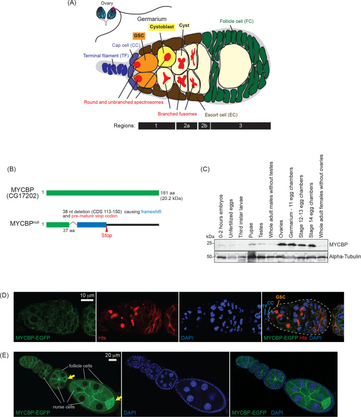
Fig 1. MYCBP expression pattern and mutant allele (A) Schematic illustration of Drosophila ovary and germarium. Each female has a pair of ovaries, each consisting of 12–16 ovarioles (cyan). The germarium (outlined in magenta) is located at the anterior tip of each ovariole and contains both germ cells and somatic cells. Germ cells include germline stem cells (GSCs), cystoblasts, cysts, and differentiating oocytes. Somatic cells include terminal filament (TF) cells, cap cells (CCc), escort cells (ECs), and follicle cells (FCs). GSCs and cystoblasts have spherical, unbranched spectrosomes, whereas cysts posesss branched fusomes. The distinct regions of the germarium—1, 2a, 2b, and 3—are indicated. (B) Drosophila MYCBP (CG17202) protein and the null mutant allele generated in this study. (C) Western blot of dissected fly tissues. (D) Confocal images of germaria from mycbp-EGFP transgenic flies. MYCBP-EGFP (green), Hts (red), and DAPI (blue). Scale bar: 10 μm. (E) Confocal images of egg chambers from mycbp-EGFP transgenic flies. MYCBP-EGFP (green) and DAPI (blue). MYCBP-EGFP is highly expressed in nurse cells, enriched in the developing oocyte (yellow arrow), and is also detectable in somatic follicle cells. Scale bar: 20 μm.
Fig 1. MYCBP expression pattern and mutant allele (A) Schematic illustration of Drosophila ovary and germarium. Each female has a pair of ovaries, each consisting of 12–16 ovarioles (cyan). The germarium (outlined in magenta) is located at the anterior tip of each ovariole and contains both germ cells and somatic cells. Germ cells include germline stem cells (GSCs), cystoblasts, cysts, and differentiating oocytes. Somatic cells include terminal filament (TF) cells, cap cells (CCc), escort cells (ECs), and follicle cells (FCs). GSCs and cystoblasts have spherical, unbranched spectrosomes, whereas cysts posesss branched fusomes. The distinct regions of the germarium—1, 2a, 2b, and 3—are indicated. (B) Drosophila MYCBP (CG17202) protein and the null mutant allele generated in this study. (C) Western blot of dissected fly tissues. (D) Confocal images of germaria from mycbp-EGFP transgenic flies. MYCBP-EGFP (green), Hts (red), and DAPI (blue). Scale bar: 10 μm. (E) Confocal images of egg chambers from mycbp-EGFP transgenic flies. MYCBP-EGFP (green) and DAPI (blue). MYCBP-EGFP is highly expressed in nurse cells, enriched in the developing oocyte (yellow arrow), and is also detectable in somatic follicle cells. Scale bar: 20 μm.CC BY · PubMed Central
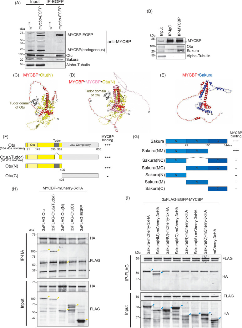
Fig 2. MYCBP interacts with Otu and Sakura (A) Co-immunoprecipitation (co-IP) using anti-GFP magnetic beads followed by Western blotting. Ovary lysates expressing MYCBP-EGFP in mycbp+/+ background and those from w1118 negative control were analyzed. (B) Co-IP using anti-MYCBP antibodies and ovary lysates from w1118. IgG was used as a negative control IP. (C-E) Structures of (C) MYCBP•Otu(N), (D) MYCBP•MYCBP•Otu(N), and (E) MYCBP•Sakura predicted using Alphafold. (F, G) Schematic diagrams of full-length and fragment constructs of (F) Otu and (G) Sakura used in co-IP assays. The binding assay results from (H) is summarized. (G) Full-length Sakura and Sakura fragments tested in co-immunoprecipitation assays. N: N-terminal, M: middle, C: C-terminal. The co-IP assay results from (H) and (I) are summarized. (H) Co-IP using anti-HA beads followed by Western blotting. S2 cell lysates co-expressing MYCBP-mCherry-3xHA and 3xFLAG-Otu (full-length or fragments) were tested. 3XFLAG-EGFPserved as a negative control. (I) Co-IP using anti-FLAG beads followed by Western blotting. S2 cell lysates co-expressing 3xFLAG-EGFP-MYCBP and Sakura-mCherry-3xHA (full-length or fragments) were tested. mCherry-3xHA served as a negative control.
Fig 2. MYCBP interacts with Otu and Sakura (A) Co-immunoprecipitation (co-IP) using anti-GFP magnetic beads followed by Western blotting. Ovary lysates expressing MYCBP-EGFP in mycbp+/+ background and those from w1118 negative control were analyzed. (B) Co-IP using anti-MYCBP antibodies and ovary lysates from w1118. IgG was used as a negative control IP. (C-E) Structures of (C) MYCBP•Otu(N), (D) MYCBP•MYCBP•Otu(N), and (E) MYCBP•Sakura predicted using Alphafold. (F, G) Schematic diagrams of full-length and fragment constructs of (F) Otu and (G) Sakura used in co-IP assays. The binding assay results from (H) is summarized. (G) Full-length Sakura and Sakura fragments tested in co-immunoprecipitation assays. N: N-terminal, M: middle, C: C-terminal. The co-IP assay results from (H) and (I) are summarized. (H) Co-IP using anti-HA beads followed by Western blotting. S2 cell lysates co-expressing MYCBP-mCherry-3xHA and 3xFLAG-Otu (full-length or fragments) were tested. 3XFLAG-EGFPserved as a negative control. (I) Co-IP using anti-FLAG beads followed by Western blotting. S2 cell lysates co-expressing 3xFLAG-EGFP-MYCBP and Sakura-mCherry-3xHA (full-length or fragments) were tested. mCherry-3xHA served as a negative control.CC BY · PubMed Central
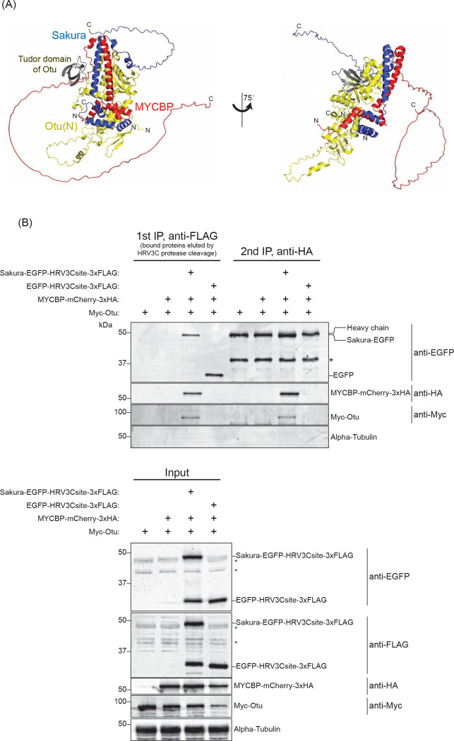
Fig 3. Sequential co-immunoprecipitation shows MYCBP, Sakura, and Otu form a ternary complex (A) MYCBP•Sakura•Otu structure predicted by AlphaFold. (B) Sequential co-IP followed by Western blotting. S2 cell lysates co-expressing Sakura-EGFP-HRV3Csite-3xFLAG, MYCBP-mCherry-3xHA, and Myc-Otu were used. EGFP-HRV3csite-3xFLAG served as a negative control. The first IP was performed using anti-FLAG beads and eluted using HRV3C protease. Second IP was performed with anti-HA beads. Non-specific bands are marked with *.
Fig 3. Sequential co-immunoprecipitation shows MYCBP, Sakura, and Otu form a ternary complex (A) MYCBP•Sakura•Otu structure predicted by AlphaFold. (B) Sequential co-IP followed by Western blotting. S2 cell lysates co-expressing Sakura-EGFP-HRV3Csite-3xFLAG, MYCBP-mCherry-3xHA, and Myc-Otu were used. EGFP-HRV3csite-3xFLAG served as a negative control. The first IP was performed using anti-FLAG beads and eluted using HRV3C protease. Second IP was performed with anti-HA beads. Non-specific bands are marked with *.CC BY · PubMed Central
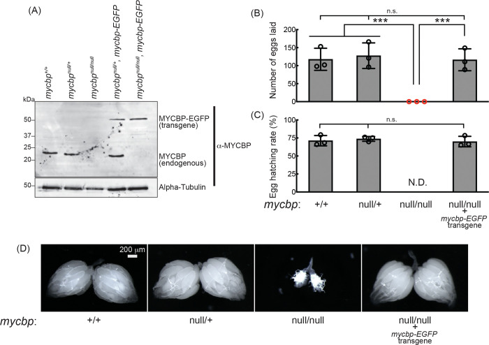
Fig 4. mycbpnull female flies are sterile and have rudimentary ovaries. (A) Western blot of ovary lysates. (B, C) Female fertility assays. (B) The number of eggs laid by test females mated with wild-type (OregonR) males. (C) Hatching rate of the eggs. Mean ± SD (n = 3). P-value < 0.001 (Student’s t-test, unpaired, two-tailed) is indicated by ***. (D) Stereomicroscope images of dissected ovaries. Scale bar: 200 μm.
Fig 4. mycbpnull female flies are sterile and have rudimentary ovaries. (A) Western blot of ovary lysates. (B, C) Female fertility assays. (B) The number of eggs laid by test females mated with wild-type (OregonR) males. (C) Hatching rate of the eggs. Mean ± SD (n = 3). P-value < 0.001 (Student’s t-test, unpaired, two-tailed) is indicated by ***. (D) Stereomicroscope images of dissected ovaries. Scale bar: 200 μm.CC BY · PubMed Central
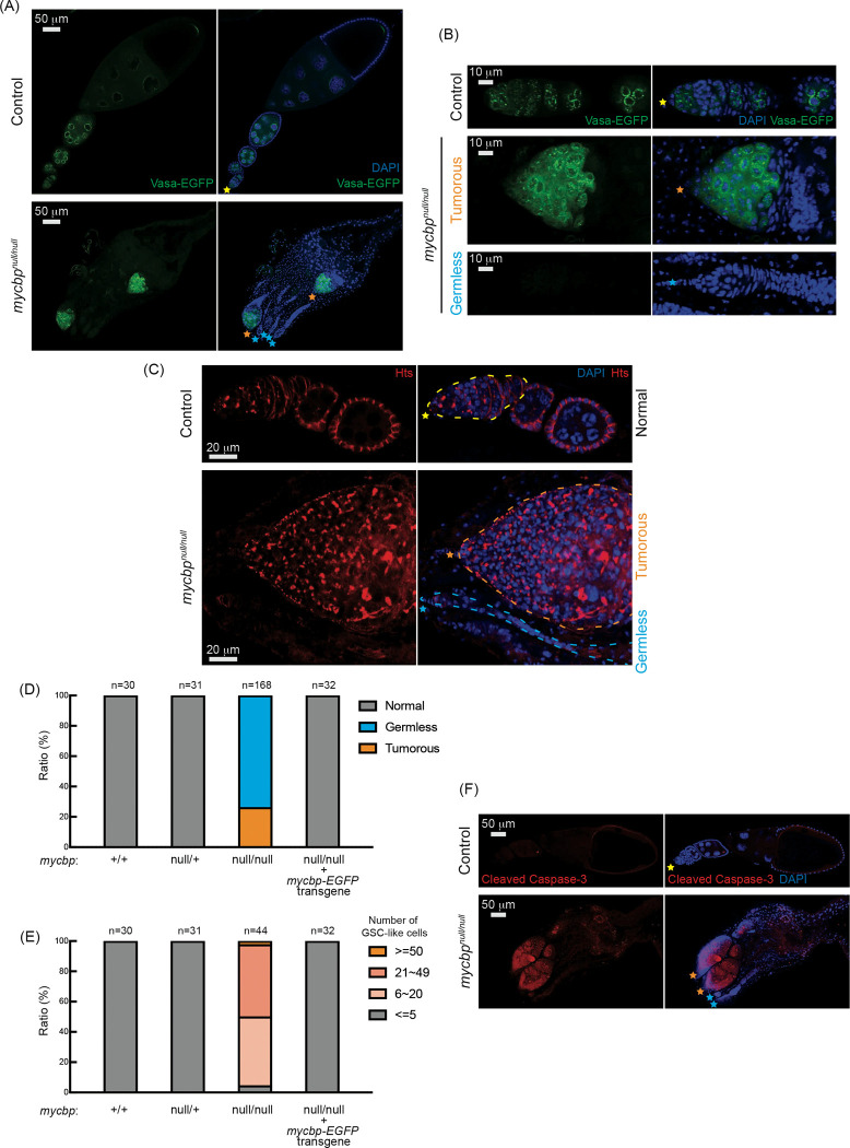
Fig 5. mycbpnull ovaries are germless and tumorous (A, B) Confocal images of ovaries from control (mycbpnull/+) and mycbpnull/null flies expressing Vasa-EGFP. Vasa-EGFP (green) and DAPI (blue). Yellow, orange, and. cyan stars mark normal ovarioles, tumorous, and germless ovarioles, respectively, in Figure 5. (B) Higher-magnification images of germaria. Scale bars: 50 μm (A), 10 μm (B). (C) Confocal images of ovaries from control (mycbpnull/+) and mycbpnull/null flies stained with anti-Hts to label spectrosomes and fusomes. Hts (red) and DAPI (blue). Germaria are outlined. Scale bars: 20 μm. (D) Percentage of normal, germless, and tumorous ovarioles in indicated genotypes (ages 2–5 days). (E) Quantification of GSC-like cells per germarium (ages 2–5 days). (F) Confocal images of ovaries stained with anti-cleaved Caspase-3. Cleaved caspase-3 (red) and DAPI (blue). Scale bars: 50 μm.
Fig 5. mycbpnull ovaries are germless and tumorous (A, B) Confocal images of ovaries from control (mycbpnull/+) and mycbpnull/null flies expressing Vasa-EGFP. Vasa-EGFP (green) and DAPI (blue). Yellow, orange, and. cyan stars mark normal ovarioles, tumorous, and germless ovarioles, respectively, in Figure 5. (B) Higher-magnification images of germaria. Scale bars: 50 μm (A), 10 μm (B). (C) Confocal images of ovaries from control (mycbpnull/+) and mycbpnull/null flies stained with anti-Hts to label spectrosomes and fusomes. Hts (red) and DAPI (blue). Germaria are outlined. Scale bars: 20 μm. (D) Percentage of normal, germless, and tumorous ovarioles in indicated genotypes (ages 2–5 days). (E) Quantification of GSC-like cells per germarium (ages 2–5 days). (F) Confocal images of ovaries stained with anti-cleaved Caspase-3. Cleaved caspase-3 (red) and DAPI (blue). Scale bars: 50 μm.CC BY · PubMed Central
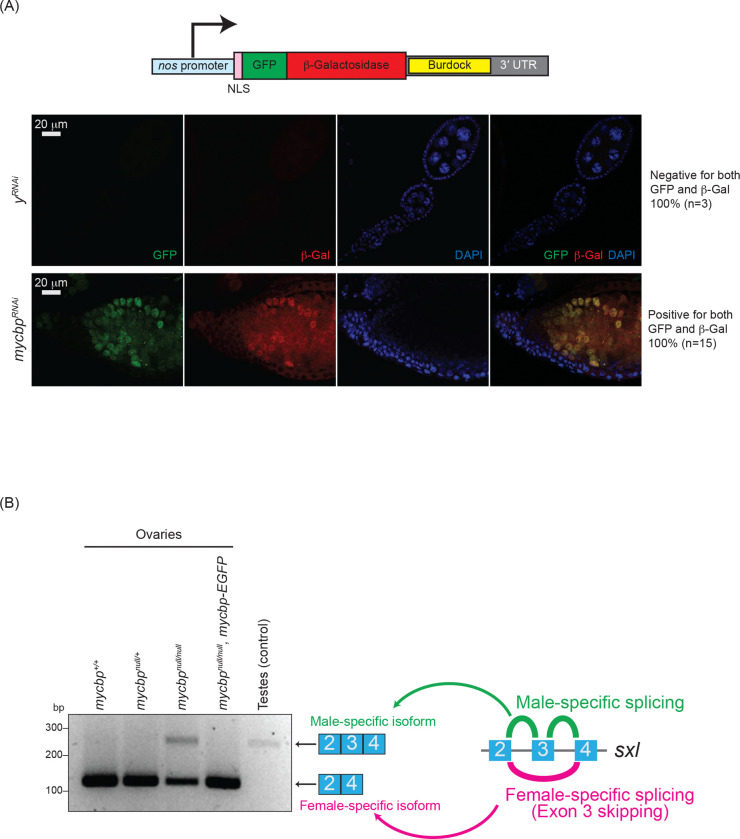
Fig 6. Loss of mycbp disrupts piRNA-mediated transposon silencing and sxl splicing (A) Schematic of the Burdock sensor construct and representative images of sensor expression in ovaries from control (yRNAi) and mycbpRNAi flies. The Burdock sensor harbors a nanos promoter, a nuclear localization signal (NLS) appended to GFP and β-gal coding sequences, and a target sequence for Burdock piRNAs in the 3’UTR. RNAi was driven in the germline using UAS-Dcr2, NGT-Gal4, and nos-Gal4-VP16. GFP (green), β-gal (red), and DAPI (blue). Scale bars: 20 μm. 0/3 control samples showed sensor activation; 15/15 mycbpRNAi samples did. (B) RT-PCR analysis of sxl alternative splicing in ovaries and testes.
Fig 6. Loss of mycbp disrupts piRNA-mediated transposon silencing and sxl splicing (A) Schematic of the Burdock sensor construct and representative images of sensor expression in ovaries from control (yRNAi) and mycbpRNAi flies. The Burdock sensor harbors a nanos promoter, a nuclear localization signal (NLS) appended to GFP and β-gal coding sequences, and a target sequence for Burdock piRNAs in the 3’UTR. RNAi was driven in the germline using UAS-Dcr2, NGT-Gal4, and nos-Gal4-VP16. GFP (green), β-gal (red), and DAPI (blue). Scale bars: 20 μm. 0/3 control samples showed sensor activation; 15/15 mycbpRNAi samples did. (B) RT-PCR analysis of sxl alternative splicing in ovaries and testes.CC BY · PubMed Central
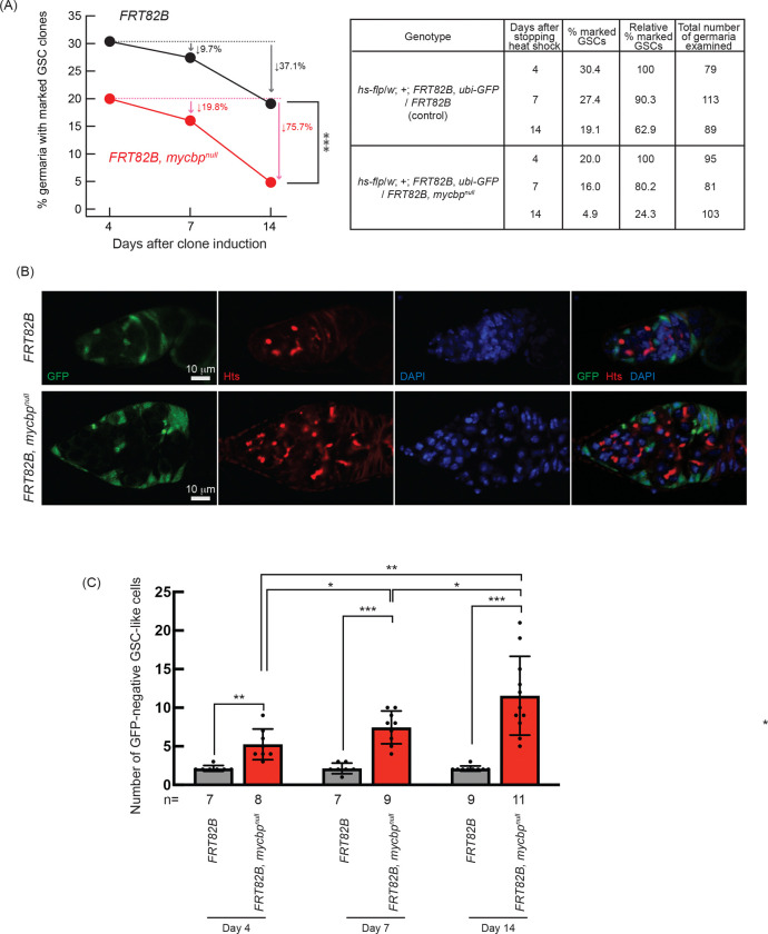
Fig 7. Germline clonal analysis of mycbpnull (A) Percentage of germaria with marked (GFP-negative) GSC clones at 4, 7, and 14 days after clone induction. P-value < 0.001 (Chi-squared test) is indicated by ***. (B) Confocal images of control and mycbpnull clones. GFP (green), Hts (red), and DAPI (blue). Scale bar: 10 μm. (C) Number of marked (GFP-negative) GSC-like cells per germarium containing marked GSCs at 4, 7, and 14 days after clone induction. GSC-like cells containing round spectrosome were identified by anti-Hts staining. P-value < 0.05, <0.01, and <0.001 indicated by *, **, and *** (Student’s t-test, unpaired, two-tailed).
Fig 7. Germline clonal analysis of mycbpnull (A) Percentage of germaria with marked (GFP-negative) GSC clones at 4, 7, and 14 days after clone induction. P-value < 0.001 (Chi-squared test) is indicated by ***. (B) Confocal images of control and mycbpnull clones. GFP (green), Hts (red), and DAPI (blue). Scale bar: 10 μm. (C) Number of marked (GFP-negative) GSC-like cells per germarium containing marked GSCs at 4, 7, and 14 days after clone induction. GSC-like cells containing round spectrosome were identified by anti-Hts staining. P-value < 0.05, <0.01, and <0.001 indicated by *, **, and *** (Student’s t-test, unpaired, two-tailed).CC BY · PubMed Central
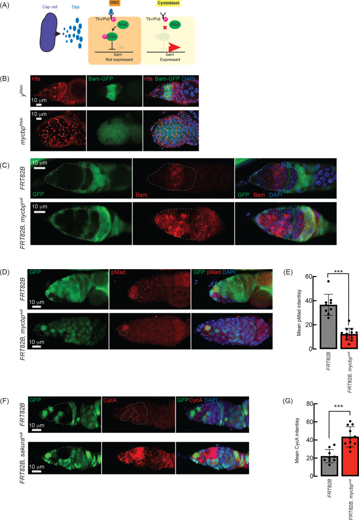
Fig 8. Loss of sakura inhibits Dpp/BMP signaling (A) Schematic of Dpp-mediated bam repression via pMad activation. Cap cells secrete diffusible Decapentaplegic (Dpp), which is received by its receptor, a heterodimer of Thick vein (Tkv) and Punt (Put), in GSCs. The activated Dpp signaling eventually phosphorylates Mother-against-dpp (Mad). The phosphorylated Mad (pMad) represses the transcription of bam. The repression of bam in GSCs is crucial for maintaining their stemness. Cystoblasts do not receive Dpp, and Bam expression is crucial for promoting cystoblast differentiation from GSCs. (B) Confocal images of bam-GFP reporter expression in control (yRNAi) and mycbpRNAi ovaries (Azlan et al. 2024). RNAi was driven in the female germline using UAS-Dcr2 and NGT-Gal4. Bam-GFP (green), Hts (red), and DAPI (blue). Germaria are outlined. Scale bar: 10 μm. (C, D) Confocal images of germaria with control and mycbpnull germline clones stained with (C) anti-Bam and (D) anti-pMad. GFP (green), Bam/pMad (red), and DAPI (blue). Scale bar: 10 μm. (E) Quantification of pMad intensity in GSC clones. Mean ± SD (n = 9 and 13 for FRT82B and FRT82B, mycbpnull, respectively). P-value < 0.001 (Student’s t-test, unpaired, two-tailed) is indicated by ***. (F) Confocal images of germaria with control and mycbpnull germline clones stained with anti-CycA. GFP (green), CycA (red), and DAPI (blue). Scale bar: 10 μm. (G) Quantification of CycA intensity in the germline clones. Mean ± SD (n = 9 and 11 for FRT82B and FRT82B, mycbpnull, respectively). P-value < 0.01 (Student’s t-test, unpaired, two-tailed) is indicated by **. Clones in C, D, and F are GFP-negative.
Fig 8. Loss of sakura inhibits Dpp/BMP signaling (A) Schematic of Dpp-mediated bam repression via pMad activation. Cap cells secrete diffusible Decapentaplegic (Dpp), which is received by its receptor, a heterodimer of Thick vein (Tkv) and Punt (Put), in GSCs. The activated Dpp signaling eventually phosphorylates Mother-against-dpp (Mad). The phosphorylated Mad (pMad) represses the transcription of bam. The repression of bam in GSCs is crucial for maintaining their stemness. Cystoblasts do not receive Dpp, and Bam expression is crucial for promoting cystoblast differentiation from GSCs. (B) Confocal images of bam-GFP reporter expression in control (yRNAi) and mycbpRNAi ovaries (Azlan et al. 2024). RNAi was driven in the female germline using UAS-Dcr2 and NGT-Gal4. Bam-GFP (green), Hts (red), and DAPI (blue). Germaria are outlined. Scale bar: 10 μm. (C, D) Confocal images of germaria with control and mycbpnull germline clones stained with (C) anti-Bam and (D) anti-pMad. GFP (green), Bam/pMad (red), and DAPI (blue). Scale bar: 10 μm. (E) Quantification of pMad intensity in GSC clones. Mean ± SD (n = 9 and 13 for FRT82B and FRT82B, mycbpnull, respectively). P-value < 0.001 (Student’s t-test, unpaired, two-tailed) is indicated by ***. (F) Confocal images of germaria with control and mycbpnull germline clones stained with anti-CycA. GFP (green), CycA (red), and DAPI (blue). Scale bar: 10 μm. (G) Quantification of CycA intensity in the germline clones. Mean ± SD (n = 9 and 11 for FRT82B and FRT82B, mycbpnull, respectively). P-value < 0.01 (Student’s t-test, unpaired, two-tailed) is indicated by **. Clones in C, D, and F are GFP-negative.CC BY · PubMed Central
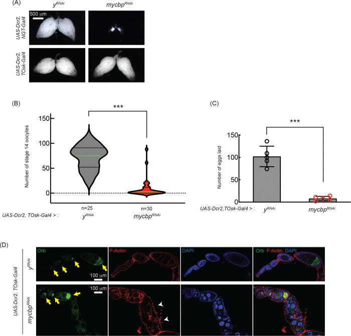
Fig 9. mycbp is important for late oogenesis (A) Stereomicroscope images of dissected ovaries. Scale bar: 500 μm. yRNAi served as a control. (B, C) Number of stage 14 oocytes per fly (B) and eggs laid (C) in mycbp RNAi knockdown usingby UAS-Dcr2 and TOsk-Gal4. Mean ± SD (n = 5). P-value < 0.001 (Student’s t-test, unpaired, two-tailed) is indicated by ***. (D) Confocal images of ovaries stained with phalloidin (F-Actin, red), anti-Orb (Green), and DAPI (blue). Yellow arrows indicate normal Orb localization; white arrowheads indicate cytoskeletal disorganization and loss of Orb localization. Scale bar: 100 μm.
Fig 9. mycbp is important for late oogenesis (A) Stereomicroscope images of dissected ovaries. Scale bar: 500 μm. yRNAi served as a control. (B, C) Number of stage 14 oocytes per fly (B) and eggs laid (C) in mycbp RNAi knockdown usingby UAS-Dcr2 and TOsk-Gal4. Mean ± SD (n = 5). P-value < 0.001 (Student’s t-test, unpaired, two-tailed) is indicated by ***. (D) Confocal images of ovaries stained with phalloidin (F-Actin, red), anti-Orb (Green), and DAPI (blue). Yellow arrows indicate normal Orb localization; white arrowheads indicate cytoskeletal disorganization and loss of Orb localization. Scale bar: 100 μm.CC BY · PubMed Central
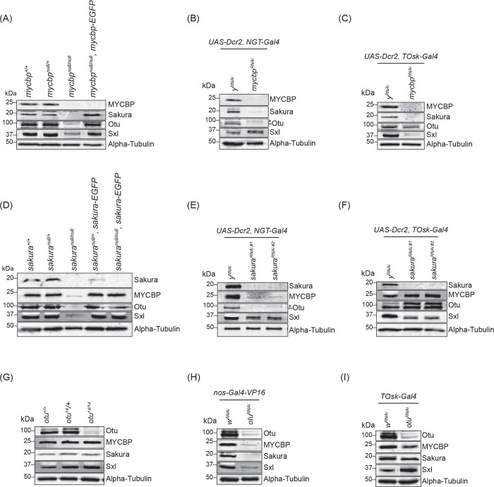
Fig 10. MYCBP is required for Sakura level Western blot of ovary lysates. The sakura RNAi #1 (VDRC: v39727) and #2 (VDRC: v103660). Non-specific bands are marked (*). Sakura and alpha-Tubulin images in (E), Sakura, Otu, and alpha-Tubulin images in (F), Otu and alpha-Tubulin images in (H), Sakura, Otu, and alpha-Tubulin images in (I), are from (Azlan et al. 2024).
Fig 10. MYCBP is required for Sakura level Western blot of ovary lysates. The sakura RNAi #1 (VDRC: v39727) and #2 (VDRC: v103660). Non-specific bands are marked (*). Sakura and alpha-Tubulin images in (E), Sakura, Otu, and alpha-Tubulin images in (F), Otu and alpha-Tubulin images in (H), Sakura, Otu, and alpha-Tubulin images in (I), are from (Azlan et al. 2024).CC BY · PubMed Central
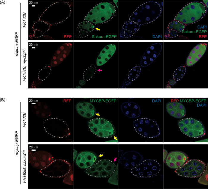
Fig 11. Mutual dependance of MYCBP and Sakura level and localization (A) Confocal images of egg chambers with control and mycbpnull germline clones expressing Sakura-EGFP. Marked clones (RFP-negative) are outlined. RFP (red), Sakura-EGFP (green), and DAPI (blue). Scale bar: 20 μm. Yellow arrow: normal Sakura-EGFP posterior enrichment; magenta arrow: reduced Sakura-EGFP signal. Fly genotypes used: hs-flp/w; sakura-EGFP/+; FRT82B, ubi-RFP/FRT82B. hs-flp/w; sakura-EGFP/+; FRT82B, ubi-RFP/FRT82B, mycbpnull. (B) Confocal images of egg chambers with control and sakuranull germline clones expressing MYCBP-EGFP. Marked clones (RFP-negative) are outlined. RFP (red), MYCBP-EGFP (green), and DAPI (blue). Scale bar: 20 μm. Yellow arrow: normal MYCBP-EGFP posterior enrichment; magenta arrow: reduced levels and loss of posterior localization of MYCBP-EGFP. Fly genotypes used: hs-flp/w; mycbp-EGFP/+; FRT82B, ubi-RFP/FRT82B. hs-flp/w; mycbp-EGFP/+; FRT82B, ubi-RFP/FRT82B, sakuranull.
Fig 11. Mutual dependance of MYCBP and Sakura level and localization (A) Confocal images of egg chambers with control and mycbpnull germline clones expressing Sakura-EGFP. Marked clones (RFP-negative) are outlined. RFP (red), Sakura-EGFP (green), and DAPI (blue). Scale bar: 20 μm. Yellow arrow: normal Sakura-EGFP posterior enrichment; magenta arrow: reduced Sakura-EGFP signal. Fly genotypes used: hs-flp/w; sakura-EGFP/+; FRT82B, ubi-RFP/FRT82B. hs-flp/w; sakura-EGFP/+; FRT82B, ubi-RFP/FRT82B, mycbpnull. (B) Confocal images of egg chambers with control and sakuranull germline clones expressing MYCBP-EGFP. Marked clones (RFP-negative) are outlined. RFP (red), MYCBP-EGFP (green), and DAPI (blue). Scale bar: 20 μm. Yellow arrow: normal MYCBP-EGFP posterior enrichment; magenta arrow: reduced levels and loss of posterior localization of MYCBP-EGFP. Fly genotypes used: hs-flp/w; mycbp-EGFP/+; FRT82B, ubi-RFP/FRT82B. hs-flp/w; mycbp-EGFP/+; FRT82B, ubi-RFP/FRT82B, sakuranull.CC BY · PubMed Central
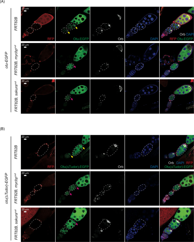
Fig 12. Otu localization to the developing oocyte depends on MYCBP and Sakura Confocal images of egg chambers with control, mycbpnull, and sakuranull germline clones expressing (A) Otu-EGFP or (B) Otu(ΔTudor)-EGFP. Marked clones (RFP-negative) are outlined. RFP (red), Otu-EGFP or Otu(ΔTudor)-EGFP (green), Orb (white), and DAPI (blue). Scale bar: 20 μm. Yellow arrows: normal posterior enrichment of Otu-EGFP and Otu(ΔTudor)-EGFP; magenta arrows: loss of localization. Orb localization remains intact in mutant clones. Fly genotypes used: hs-flp/w; otu-EGFP/+; FRT82B, ubi-RFP/FRT82B. hs-flp/w; otu-EGFP/+; FRT82B, ubi-RFP/FRT82B, mycbpnull. hs-flp/w; otu-EGFP/+; FRT82B, ubi-RFP/FRT82B, sakuranull. hs-flp/w; otu(ΔTudor)-EGFP/+; FRT82B, ubi-RFP/FRT82B. hs-flp/w; otu(ΔTudor)-EGFP /+; FRT82B, ubi-RFP/FRT82B, mycbpnull. hs-flp/w; otu(ΔTudor)-EGFP /+; FRT82B, ubi-RFP/FRT82B, sakuranull.
Fig 12. Otu localization to the developing oocyte depends on MYCBP and Sakura Confocal images of egg chambers with control, mycbpnull, and sakuranull germline clones expressing (A) Otu-EGFP or (B) Otu(ΔTudor)-EGFP. Marked clones (RFP-negative) are outlined. RFP (red), Otu-EGFP or Otu(ΔTudor)-EGFP (green), Orb (white), and DAPI (blue). Scale bar: 20 μm. Yellow arrows: normal posterior enrichment of Otu-EGFP and Otu(ΔTudor)-EGFP; magenta arrows: loss of localization. Orb localization remains intact in mutant clones. Fly genotypes used: hs-flp/w; otu-EGFP/+; FRT82B, ubi-RFP/FRT82B. hs-flp/w; otu-EGFP/+; FRT82B, ubi-RFP/FRT82B, mycbpnull. hs-flp/w; otu-EGFP/+; FRT82B, ubi-RFP/FRT82B, sakuranull. hs-flp/w; otu(ΔTudor)-EGFP/+; FRT82B, ubi-RFP/FRT82B. hs-flp/w; otu(ΔTudor)-EGFP /+; FRT82B, ubi-RFP/FRT82B, mycbpnull. hs-flp/w; otu(ΔTudor)-EGFP /+; FRT82B, ubi-RFP/FRT82B, sakuranull.CC BY · PubMed Central

Verification

Claims the paper makes

The claims and statistics of this paper haven’t been checked yet.

Run the check — free, no account needed. It reads the open-access text and checks the sources it cites.

A few free checks a day · papers already verified open instantly · results are public

Self-check

Percentages, sums and reported differences re-computed from the paper’s own numbers.

The self-check needs the paper’s full text.

Citations

Cites

Show all 1 references

Research works printed but not linked: 70

  1. Azlan A , Zhu L , Fukunaga R. 2024. Female-germline specific protein Sakura interacts with Otu and is crucial for germline stem cell renewal and differentiation and oogenesis. bioRxiv. · DOI
  2. 10.1016/0092-8674(91)90157-T · DOI · Paper page
  3. 10.1242/dev.118.3.797 · DOI
  4. 10.1073/pnas.1120473109 · DOI
  5. 10.1016/j.cub.2003.09.033 · DOI
  6. 10.1242/dev.00325 · DOI · Paper page
  7. 10.1242/dev.039032 · DOI
  8. 10.1016/j.devcel.2014.01.027 · DOI
  9. 10.1101/gad.12.23.3715 · DOI
  10. 10.1242/dev.056242 · DOI · Paper page
  11. 10.1073/pnas.1015874108 · DOI
  12. ElMaghraby MF , Tirian L , Senti KA , Meixner K , Brennecke J . 2022. A genetic toolkit for studying transposon control in the Drosophila melanogaster ovary. Genetics 220. · DOI
  13. 10.1038/emboj.2009.2 · DOI
  14. 10.1016/j.cell.2012.09.027 · DOI
  15. 10.1093/genetics/81.4.683 · DOI
  16. 10.1387/ijdb.8735924 · DOI
  17. 10.1016/S0925-4773(01)00314-8 · DOI
  18. 10.1002/dvg.1020020206 · DOI
  19. 10.1159/000521235 · DOI
  20. 10.1016/j.molcel.2013.04.031 · DOI
  21. 10.1016/j.cois.2019.10.006 · DOI
  22. 10.3389/fcell.2020.00019 · DOI
  23. 10.1016/j.tig.2017.09.002 · DOI
  24. 10.1038/344461a0 · DOI
  25. 10.1073/pnas.1619188114 · DOI
  26. 10.1016/j.molcel.2019.02.004 · DOI
  27. 10.1016/j.cub.2013.06.021 · DOI
  28. 10.1016/j.ceb.2019.03.008 · DOI
  29. 10.1073/pnas.1612393113 · DOI
  30. 10.1261/rna.059915.116 · DOI
  31. 10.1038/s41598-018-25483-9 · DOI
  32. 10.1002/dvg.1020030107 · DOI
  33. 10.1038/sj.cr.7310123 · DOI
  34. 10.1038/nrg3030 · DOI · Paper page
  35. 10.1073/pnas.0901452106 · DOI
  36. 10.1371/journal.pone.0058301 · DOI
  37. Liao SE , Kandasamy SK , Zhu L , Fukunaga R . 2019. DEAD-box RNA helicase Belle post-transcriptionally promotes gene expression in an ATPase activity-dependent manner. Rna. · DOI
  38. 10.1146/annurev.genet.31.1.455 · DOI
  39. 10.1006/dbio.1993.1228 · DOI
  40. 10.1016/j.devcel.2011.06.018 · DOI · Paper page
  41. 10.1101/gad.4.12b.2242 · DOI
  42. 10.1073/pnas.2426524122 · DOI
  43. 10.1016/j.devcel.2018.10.011 · DOI
  44. 10.1093/genetics/155.4.1809 · DOI
  45. 10.1038/s42003-020-0923-3 · DOI
  46. 10.1242/dev.119.1.123 · DOI
  47. 10.1016/j.devcel.2009.07.017 · DOI
  48. 10.1128/MMBR.67.3.343-359.2003 · DOI
  49. 10.1006/dbio.1997.8697 · DOI
  50. 10.1007/978-1-4939-2851-4_3 · DOI
  51. 10.4161/fly.4.1.11277 · DOI · Paper page
  52. 1966, Studies on Fused a Mutant Gene Producing Ovarian Tumors in Drosophila Melanogaster, Genetics, 54, 363
  53. 10.1101/gad.6.2.233 · DOI
  54. 10.1002/dvg.1020090203 · DOI
  55. 10.1046/j.1365-2443.1998.00206.x · DOI
  56. 10.1261/rna.080311.124 · DOI
  57. 10.1038/362171a0 · DOI · Paper page
  58. 10.1126/science.1093983 · DOI
  59. 10.1016/j.cell.2010.11.022 · DOI
  60. 10.1016/S0092-8674(00)81424-5 · DOI · Paper page
  61. 10.1126/science.1197142 · DOI · Paper page
  62. 10.1021/acs.chemrev.7b00393 · DOI
  63. Yang F , Quan Z , Huang H , He M , Liu X , Cai T , Xi R . 2019. Ovaries absent links dLsd1 to HP1a for local H3K4 demethylation required for heterochromatic gene silencing. eLife 8. · DOI
  64. 10.1242/dev.009159 · DOI
  65. 10.1038/s41392-023-01381-z · DOI · Paper page
  66. 10.1371/journal.pgen.1009655 · DOI
  67. Zhu L , Kandasamy SK , Fukunaga R . 2018a. Dicer partner protein tunes the length of miRNAs using base-mismatch in the pre-miRNA stem. Nucleic acids research. · DOI
  68. 10.1038/s41467-018-06404-w · DOI · Paper page
  69. 10.1371/journal.pone.0217603 · DOI
  70. 10.1080/15476286.2019.1630799 · DOI

Metadata

Authors
Azlan, A., & Fukunaga, R.
Affiliations
Johns Hopkins University (US)
Corresponding author
Fukunaga, Ryuya
Venue
PLoS Genetics
Publisher
openRxiv
Published
2025-07-02
Volume
21
Issue
12
Pages
e1011792-e1011792
ISSN
1553-7390 (ISSN-L)
OpenAlex
W4416873099
Cited by
0
References
1
FWCI
0.00
Type
journal article
Language
en
Open access
Locations
  • PLoS Genetics (publishedVersion, cc-by)
  • bioRxiv (Cold Spring Harbor Laboratory) (acceptedVersion, cc-by)
  • PubMed (publishedVersion)
  • PubMed Central (submittedVersion, cc-by)
  • DOAJ (DOAJ: Directory of Open Access Journals) (submittedVersion, cc-by-sa)
Funders
  • Division of Intramural Research, National Institute of Allergy and Infectious Diseases (R03AI178064)
  • Johns Hopkins University
  • National Institutes of Health (1s10od016374-01)
  • National Institute of General Medical Sciences (R35GM145352)
  • National Institute of Allergy and Infectious Diseases (R03AI178064)
Topics
  • Life Sciences › Biochemistry, Genetics and Molecular Biology › Molecular Biology › Developmental Biology and Gene Regulation
  • Health Sciences › Medicine › Reproductive Medicine › Reproductive Biology and Fertility
  • Life Sciences › Biochemistry, Genetics and Molecular Biology › Genetics › Genetic and Clinical Aspects of Sex Determination and Chromosomal Abnormalities
Keywords
cherry blossom, ovarian neoplasms, germline stem cells, oogenesis, germline stem cell differentiation, bag of marbles, sex determination, protein complex formation, Drosophila ovary, sterility

About these checks

Registry, retraction, Cites and statistics results are computed, not judged, with one exception: a printed reference that no index lists is read once by a model before it counts as a citation error. Claims are extracted by a model and kept only when their quote appears verbatim in the text; each cited source is then checked, with a model’s help, for whether it says what the claim relies on. Those verdicts can be wrong — read the claim’s quote and the cited source before relying on one.

Statistics and arithmetic are recomputed, not judged.

Confidence bands are shown only for fields calibrated on our annotated test set. None is calibrated yet, so no band is shown.

What counts as a claim ›How we measure confidence ›How we check this ›

Are you an author, or do you see a problem with this page? Request a review

How to cite

Cite CoChat for the checks on this page, and the paper for its own findings.

This page
CoChat. (2026). Citation check: MYCBP interacts with Sakura and Otu and is essential for germline stem cell renewal and differentiation and oogenesis. https://app.cochat.ai/papers/10.1101/2025.07.01.662550
The paper
Azlan, A., & Fukunaga, R. (2025). MYCBP interacts with Sakura and Otu and is essential for germline stem cell renewal and differentiation and oogenesis. PLoS Genetics. https://doi.org/10.1101/2025.07.01.662550

Data sources

Bibliographic data: Semantic Scholar, OpenAlex, Crossref.

Contains information from the Semantic Scholar Academic Graph Datasets, made available under the ODC Attribution License (ODC-BY 1.0).

Semantic Scholar’s data is described in: Kinney, R., et al. (2023). The Semantic Scholar Open Data Platform. arXiv:2301.10140.