

The DOI matches its Crossref record; 47 of the 47 references it cites were found in a registry or an index.
A Graph-Transformer for Whole Slide Image Classification
Open accessPublished version of a preprint
Abstract
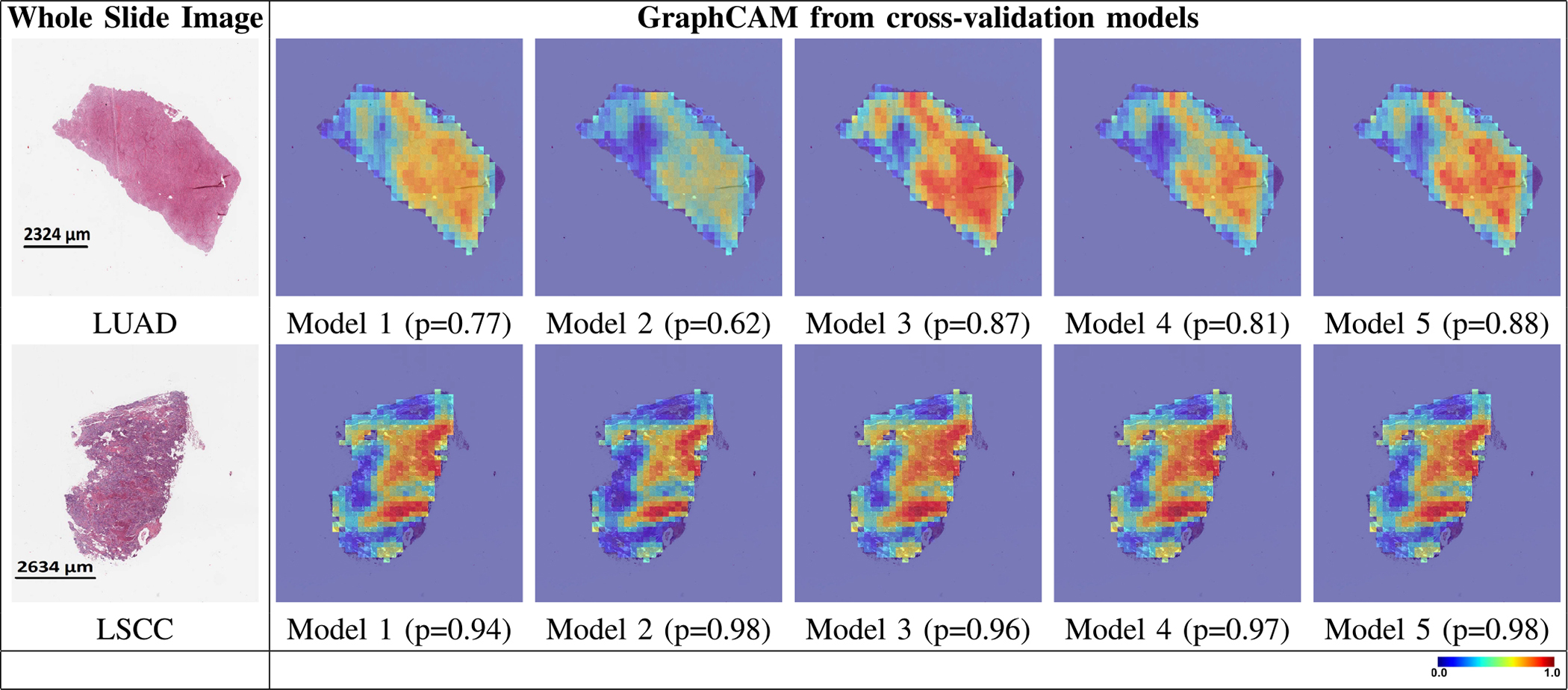
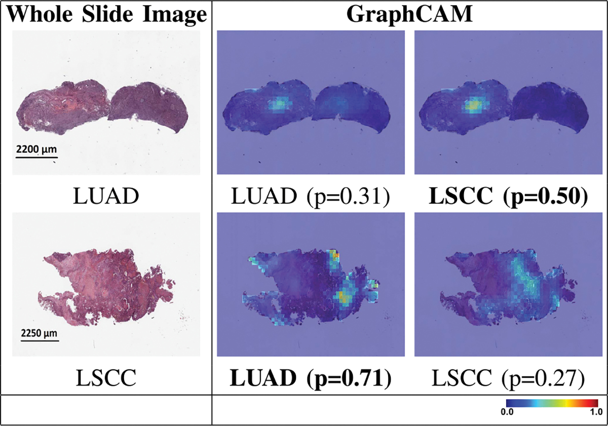
Deep learning is a powerful tool for whole slide image (WSI) analysis. Typically, when performing supervised deep learning, a WSI is divided into small patches, trained and the outcomes are aggregated to estimate disease grade. However, patch-based methods introduce label noise during training by assuming that each patch is independent with the same label as the WSI and neglect overall WSI-level information that is significant in disease grading. Here we present a Graph-Transformer (GT) that fuses a graph-based representation of an WSI and a vision transformer for processing pathology images, called GTP, to predict disease grade. We selected 4,818 WSIs from the Clinical Proteomic Tumor Analysis Consortium (CPTAC), the National Lung Screening Trial (NLST), and The Cancer Genome Atlas (TCGA), and used GTP to distinguish adenocarcinoma (LUAD) and squamous cell carcinoma (LSCC) from adjacent non-cancerous tissue (normal). First, using NLST data, we developed a contrastive learning framework to generate a feature extractor. This allowed us to compute feature vectors of individual WSI patches, which were used to represent the nodes of the graph followed by construction of the GTP framework. Our model trained on the CPTAC data achieved consistently high performance on three-label classification (normal versus LUAD versus LSCC: mean accuracy = 91.2 ± 2.5%) based on five-fold cross-validation, and mean accuracy = 82.3 ± 1.0% on external test data (TCGA). We also introduced a graph-based saliency mapping technique, called GraphCAM, that can identify regions that are highly associated with the class label. Our findings demonstrate GTP as an interpretable and effective deep learning framework for WSI-level classification.
Overview
The claims and statistics of this paper haven’t been checked yet.
Run the check — free, no account needed. It reads the open-access text and checks the sources it cites.
Integrity signals
Registry
Cites
Statistics
Figures
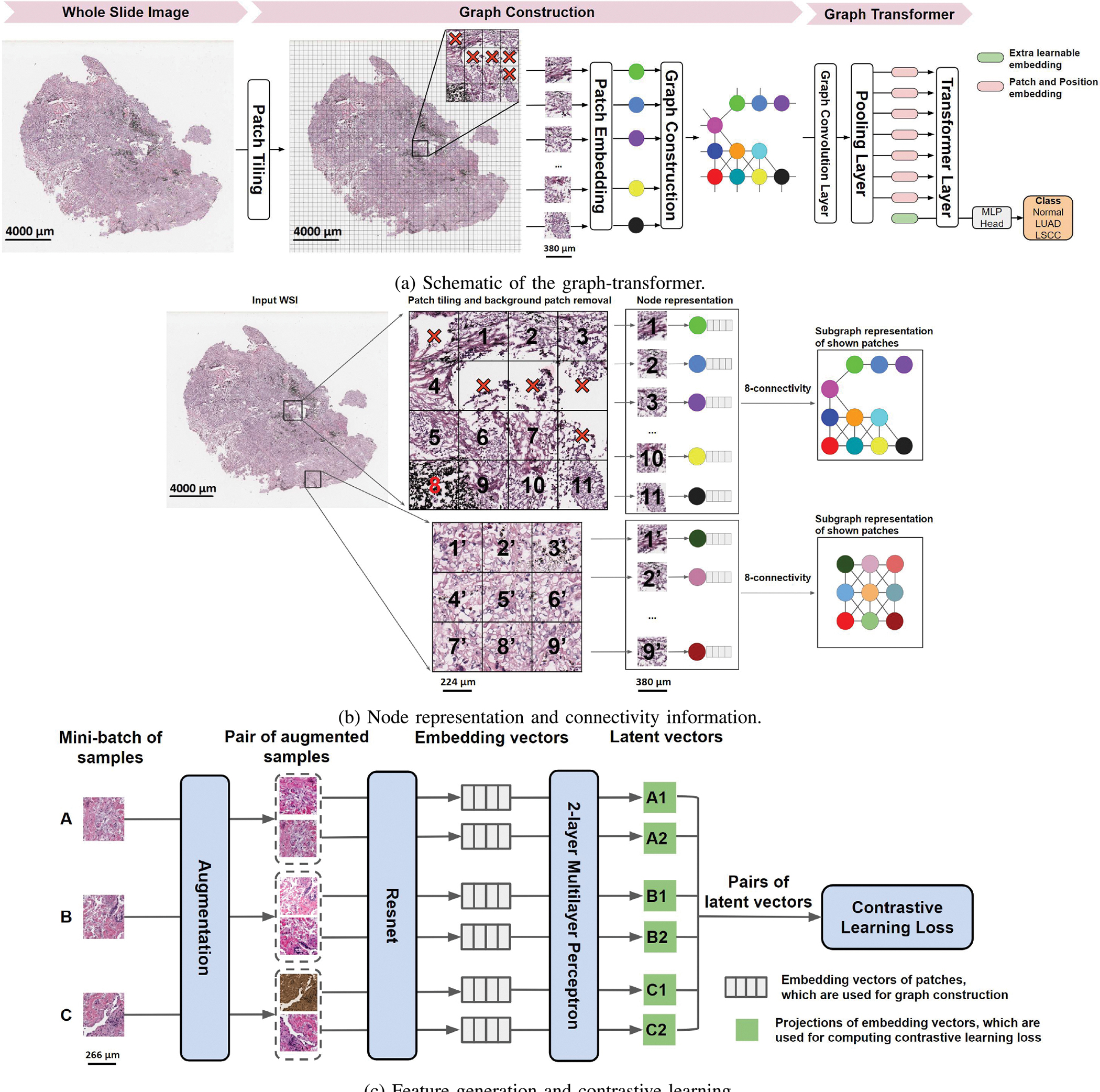
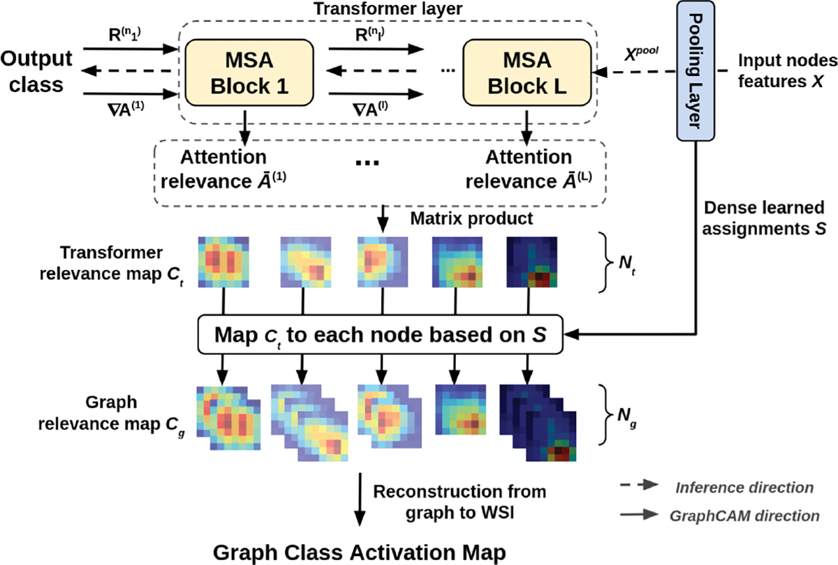
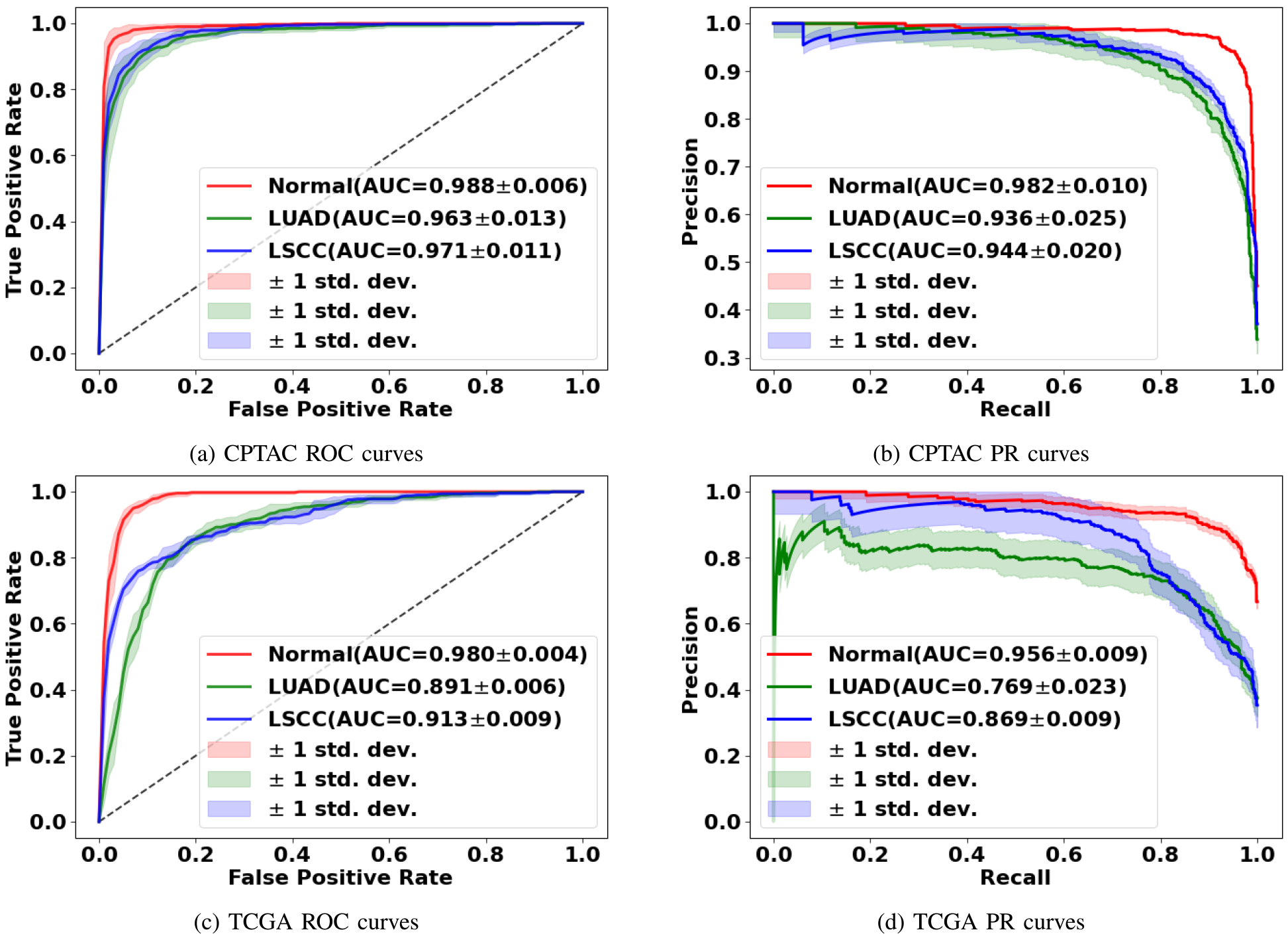
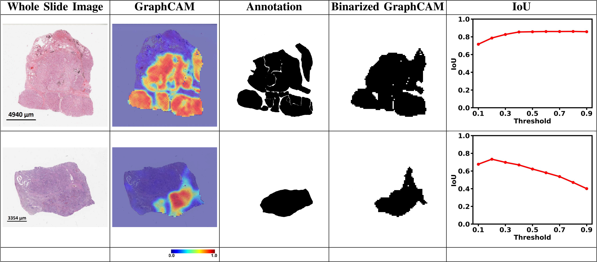
Verification
Claims the paper makes
The claims and statistics of this paper haven’t been checked yet.
Run the check — free, no account needed. It reads the open-access text and checks the sources it cites.
Claims other papers make about this one
Each is checked against this paper’s own text: does it say what the citing paper says it does?
In the citing paper
“While other researchers have previously explored the development of mid-level-fusion and mixer architectures [6], [7] as well as the use of graph-based representations of WSIs [8], our work is unique in mixing node and edge embeddings along with fusion of bulk gene expression embeddings to learn a multimodal topographic mapping to predict survival.”
From Graph attention-based fusion of pathology images and gene expression for prediction of cancer survival (2024) · cites this paper as [8]
In the citing paper
“We used NLST for generating node-level features for graph construction [8], TCGA for training the models using 5-fold cross-validation, and CPTAC as an independent dataset for testing.”
From Graph attention-based fusion of pathology images and gene expression for prediction of cancer survival (2024) · cites this paper as [8]
In the citing paper
“The ISM model’s performance exceeded that of other recent methodologies [8], [21], [25]-[27], as illustrated in Table II.”
From Graph attention-based fusion of pathology images and gene expression for prediction of cancer survival (2024) · cites this paper as [8]
Self-check
Percentages, sums and reported differences re-computed from the paper’s own numbers.
The self-check needs the paper’s full text.
Citations
Cites
Deep Residual Learning for Image Recognition
He, K., Zhang, X., Ren, S., & Sun, J. (2016) · 2016 IEEE Conference on Computer Vision and Pattern Recognition (CVPR) · doi:10.1109/cvpr.2016.90 · Cited by 235,474
Vaswani, A., Shazeer, N., Parmar, N., Uszkoreit, J., Jones, L., Gomez, A. N., Kaiser, L., & Polosukhin, I. (2017) · arXiv · doi:10.48550/arxiv.1706.03762 · Cited by 120,404
Adam: A Method for Stochastic Optimization
Kingma, D. P., & Ba, J. (2014) · arXiv · doi:10.48550/arxiv.1412.6980 · Cited by 98,946
ImageNet: A large-scale hierarchical image database
Deng, J., Dong, W., Socher, R., Li, L.-J., Li, K., & Fei-Fei, L. (2009) · 2009 IEEE Conference on Computer Vision and Pattern Recognition · doi:10.1109/cvpr.2009.5206848 · Cited by 65,333
Semi-supervised classification with graph convolutional networks
Kipf, T. N., & Welling, M. (2016) · arXiv · doi:10.48550/arxiv.1609.02907 · Cited by 24,853
Reduced Lung-Cancer Mortality with Low-Dose Computed Tomographic Screening
The National Lung Screening Trial Research Team (2011) · New England Journal of Medicine · doi:10.1056/nejmoa1102873 · Cited by 11,064
A simple framework for contrastive learning of visual representations
Chen, T., Kornblith, S., Norouzi, M., & Hinton, G. (2020) · arXiv · doi:10.48550/arxiv.2002.05709 · Cited by 10,857
Deformable DETR: Deformable Transformers for End-to-End Object Detection
Zhu, X., Su, W., Lu, L., Li, B., Wang, X., & Dai, J. (2020) · arXiv · doi:10.48550/arxiv.2010.04159 · Cited by 6,027 · cited 1×
“row normalization function in transformers for computer vision tasks [36], [37].”
BackgroundSGDR: Stochastic Gradient Descent with Warm Restarts
Loshchilov, I., & Hutter, F. (2016) · arXiv · doi:10.48550/arxiv.1608.03983 · Cited by 5,309
TransUNet: Transformers Make Strong Encoders for Medical Image Segmentation
Chen, J., Lu, Y., Yu, Q., Luo, X., Adeli, E., Wang, Y., Lu, L., Yuille, A. L., & Zhou, Y. (2021) · arXiv · doi:10.48550/arxiv.2102.04306 · Cited by 4,206
Research works printed but not linked: 2
- Dosovitskiy, An image is worth 16×16 words: Transformers for image recognition at scale
- 2021, The Cancer Genome Atlas Program
Cited by
Sorted by citations
Transformers in medical imaging: A survey
Shamshad, F., Khan, S., Zamir, S. W., Khan, M. H., Hayat, M., Khan, F. S., & Fu, H. (2023) · Medical Image Analysis · doi:10.1016/j.media.2023.102802 · Cited by 1,523
Advances in medical image analysis with vision Transformers: A comprehensive review
Azad, R., Kazerouni, A., Heidari, M., Aghdam, E. K., Molaei, A., Jia, Y., Jose, A., Roy, R., & Merhof, D. (2023) · Medical Image Analysis · doi:10.1016/j.media.2023.103000 · Cited by 557
McGenity, C., Clarke, E. L., Jennings, C., Matthews, G., Cartlidge, C., Freduah-Agyemang, H., Stocken, D. D., & Treanor, D. (2024) · npj Digital Medicine · doi:10.1038/s41746-024-01106-8 · Cited by 290
Jiang, R., Yin, X., Yang, P., Cheng, L., Hu, J., Jiao, Y., Wang, Y., Fu, X., Shang, L., Li, L., et al. (2024) · Nature Machine Intelligence · doi:10.1038/s42256-024-00868-w · Cited by 165
A multimodal whole-slide foundation model for pathology
Ding, T., Wagner, S. J., Song, A. H., Chen, R. J., Lu, M. Y., Zhang, A., Vaidya, A. J., Jaume, G., Shaban, M., Kim, A., et al. (2025) · Nature Medicine · doi:10.1038/s41591-025-03982-3 · Cited by 171
Metadata
- Authors
- Zheng, Y., Gindra, R. H., Green, E. J., Burks, E. J., Betke, M., Beane, J. E., & Kolachalama, V. B.
- Affiliations
- Boston University (US)
- ORCID
- Venue
- IEEE Transactions on Medical Imaging
- Published
- 2022-11
- Volume
- 41
- Issue
- 11
- Pages
- 3003-3015
- ISSN
- 0278-0062 (ISSN-L)
- 1558-254X
- PMC
- PMC9670036
- OpenAlex
- W4281257868
- Cited by
- 255
- References
- 47
- Citations by year
- 2026: 54 · 2025: 81 · 2024: 73 · 2023: 42 · 2022: 5
- FWCI
- 16.55
- Type
- journal article
- Language
- en
- Open access
- hybrid
- doi.org
- Licence
- Locations
- IEEE Transactions on Medical Imaging (publishedVersion, cc-by)
- medRxiv (acceptedVersion, cc-by)
- PubMed (publishedVersion)
- PubMed Central (acceptedVersion, cc-by)
- arXiv (Cornell University) (submittedVersion)
- arXiv (Cornell University) (cc-by)
- Funders
- Grants from the National Institutes of Heath (R21-CA253498, R01-HL159620)
- Johnson & Johnson Enterprise Innovation, Inc.
- American Heart Association (20SFRN35460031)
- Karen Toffler Charitable Trust
- National Science Foundation (1551572, 1838193)
- Topics
- Physical Sciences › Computer Science › Artificial Intelligence › AI in cancer detection
- Health Sciences › Medicine › Radiology, Nuclear Medicine and Imaging › Radiomics and Machine Learning in Medical Imaging
- Physical Sciences › Computer Science › Computer Vision and Pattern Recognition › Advanced Neural Network Applications
- Keywords
- whole slide image classification, graph transformer, vision transformer, lung adenocarcinoma, squamous cell carcinoma, lung cancer histopathology, contrastive learning, interpretability
- MeSH terms
- Guanosine Triphosphate, Image Processing, Computer-Assisted, Proteomics
About these checks
Registry, retraction, Cites and statistics results are computed, not judged, with one exception: a printed reference that no index lists is read once by a model before it counts as a citation error. Claims are extracted by a model and kept only when their quote appears verbatim in the text; each cited source is then checked, with a model’s help, for whether it says what the claim relies on. Those verdicts can be wrong — read the claim’s quote and the cited source before relying on one.
Statistics and arithmetic are recomputed, not judged.
Confidence bands are shown only for fields calibrated on our annotated test set. None is calibrated yet, so no band is shown.
What counts as a claim ›How we measure confidence ›How we check this ›
Are you an author, or do you see a problem with this page? Request a review
