

The DOI matches its Crossref record; 47 of the 47 references it cites were found in a registry or an index.
A Graph-Transformer for Whole Slide Image Classification
Open accessPublished version of a preprint
Abstract
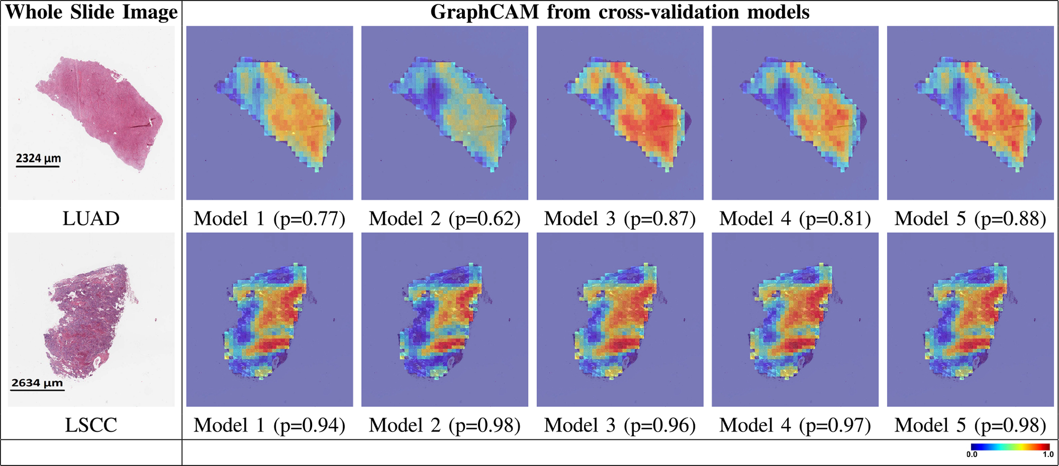
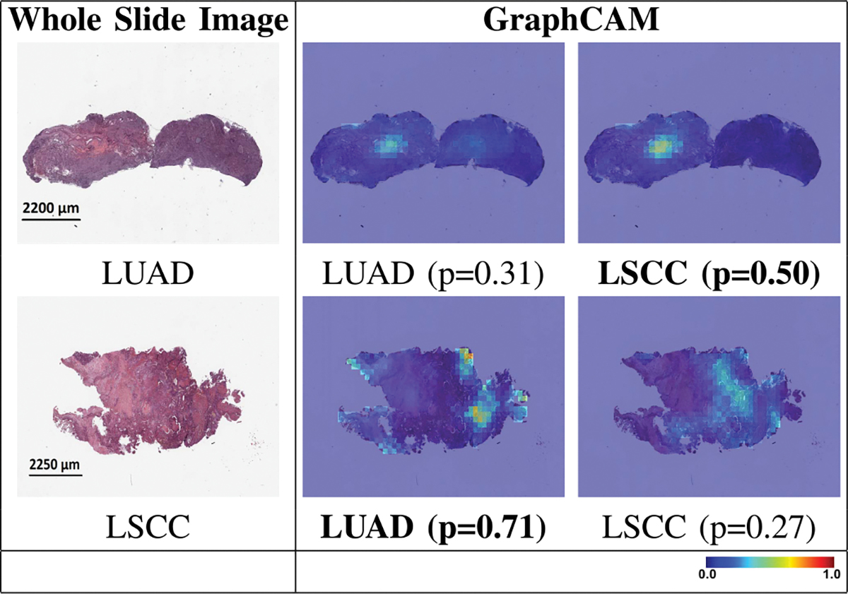
Deep learning is a powerful tool for whole slide image (WSI) analysis. Typically, when performing supervised deep learning, a WSI is divided into small patches, trained and the outcomes are aggregated to estimate disease grade. However, patch-based methods introduce label noise during training by assuming that each patch is independent with the same label as the WSI and neglect overall WSI-level information that is significant in disease grading. Here we present a Graph-Transformer (GT) that fuses a graph-based representation of an WSI and a vision transformer for processing pathology images, called GTP, to predict disease grade. We selected 4,818 WSIs from the Clinical Proteomic Tumor Analysis Consortium (CPTAC), the National Lung Screening Trial (NLST), and The Cancer Genome Atlas (TCGA), and used GTP to distinguish adenocarcinoma (LUAD) and squamous cell carcinoma (LSCC) from adjacent non-cancerous tissue (normal). First, using NLST data, we developed a contrastive learning framework to generate a feature extractor. This allowed us to compute feature vectors of individual WSI patches, which were used to represent the nodes of the graph followed by construction of the GTP framework. Our model trained on the CPTAC data achieved consistently high performance on three-label classification (normal versus LUAD versus LSCC: mean accuracy = 91.2 ± 2.5%) based on five-fold cross-validation, and mean accuracy = 82.3 ± 1.0% on external test data (TCGA). We also introduced a graph-based saliency mapping technique, called GraphCAM, that can identify regions that are highly associated with the class label. Our findings demonstrate GTP as an interpretable and effective deep learning framework for WSI-level classification.
Overview
The claims and statistics of this paper haven’t been checked yet.
Run the check — free, no account needed. It reads the open-access text and checks the sources it cites.
Integrity signals
Registry
Cites
Statistics
Figures
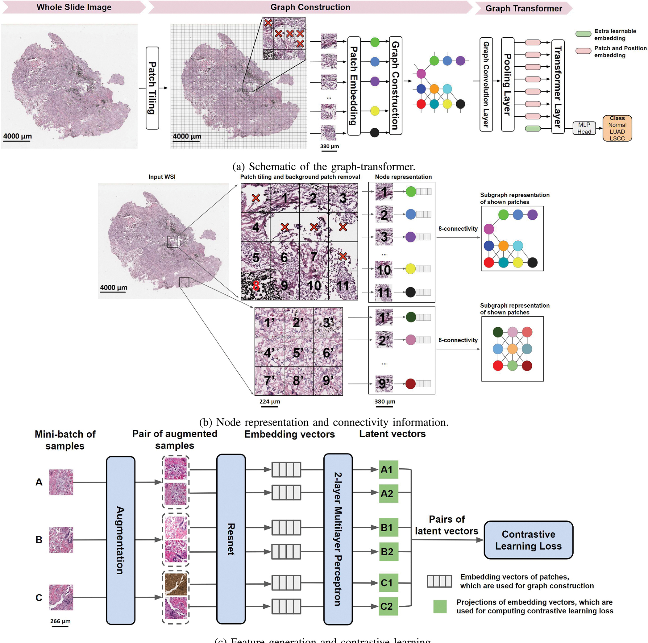
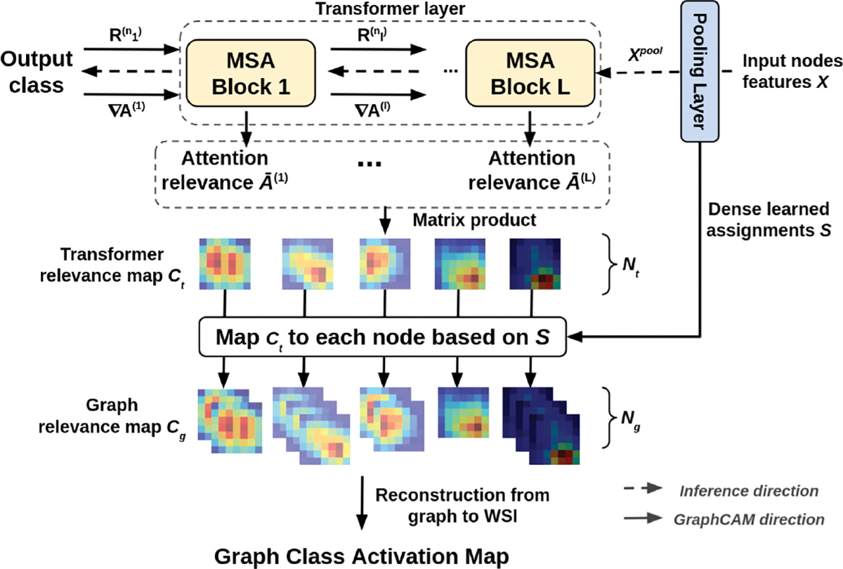
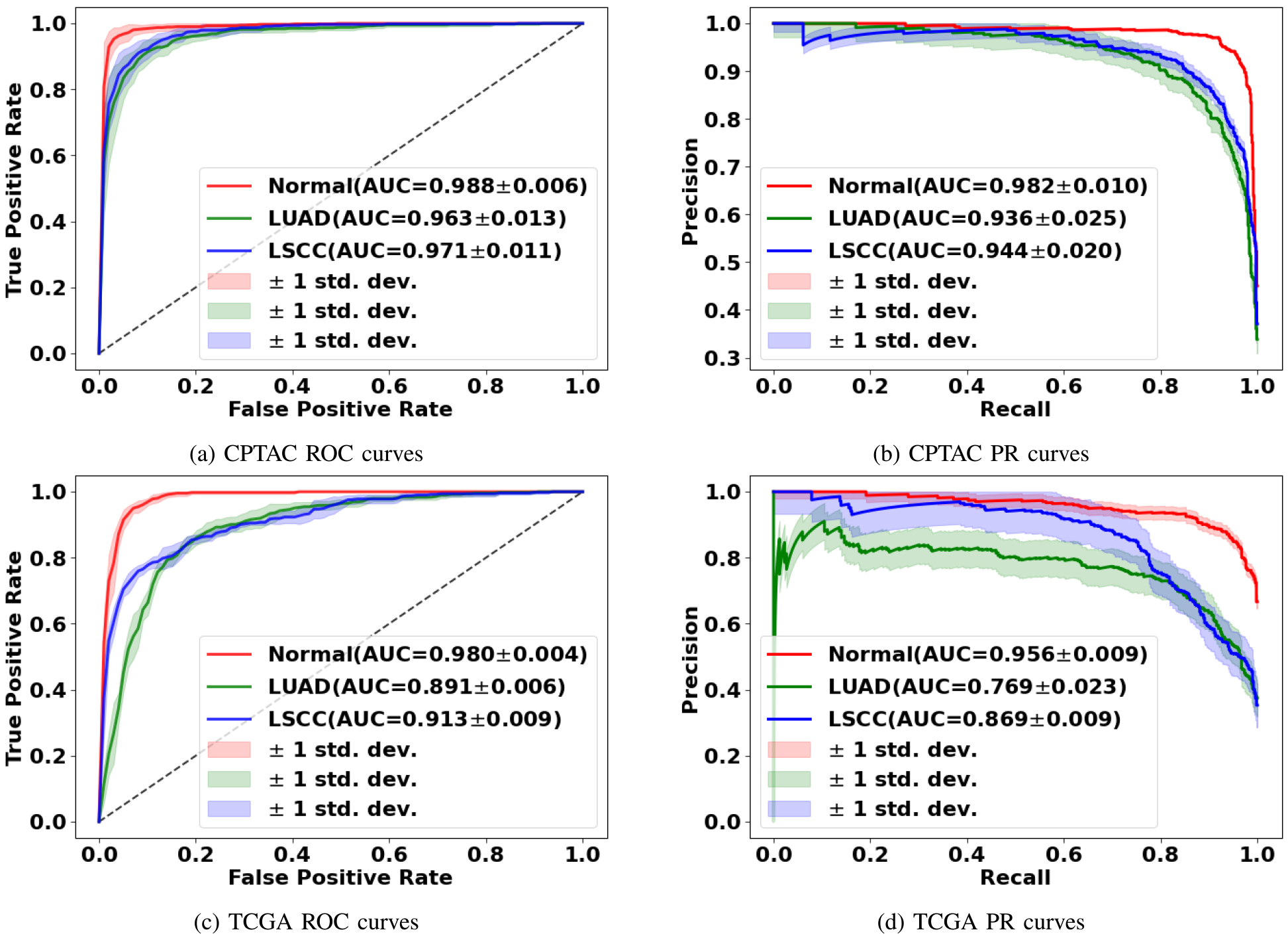
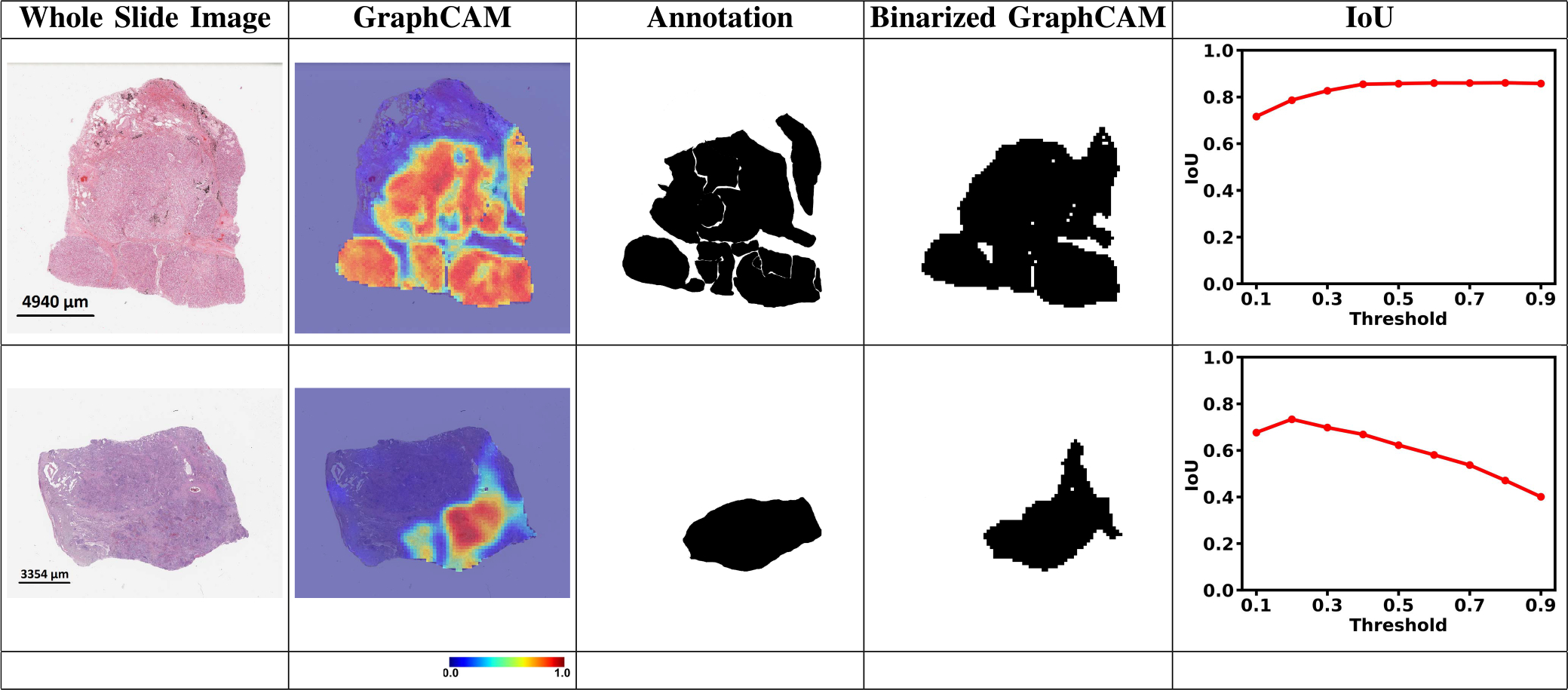
Verification
Claims the paper makes
The claims and statistics of this paper haven’t been checked yet.
Run the check — free, no account needed. It reads the open-access text and checks the sources it cites.
Claims other papers make about this one
Each is checked against this paper’s own text: does it say what the citing paper says it does?
In the citing paper
“While other researchers have previously explored the development of mid-level-fusion and mixer architectures [6], [7] as well as the use of graph-based representations of WSIs [8], our work is unique in mixing node and edge embeddings along with fusion of bulk gene expression embeddings to learn a multimodal topographic mapping to predict survival.”
From Graph attention-based fusion of pathology images and gene expression for prediction of cancer survival (2024) · cites this paper as [8]
In the citing paper
“We used NLST for generating node-level features for graph construction [8], TCGA for training the models using 5-fold cross-validation, and CPTAC as an independent dataset for testing.”
From Graph attention-based fusion of pathology images and gene expression for prediction of cancer survival (2024) · cites this paper as [8]
In the citing paper
“The ISM model’s performance exceeded that of other recent methodologies [8], [21], [25]-[27], as illustrated in Table II.”
From Graph attention-based fusion of pathology images and gene expression for prediction of cancer survival (2024) · cites this paper as [8]
Self-check
Percentages, sums and reported differences re-computed from the paper’s own numbers.
The self-check needs the paper’s full text.
Citations
Cites
Deep Residual Learning for Image Recognition
He, K., Zhang, X., Ren, S., & Sun, J. (2016) · 2016 IEEE Conference on Computer Vision and Pattern Recognition (CVPR) · doi:10.1109/cvpr.2016.90 · Cited by 235,474
Vaswani, A., Shazeer, N., Parmar, N., Uszkoreit, J., Jones, L., Gomez, A. N., Kaiser, L., & Polosukhin, I. (2017) · arXiv · doi:10.48550/arxiv.1706.03762 · Cited by 120,404
Adam: A Method for Stochastic Optimization
Kingma, D. P., & Ba, J. (2014) · arXiv · doi:10.48550/arxiv.1412.6980 · Cited by 98,946
ImageNet: A large-scale hierarchical image database
Deng, J., Dong, W., Socher, R., Li, L.-J., Li, K., & Fei-Fei, L. (2009) · 2009 IEEE Conference on Computer Vision and Pattern Recognition · doi:10.1109/cvpr.2009.5206848 · Cited by 65,333
Semi-supervised classification with graph convolutional networks
Kipf, T. N., & Welling, M. (2016) · arXiv · doi:10.48550/arxiv.1609.02907 · Cited by 24,853
Reduced Lung-Cancer Mortality with Low-Dose Computed Tomographic Screening
The National Lung Screening Trial Research Team (2011) · New England Journal of Medicine · doi:10.1056/nejmoa1102873 · Cited by 11,064
A simple framework for contrastive learning of visual representations
Chen, T., Kornblith, S., Norouzi, M., & Hinton, G. (2020) · arXiv · doi:10.48550/arxiv.2002.05709 · Cited by 10,857
Deformable DETR: Deformable Transformers for End-to-End Object Detection
Zhu, X., Su, W., Lu, L., Li, B., Wang, X., & Dai, J. (2020) · arXiv · doi:10.48550/arxiv.2010.04159 · Cited by 6,027 · cited 1×
“row normalization function in transformers for computer vision tasks [36], [37].”
BackgroundSGDR: Stochastic Gradient Descent with Warm Restarts
Loshchilov, I., & Hutter, F. (2016) · arXiv · doi:10.48550/arxiv.1608.03983 · Cited by 5,309
TransUNet: Transformers Make Strong Encoders for Medical Image Segmentation
Chen, J., Lu, Y., Yu, Q., Luo, X., Adeli, E., Wang, Y., Lu, L., Yuille, A. L., & Zhou, Y. (2021) · arXiv · doi:10.48550/arxiv.2102.04306 · Cited by 4,206
Research works printed but not linked: 2
- Dosovitskiy, An image is worth 16×16 words: Transformers for image recognition at scale
- 2021, The Cancer Genome Atlas Program
Cited by
Sorted by citations
Transformers in medical imaging: A survey
Shamshad, F., Khan, S., Zamir, S. W., Khan, M. H., Hayat, M., Khan, F. S., & Fu, H. (2023) · Medical Image Analysis · doi:10.1016/j.media.2023.102802 · Cited by 1,523
Advances in medical image analysis with vision Transformers: A comprehensive review
Azad, R., Kazerouni, A., Heidari, M., Aghdam, E. K., Molaei, A., Jia, Y., Jose, A., Roy, R., & Merhof, D. (2023) · Medical Image Analysis · doi:10.1016/j.media.2023.103000 · Cited by 557
McGenity, C., Clarke, E. L., Jennings, C., Matthews, G., Cartlidge, C., Freduah-Agyemang, H., Stocken, D. D., & Treanor, D. (2024) · npj Digital Medicine · doi:10.1038/s41746-024-01106-8 · Cited by 290
Jiang, R., Yin, X., Yang, P., Cheng, L., Hu, J., Jiao, Y., Wang, Y., Fu, X., Shang, L., Li, L., et al. (2024) · Nature Machine Intelligence · doi:10.1038/s42256-024-00868-w · Cited by 165
A multimodal whole-slide foundation model for pathology
Ding, T., Wagner, S. J., Song, A. H., Chen, R. J., Lu, M. Y., Zhang, A., Vaidya, A. J., Jaume, G., Shaban, M., Kim, A., et al. (2025) · Nature Medicine · doi:10.1038/s41591-025-03982-3 · Cited by 171
Bhati, D., Neha, F., & Amiruzzaman, M. (2024) · Journal of Imaging · doi:10.3390/jimaging10100239 · Cited by 177
Vision Transformers for Computational Histopathology
Xu, H., Xu, Q., Cong, F., Kang, J., Han, C., Liu, Z., Madabhushi, A., & Lu, C. Y. (2023) · IEEE Reviews in Biomedical Engineering · doi:10.1109/rbme.2023.3297604 · Cited by 153
Artificial intelligence to identify genetic alterations in conventional histopathology
Çifçi, D., Foersch, S., & Kather, J. N. (2022) · The Journal of Pathology · doi:10.1002/path.5898 · Cited by 140
Li, J., Chen, Y., Chu, H., Sun, Q., Guan, T. M., Han, A., & He, Y. (2024) · IEEE Computer Society Conference on Computer Vision and Pattern Recognition/Proceedings - IEEE Computer Society Conference on Computer Vision and Pattern Recognition/Proceedings · doi:10.1109/cvpr52733.2024.01076 · Cited by 118
“Unlike conventional methods that construct graphs using spatial relationships [6, 18, 44], our approach is dedicated to quantifying positional relationships between patches through learnable implicit features.”
BackgroundMethodAtabansi, C. C., Nie, J., Liu, H., Song, Q., Yan, L., & Zhou, X. (2023) · BioMedical Engineering OnLine · doi:10.1186/s12938-023-01157-0 · Cited by 114
“9(a) GTP).”
BackgroundInfluentialKim, G., Hugonnet, H. J., Kim, K., Lee, J.-H., Lee, S. S., Ha, J., Lee, C., Park, H., Yoon, K., Shin, Y., et al. (2024) · Nature Reviews Methods Primers · doi:10.1038/s43586-024-00327-1 · Cited by 117
“Past research on established medical imaging modalities has showcased the feasibility of storing and profiling large-scale images, enabling global-scale attributes to be factored into analyses 213,214 .”
Pati, P., Karkampouna, S., Bonollo, F., Compérat, É., Radić, M., Spahn, M., Martinelli, A. L., Wartenberg, M., Julio, M. K., & Rapsomaniki, M. (2024) · Nature Machine Intelligence · doi:10.1038/s42256-024-00889-5 · Cited by 96
“41, fuses a graph neural network and a vision transformer (ViT) to process histopathology images.”
BackgroundHistopathology Whole Slide Image Analysis with Heterogeneous Graph Representation Learning
Chan, T. H., Cendra, F. J., Ma, L., Yin, G., & Yu, L. (2023) · IEEE Computer Society Conference on Computer Vision and Pattern Recognition/Proceedings - IEEE Computer Society Conference on Computer Vision and Pattern Recognition/Proceedings · doi:10.1109/cvpr52729.2023.01503 · Cited by 87
“Existing WSI analysis approaches generally adopt MIL [5,7,12,26,30,33,41], which first divide the WSI into fixed-size patches and then compress the information of these patches into low-dimensional vectors.”
MethodInfluentialDavri, A. S., Birbas, E., Kanavos, T., Ntritsos, G., Γιαννακέας, Ν., Tzallas, A. T., & Batistatou, A. K. (2023) · Cancers · doi:10.3390/cancers15153981 · Cited by 94
“The majority of these mainly include ADC and SCC WSIs, mostly from the TCGA dataset, whereas the classification task is performed by a CNN or a combination of the state-of-the-art CNN architectures with varying approaches and techniques [37,47,51–56, 58, 59 ].”
MethodHu, W., Li, X., Li, C., Li, R., Jiang, T., Sun, H., Huang, X., Grzegorzek, M., & Li, X. (2023) · Computers in Biology and Medicine · doi:10.1016/j.compbiomed.2023.107034 · Cited by 86
Recent advances of Transformers in medical image analysis: A comprehensive review
Xia, K., & Wang, J. (2023) · MedComm – Future Medicine · doi:10.1002/mef2.38 · Cited by 81
A Survey on Attention Mechanisms for Medical Applications: are we Moving Toward Better Algorithms?
Gonçalves, T., Rio-Torto, I., Teixeira, L. F., & Cardoso, J. S. (2022) · IEEE Access · doi:10.1109/access.2022.3206449 · Cited by 83
Improving diagnosis and prognosis of lung cancer using vision transformers: a scoping review
Ali, H., Mohsen, F., & Shah, Z. (2023) · BMC Medical Imaging · doi:10.1186/s12880-023-01098-z · Cited by 76
“Both precision and recall [43, 45, 47, 48, 50, 57] Dimension-ality of data 2D models (2D data) (n = 23) [24], [26–28], [30–32], [34–37], [40, 41], [47–50], [52–57] 3D models (volumetric data) (n = 11) [25, 29, 33, 38, 39], [42–46 Similarly, eight studies used a 5-fold cross-validation scheme, while…”
BackgroundInfluentialLai, T. (2024) · BioMedInformatics · doi:10.3390/biomedinformatics4010008 · Cited by 74
Xiong, C., Chen, H., Sung, J. J. �., & King, I. (2023) · International Joint Conference on Artificial Intelligence · doi:10.24963/ijcai.2023/176 · Cited by 73
Wong, A. N. N., He, Z., Leung, K. L., To, C. C. K., Wong, C. Y., Wong, S. C. C., Yoo, J. S., Chan, C. K. R., Chan, A. Z., Lacambra, M. D., et al. (2022) · Cancers · doi:10.3390/cancers14153780 · Cited by 64
“97 in the grading of lung carcinoma [111].”
BackgroundQu, L., Ma, Y., Luo, X., Guo, Q., Wang, M., & Song, Z. (2024) · IEEE Transactions on Circuits and Systems for Video Technology · doi:10.1109/tcsvt.2024.3400876 · Cited by 61
“Some methods have also been proposed to accomplish WSI classification using reinforcement learning [33], graph learning [34], [35], and bayesian learning [36].”
MethodMarsGT: Multi-omics analysis for rare population inference using single-cell graph transformer
Wang, X., Duan, M., Li, J., Ma, A., Xin, G., Xu, D. H., Li, Z., Liu, B., & Ma, Q. (2024) · Nature Communications · doi:10.1038/s41467-023-44570-8 · Cited by 53
“In addition, it could be more useful to have something simple like the adjusted Rand index against the original labels.”
Liang, M., Chen, Q., Li, B., Wang, L., Wang, Y., Zhang, Y., Wang, R., Jiang, X. Y., & Zhang, C. (2022) · Computer Methods and Programs in Biomedicine · doi:10.1016/j.cmpb.2022.107268 · Cited by 55
An aggregation of aggregation methods in computational pathology
Bilal, M., Jewsbury, R., Wang, R., AlGhamdi, H. M., Asif, A., Eastwood, M., & Rajpoot, N. (2023) · Medical Image Analysis · doi:10.1016/j.media.2023.102885 · Cited by 52
Zheng, Y., Conrad, R. D., Green, E. J., Burks, E. J., Betke, M., Beane, J. E., & Kolachalama, V. B. (2024) · IEEE Transactions on Medical Imaging · doi:10.1109/tmi.2024.3386108 · Cited by 44
“The ISM model’s performance exceeded that of other recent methodologies [8], [21], [25], [26], [27], as illustrated in Table.”
BackgroundMethodInfluentialSelf-citationCGAM: An end-to-end causality graph attention Mamba network for esophageal pathology grading
Qu, Y., Zhou, X., Huang, P., Liu, Y., Mercaldo, F., Santone, A., & Deng, X. (2025) · Biomedical Signal Processing and Control · doi:10.1016/j.bspc.2024.107452 · Cited by 41
Mir, A. N., Rizvi, D. R., & Ahmad, M. (2025) · Engineering Applications of Artificial Intelligence · doi:10.1016/j.engappai.2025.110519 · Cited by 44
Vision transformer promotes cancer diagnosis: A comprehensive review
Jiang, X., Wang, S., & Zhang, Y. (2024) · Expert Systems with Applications · doi:10.1016/j.eswa.2024.124113 · Cited by 41
Bazargani, R., Fazli, L., Gleave, M. E., Goldenberg, L., Bashashati, A., & Salcudean, S. E. (2024) · Medical Image Analysis · doi:10.1016/j.media.2024.103197 · Cited by 42
Shi, J., Tang, L., Li, Y., Zhang, X., Gao, Z., Zheng, Y., Wang, C., Gong, T., & Li, C. (2023) · IEEE Transactions on Medical Imaging · doi:10.1109/tmi.2023.3273236 · Cited by 40
“GT [19] utilizes a graph-based representation of a WSI and a vision Transformer for processing pathological images; • Multi-scale-based methods : MFNet [35] proposes a multi-scale input and multi-feature network.”
InfluentialSelf-citationBrahmareddy, A., & Selvan, M. P. (2025) · Scientific Reports · doi:10.1038/s41598-025-19173-6 · Cited by 37
Weakly supervised joint whole-slide segmentation and classification in prostate cancer
Pati, P., Jaume, G., Ayadi, Z., Thandiackal, K., Bozorgtabar, B., Gabrani, M., & Goksel, O. (2023) · Medical Image Analysis · doi:10.1016/j.media.2023.102915 · Cited by 37
Novel tools for early diagnosis and precision treatment based on artificial intelligence
Shao, J., Feng, J., Li, J., Liang, S., Li, W., & Wang, C. (2023) · Chinese Medical Journal - Pulmonary and Critical Care Medicine · doi:10.1016/j.pccm.2023.05.001 · Cited by 41
Generating dermatopathology reports from gigapixel whole slide images with HistoGPT
Tran, M., Schmidle, P., Guo, R. R., Wagner, S. J., Koch, V., Lupperger, V., Novotny, B., Murphree, D. H., Hardway, H. D., D’Amato, M., et al. (2025) · Nature Communications · doi:10.1038/s41467-025-60014-x · Cited by 36
“They excel at image processing tasks such as cancer classi fi cation 6 , tissue segmentation 7 , survival prediction 8 , and biomarker detection 9 .”
Shi, J., Tang, L., Gao, Z., Li, Y., Wang, C., Gong, T., Li, C., & Fu, H. (2023) · IEEE Transactions on Medical Imaging · doi:10.1109/tmi.2023.3313252 · Cited by 36
“2) Effect of PAM and Multi-Scale Learning: To evaluate the effect of PAM and the Multi-scale Learning method on the baseline model, we select three baseline methods ( i.e. , ABMIL [3], TransMIL [7], and GT [11]).”
BackgroundMethodInfluentialCai, L., Huang, S., Zhang, Y., Lu, J., & Zhang, Y. (2025) · Medical Image Analysis · doi:10.1016/j.media.2025.103631 · Cited by 33
Yacob, F., Siarov, J., Villiamsson, K., Suvilehto, J. T., Sjöblom, L., Kjellberg, M., & Neittaanmäki, N. (2023) · Scientific Reports · doi:10.1038/s41598-023-33863-z · Cited by 33
Elastic Graph Transformer Networks for EEG-Based Emotion Recognition
Jiang, W.-B., Yan, X., Zheng, W., & Lu, B. (2023) · IEEE International Conference on Acoustics Speech and Signal Processing · doi:10.1109/icassp49357.2023.10096511 · Cited by 32
“Note that in computer vision, Zheng et al. [14] combined GCN and Transformer for whole slide image classification.”
BackgroundSun, X., Li, W., Fu, B., Peng, Y., He, J., Wang, L., Yang, T., Meng, X., Li, J., Wang, J., et al. (2023) · Computer Methods and Programs in Biomedicine · doi:10.1016/j.cmpb.2023.107789 · Cited by 33
Yao, I. Z., Dong, M., & Hwang, W. Y. K. (2025) · Mayo Clinic Proceedings Digital Health · doi:10.1016/j.mcpdig.2025.100253 · Cited by 44
“45 These advancements can actually solve the problem of some rare cancers with deep-infiltrating cells, which are difficult to separate from healthy tissue.”
Li, L. K., Pan, H., Liang, Y., Shao, M., Xie, S., Lu, S., & Liao, S. (2024) · Knowledge-Based Systems · doi:10.1016/j.knosys.2024.111502 · Cited by 30
Chikontwe, P., Kim, M., Jeong, J., Sung, H. J., Go, H., Nam, S. J., & Park, S. H. (2024) · IEEE Transactions on Medical Imaging · doi:10.1109/tmi.2024.3446716 · Cited by 31
“Existing methods applied to WSI employ either graph-based learning or multi-scale multi-stage pre-training to model the spatial relationships among different entities [20], [51], [52], [53], [54], [55].”
Xu, H., Wang, M., Shi, D., Qin, H., Zhang, Y., Liu, Z., Madabhushi, A., Gao, P., Cong, F., & Lu, C. Y. (2025) · Medical Image Analysis · doi:10.1016/j.media.2025.103456 · Cited by 41
Bayesian Collaborative Learning for Whole-Slide Image Classification
Yu, J.-G., Wu, Z., Ming, Y., Deng, S., Wu, Q., Xiong, Z., Yu, T., Xia, G.-S., Jiang, Q., & Li, Y. (2023) · IEEE Transactions on Medical Imaging · doi:10.1109/tmi.2023.3241204 · Cited by 29
“Recently, vision transformers have been utilized to address WSI classification [16], [34], [35], [36], [37].”
MethodMulti-Scale Efficient Graph-Transformer for Whole Slide Image Classification
Ding, S., Li, J., Wang, J., Ying, S., & Shi, J. (2023) · IEEE Journal of Biomedical and Health Informatics · doi:10.1109/jbhi.2023.3317067 · Cited by 32
“4) The Cluster-based MIL, Re-Mix [40] 5) The Graph-based MIL, GCN-MIL [23], and GTP [41].”
InfluentialA Graph-Based Multi-Scale Approach With Knowledge Distillation for WSI Classification
Bontempo, G., Bolelli, F., Porrello, A., Calderara, S., & Ficarra, E. (2023) · IEEE Transactions on Medical Imaging · doi:10.1109/tmi.2023.3337549 · Cited by 31
“Other very recent approaches [9], [15], [16] resort to more complex hierarchical structures by adapting Vision Transformers (ViT) [17] and Graph Neural Networks (GNN) [18].”
Liu, M., Liu, Y., Xu, P., Cui, H., Ke, J., & Ma, J. (2024) · IEEE Transactions on Medical Imaging · doi:10.1109/tmi.2024.3381994 · Cited by 27
“Many studies [20], [39], [40], [41], [42] focus on pathological image classification by introducing GCNs into their models.”
BackgroundWang, Y., Luo, F., Yang, X., Wang, Q., Sun, Y., Tian, S., Feng, P., Huang, P., & Xiao, H. (2023) · Journal of Cancer Research and Clinical Oncology · doi:10.1007/s00432-023-04795-y · Cited by 24
“Previous studies have explored the use of Transformer-based models for classification, with some achieving good results (Hu et al. 2022; Maleki and Tizhoosh 2022; Zheng et al. 2022).”
Multi-scale Prototypical Transformer for Whole Slide Image Classification
Ding, S., Wang, J., Li, J., & Shi, J. (2023) · Lecture notes in computer science · doi:10.1007/978-3-031-43987-2_58 · Cited by 25
Abdullahi, M., Oyelade, O. N., Kana, A. F. D., Bagiwa, M. A., Abdullahi, F. B., Junaidu, S. B., Iliyasu, I., Ore-Ofe, A., & Chiroma, H. (2024) · Multimedia Tools and Applications · doi:10.1007/s11042-024-19823-3 · Cited by 31
MSCPT: Few-Shot Whole Slide Image Classification With Multi-Scale and Context-Focused Prompt Tuning
Han, M., Qu, L., Yang, D., Zhang, X., Wang, X., & Zhang, L. (2025) · IEEE Transactions on Medical Imaging · doi:10.1109/tmi.2025.3564976 · Cited by 30
“Furthermore, MIL methods based on Graph Neural Networks (GNNs) [26], [27] and Transformers [1], [31] have also been proposed to capture both local and global contextual information of WSIs.”
Abbaker, N., Minervini, F., Guttadauro, A., Solli, P., Cioffi, U., & Scarci, M. (2024) · Frontiers in Oncology · doi:10.3389/fonc.2024.1347464 · Cited by 25
“These studies predominantly feature ADC and SCC Whole Slide Images (WSIs), often sourced from The Cancer Genome Atlas dataset (12 – 15).”
Structure Embedded Nucleus Classification for Histopathology Images
Lou, W., Wan, X., Li, G., Lou, X., Li, C., Gao, F., & Li, H. (2024) · IEEE Transactions on Medical Imaging · doi:10.1109/tmi.2024.3388328 · Cited by 25
“Most GNN methods are to classify a whole image [48], [49], [50] or tissue patches [51].”
MethodGTP-4o: Modality-Prompted Heterogeneous Graph Learning for Omni-Modal Biomedical Representation
Li, C., Liu, X., Wang, C., Liu, Y., Yu, W., Shao, J., & Yuan, Y. (2024) · Lecture notes in computer science · doi:10.1007/978-3-031-73235-5_10 · Cited by 27
Multimodal integration strategies for clinical application in oncology
Zhang, B., Wan, Z., Luo, Y., Zhao, X., Samayoa, J. A., Zhao, W., & Wu, S. (2025) · Frontiers in Pharmacology · doi:10.3389/fphar.2025.1609079 · Cited by 28
A survey on artificial intelligence in pulmonary imaging
Saha, P. K., Nadeem, S. A., & Comellas, A. P. (2023) · Wiley Interdisciplinary Reviews Data Mining and Knowledge Discovery · doi:10.1002/widm.1510 · Cited by 23
González, R., Saha, A., Campbell, C. J. V., Nejat, P., Lokker, C., & Norgan, A. P. (2023) · Journal of Pathology Informatics · doi:10.1016/j.jpi.2023.100347 · Cited by 24
SI-MIL: Taming Deep MIL for Self-Interpretability in Gigapixel Histopathology
Kapse, S., Pati, P., Das, S., Zhang, J., Chen, C., Vakalopoulou, M., Saltz, J. H., Samaras, D., Gupta, R., & Prasanna, P. (2024) · IEEE Computer Society Conference on Computer Vision and Pattern Recognition/Proceedings - IEEE Computer Society Conference on Computer Vision and Pattern Recognition/Proceedings · doi:10.1109/cvpr52733.2024.01067 · Cited by 24
“Additionally, few methods, such as those by [47, 70], use segmentation maps or gradient-based techniques to localize significant areas.”
Shi, J., Li, C., Gong, T., & Fu, H. (2024) · Medical Image Analysis · doi:10.1016/j.media.2024.103294 · Cited by 20
Aligning knowledge concepts to whole slide images for precise histopathology image analysis
Zhao, W., Guo, Z., Fan, Y., Jiang, Y., Yeung, M. C. F., & Yu, L. (2024) · npj Digital Medicine · doi:10.1038/s41746-024-01411-2 · Cited by 23
Digital pathology, deep learning, and cancer: a narrative review
Williams, D. K. A., Graifman, G., Hussain, N., Amiel, M., Priscilla, T., Reddy, A., Haider, A., Kavitesh, B. K., Li, A., Alishahian, L., et al. (2024) · Translational Cancer Research · doi:10.21037/tcr-23-964 · Cited by 20
“GTP distinguishes adenocarcinoma and squamous cell carcinoma from normal histology (40).”
BackgroundMethodInfluentialFOCUS: Knowledge-enhanced Adaptive Visual Compression for Few-shot Whole Slide Image Classification
Guo, Z., Xiong, C., Ma, J., Sun, Q., Feng, L., Wang, J., & Chen, H. (2025) · IEEE Computer Society Conference on Computer Vision and Pattern Recognition/Proceedings - IEEE Computer Society Conference on Computer Vision and Pattern Recognition/Proceedings · doi:10.1109/cvpr52734.2025.01453 · Cited by 27
“While MIL framework has been proven highly effective in the field of CPath [5– 7, 9, 14, 18, 20, 21, 24–28, 34, 37, 42, 44, 52–54, 57], training current approaches typically demands large annotated datasets that are labor-intensive and time-consuming to obtain, particularly for rare pathological…”
HIGT: Hierarchical Interaction Graph-Transformer for Whole Slide Image Analysis
Guo, Z., Zhao, W., Wang, S., & Yu, L. (2023) · Lecture notes in computer science · doi:10.1007/978-3-031-43987-2_73 · Cited by 22
GraphMamba: Whole slide image classification meets graph-driven selective state space model
Zheng, T., Yao, H., Zhao, S., Jiang, K., & Xiao, Y. (2025) · Pattern Recognition · doi:10.1016/j.patcog.2025.111768 · Cited by 19
MGCM: Multi-modal graph convolutional mamba for cancer survival prediction
Cui, J., Li, Y., Shen, D. G., & Wang, Y. (2025) · Pattern Recognition · doi:10.1016/j.patcog.2025.111991 · Cited by 20
Unsupervised mutual transformer learning for multi-gigapixel Whole Slide Image classification
Javed, S., Mahmood, A., Qaiser, T., Werghi, N., & Rajpoot, N. (2024) · Medical Image Analysis · doi:10.1016/j.media.2024.103203 · Cited by 16
Bui, D. C., Song, B., Kim, K., & Kwak, J. T. (2024) · Computer Methods and Programs in Biomedicine · doi:10.1016/j.cmpb.2024.108112 · Cited by 15
Lai, B., Fu, J., Zhang, Q., Deng, N., Jiang, Q., & Peng, J. (2023) · International Journal of Oncology · doi:10.3892/ijo.2023.5555 · Cited by 16
Gallo, M., Gallo, M., Nenutil, R., Němeček, M., Holub, P., & Brázdil, T. (2023) · New Biotechnology · doi:10.1016/j.nbt.2023.09.008 · Cited by 16
Histopathologic brain age estimation via multiple instance learning
Marx, G. A., Kauffman, J., McKenzie, A. T., Koenigsberg, D., McMillan, C. T., Morgello, S., Karlovich, E., Insausti, R., Richardson, T. E., Walker, J. M., et al. (2023) · Acta Neuropathologica · doi:10.1007/s00401-023-02636-3 · Cited by 19
Chen, Y., Zhang, F., Wang, M., Zekelman, L., Karayumak, S. C., Xue, T., Zhang, C., Song, Y., Rushmore, J., Makris, N., et al. (2025) · Medical Image Analysis · doi:10.1016/j.media.2025.103476 · Cited by 14
Few-Shot Classification for ISAR Images of Space Targets by Complex-Valued Patch Graph Transformer
Yuan, H., Li, H., Zhang, Y., Li, M., & Wei, C. (2024) · IEEE Transactions on Aerospace and Electronic Systems · doi:10.1109/taes.2024.3382222 · Cited by 13
Cell Graph Transformer for Nuclei Classification
Lou, W., Li, G., Wan, X., & Li, H. (2024) · Proceedings of the AAAI Conference on Artificial Intelligence · doi:10.1609/aaai.v38i4.28179 · Cited by 12
Yu, J., Li, S., Tan, L., Zhou, H., Li, J., & Li, J. (2025) · Expert Systems with Applications · doi:10.1016/j.eswa.2025.127164 · Cited by 14
Zhao, W., Wang, S., Yeung, M. C. F., Niu, T., & Yu, L. (2023) · Proceedings of the AAAI Conference on Artificial Intelligence · doi:10.1609/aaai.v37i3.25471 · Cited by 12
Tang, L., Diao, S., Li, C., He, M., Ru, K., & Qin, W. (2024) · Computerized Medical Imaging and Graphics · doi:10.1016/j.compmedimag.2024.102378 · Cited by 11
mBGT: Encoding Brain Signals With Multimodal Brain Graph Transformer
Peng, C., Guo, T., Xie, C., Bai, X., Zhou, J., Zhao, X., He, E., & Xia, F. (2024) · IEEE Transactions on Consumer Electronics · doi:10.1109/tce.2024.3425478 · Cited by 11
“With Transformer showing great superiority in both natural language processing (NLP) [37] and computer vision (CV) [38], [39], many recent works attempt to generalize Transformer models to graph data [40], [41].”
Shi, Z., Zhang, J., Kong, J., & Wang, F. (2024) · Lecture notes in computer science · doi:10.1007/978-3-031-72120-5_32 · Cited by 14
Li, Q., Teodoro, G., Jiang, Y., & Kong, J. (2024) · Computerized Medical Imaging and Graphics · doi:10.1016/j.compmedimag.2024.102467 · Cited by 12
Jin, H., Shen, J., Cui, L., Shi, X., Li, K., & Zhu, X. (2025) · Medical Image Analysis · doi:10.1016/j.media.2025.103468 · Cited by 11
Xia, Z., Li, H., & Lan, L. (2025) · Physics in Medicine and Biology · doi:10.1088/1361-6560/ae07a1 · Cited by 11
“…signal-to-noise ratio and artifacts), most existing vision transformer architectures are typically tailored for task-specific scenarios, such as disease diagnosis [5], [6], [7], [8], anatomical structure and lesion segmentation [9], [10], [11], [12], [13], and abnormality detection [14], [15].”
SliDL: A toolbox for processing whole-slide images in deep learning
Berman, A. G., Orchard, W. R., Gehrung, M., & Markowetz, F. M. (2023) · PLoS ONE · doi:10.1371/journal.pone.0289499 · Cited by 16
“Researchers have more recently begun applying vision transformers to whole-slide images and histopathological problems, but they form a promising new avenue [11].”
A survey on attention mechanisms for medical applications: are we moving towards better algorithms?Preprint
Gonçalves, T., Rio-Torto, I., Teixeira, L. F., & Cardoso, J. S. (2022) · Research Square · doi:10.21203/rs.3.rs-1594205/v1 · Cited by 11
A critical review of explainable deep learning in lung cancer diagnosis
Koutoulakis, E., Trivizakis, E., Markodimitrakis, E., Agelaki, S., Tsiknakis, M., & Kostas Marias, A. (2025) · Artificial Intelligence Review · doi:10.1007/s10462-025-11445-x · Cited by 11
Transformers in pathological image analysis: A survey
Liu, L., Liu, Z., Xie, J., Zhang, H., Chang, J., & Qiao, H. (2025) · Engineering Applications of Artificial Intelligence · doi:10.1016/j.engappai.2025.111114 · Cited by 11
Rethinking Attention Mechanisms in Vision Transformers with Graph Structures
Kim, H.-J., & Ko, B. C. (2024) · Sensors · doi:10.3390/s24041111 · Cited by 10
“It was proven that both the graph convolution Zheng et al. [18] proposed a graph transformer network, which is a graph representation of an entire slide image, as well as a method for fusing transformers. and grid features of Mesh The image patches become a set of nodes in the graph, and each node is connected by an e…”
BackgroundGindra, R. H., Zheng, Y., Green, E. J., Reid, M. E., Mazzilli, S. A., Merrick, D., Burks, E., Kolachalama, V. B., & Beane, J. (2024) · American Journal Of Pathology · doi:10.1016/j.ajpath.2024.03.009 · Cited by 9
Self-citationAnoop, M., Mary, L. W., Wilson, A. J., & Kiran, W. S. (2024) · Multimedia Tools and Applications · doi:10.1007/s11042-024-19516-x · Cited by 9
Perera, R. D., Savas, P. S., Senanayake, D., Salgado, R., Joensuu, H., O’Toole, S. A., Li, J., Loi, S., & Halgamuge, S. K. (2024) · Communications Engineering · doi:10.1038/s44172-024-00246-9 · Cited by 9
Re-identification from histopathology images
Ganz, J., Ammeling, J., Jabari, S., Breininger, K., & Aubreville, M. (2024) · Medical Image Analysis · doi:10.1016/j.media.2024.103335 · Cited by 14
cDP-MIL: Robust Multiple Instance Learning via Cascaded Dirichlet Process
Chen, Y., Chan, T. H., Yin, G., Jiang, Y., & Yu, L. (2024) · Lecture notes in computer science · doi:10.1007/978-3-031-72949-2_14 · Cited by 9
Ding, R.-W., Luong, K.-D., Rodriguez, E., da Silva, A. C. A. L., & Hsu, W. (2024) · Scientific Reports · doi:10.1038/s41598-025-99042-4 · Cited by 13
“Most of the graph-based works in computational pathology use either hand-crafted features [11] or convolutional neural network (CNN)-extracted features based on either ImageNet transfer learning [10] [12] [14] or self-supervised pretraining [13] on relatively small-scale data.”
InfluentialDeep learning, transformers and graph neural networks: a linear algebra perspective
Baggag, A., & Saad, Y. (2025) · Numerical Algorithms · doi:10.1007/s11075-025-02218-2 · Cited by 10
“To conclude, this step-by-step approach allows efficient and robust analysis of whole slide images, enhancing tasks like disease detection and classification in computational pathology, e.g. see [98–100].”
Generating clinical-grade pathology reports from gigapixel whole slide images with HistoGPTPreprint
Tran, M., Schmidle, P., Wagner, S. J., Koch, V., Novotny, B. C., Lupperger, V., Feuchtinger, A., Böhner, A., Kaczmarczyk, R., Biedermann, T., et al. (2024) · medRxiv · doi:10.1101/2024.03.15.24304211 · Cited by 9
“Modern deep learning (DL) techniques allow to effectively automate several tasks, including cancer classification 6 , tissue segmentation 7 , survival prediction 8 , and biomarker detection 9 .”
BackgroundLiang, M., Jiang, X. Y., Cao, J., Li, B., Wang, L., Chen, Q., Zhang, C., & Zhao, Y. (2024) · The Visual Computer · doi:10.1007/s00371-024-03269-7 · Cited by 8
“In 2021, Zheng et al. [42] proposed a graph-transformer, which introduces the transformer into a graph network to obtain a graph representation of WSI for further processing.”
Liang, M., Jiang, X. Y., Cao, J., Zhang, S., Liu, H., Li, B., Wang, L., Zhang, C., & Jia, X. (2024) · Computer Methods and Programs in Biomedicine · doi:10.1016/j.cmpb.2024.108099 · Cited by 7
Luan, H., Hu, T., Hu, J., Li, R., Ji, D., He, J., Duan, X., Yang, C., Gao, Y., Chen, F., et al. (2023) · Lecture notes in computer science · doi:10.1007/978-981-99-7074-2_12 · Cited by 7
FCDS-DETR: detection transformer based on feature correction and double sampling
Wang, M., Jiao, Z., Huang, Z., & Yu, S. (2024) · Neural Computing and Applications · doi:10.1007/s00521-024-09430-6 · Cited by 7
Raswa, F. H., Lu, C.-S., & Wang, J. (2024) · IEEE Winter Conference on Applications of Computer Vision · doi:10.1109/wacv57701.2024.00745 · Cited by 9
“(8) from the last layer of Transformer-MIL module [28] [41].”
BackgroundMethodInfluential
Metadata
- Authors
- Zheng, Y., Gindra, R. H., Green, E. J., Burks, E. J., Betke, M., Beane, J. E., & Kolachalama, V. B.
- Affiliations
- Boston University (US)
- ORCID
- Venue
- IEEE Transactions on Medical Imaging
- Published
- 2022-11
- Volume
- 41
- Issue
- 11
- Pages
- 3003-3015
- ISSN
- 0278-0062 (ISSN-L)
- 1558-254X
- PMC
- PMC9670036
- OpenAlex
- W4281257868
- Cited by
- 255
- References
- 47
- Citations by year
- 2026: 54 · 2025: 81 · 2024: 73 · 2023: 42 · 2022: 5
- FWCI
- 16.55
- Type
- journal article
- Language
- en
- Open access
- hybrid
- doi.org
- Licence
- Locations
- IEEE Transactions on Medical Imaging (publishedVersion, cc-by)
- medRxiv (acceptedVersion, cc-by)
- PubMed (publishedVersion)
- PubMed Central (acceptedVersion, cc-by)
- arXiv (Cornell University) (submittedVersion)
- arXiv (Cornell University) (cc-by)
- Funders
- Grants from the National Institutes of Heath (R21-CA253498, R01-HL159620)
- Johnson & Johnson Enterprise Innovation, Inc.
- American Heart Association (20SFRN35460031)
- Karen Toffler Charitable Trust
- National Science Foundation (1551572, 1838193)
- Topics
- Physical Sciences › Computer Science › Artificial Intelligence › AI in cancer detection
- Health Sciences › Medicine › Radiology, Nuclear Medicine and Imaging › Radiomics and Machine Learning in Medical Imaging
- Physical Sciences › Computer Science › Computer Vision and Pattern Recognition › Advanced Neural Network Applications
- Keywords
- whole slide image classification, graph transformer, vision transformer, lung adenocarcinoma, squamous cell carcinoma, lung cancer histopathology, contrastive learning, interpretability
- MeSH terms
- Guanosine Triphosphate, Image Processing, Computer-Assisted, Proteomics
About these checks
Registry, retraction, Cites and statistics results are computed, not judged, with one exception: a printed reference that no index lists is read once by a model before it counts as a citation error. Claims are extracted by a model and kept only when their quote appears verbatim in the text; each cited source is then checked, with a model’s help, for whether it says what the claim relies on. Those verdicts can be wrong — read the claim’s quote and the cited source before relying on one.
Statistics and arithmetic are recomputed, not judged.
Confidence bands are shown only for fields calibrated on our annotated test set. None is calibrated yet, so no band is shown.
What counts as a claim ›How we measure confidence ›How we check this ›
Are you an author, or do you see a problem with this page? Request a review
