

The DOI matches its Crossref record; 1 of the 1 references it cites were found in a registry or an index.
Immunological significance of the accumulation of autophagy components in oral squamous cell carcinoma
Abstract
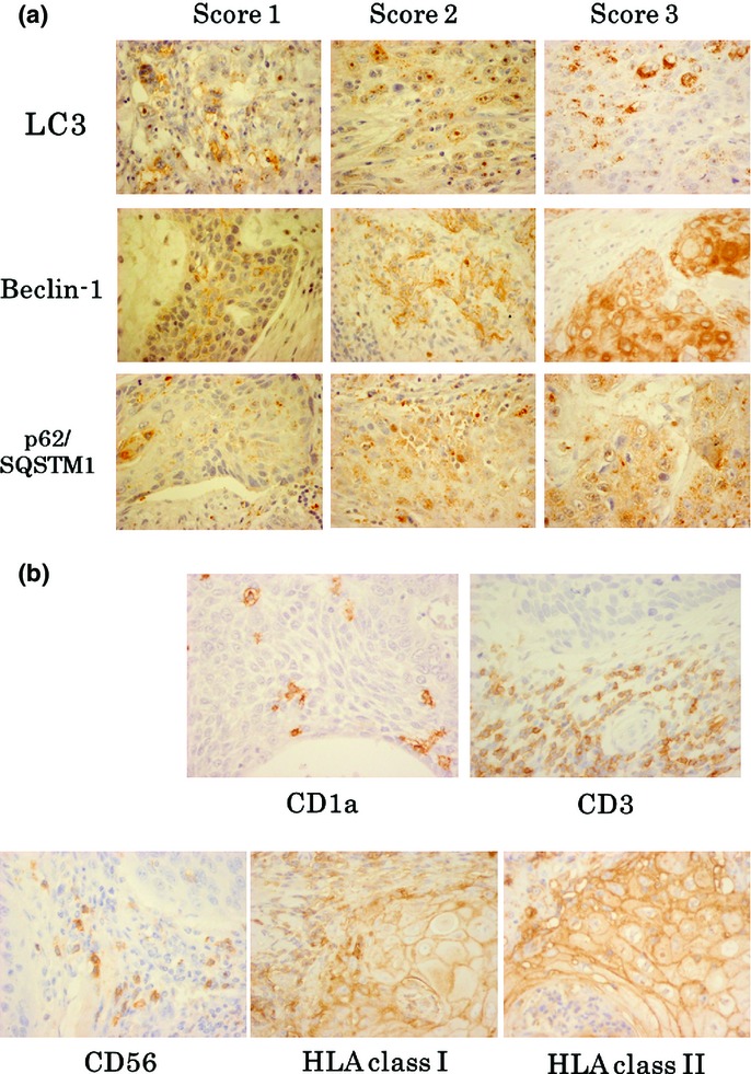
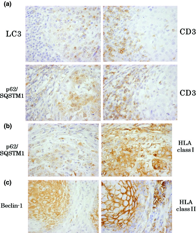
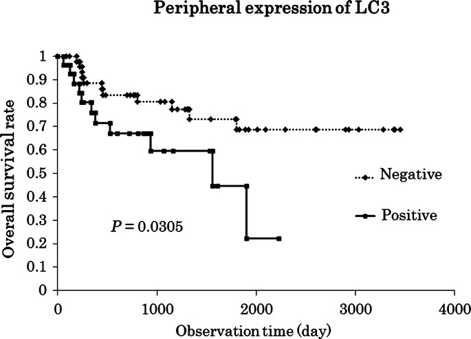
The immunological significance of autophagy in the tumor microenvironment remains unclear. To explore the relationship between autophagy and anti‐tumor immune responses, we investigated the expression of autophagy‐related proteins and infiltration of immune cells using immunohistochemistry (IHC). The expression of three representative autophagy components, LC 3, Beclin‐1 and p62/SQSTM 1, as well as the number of dendritic cells (DC), T cells and NK cells were examined by IHC in 74 patients with oral squamous cell carcinoma (OSCC). The relationship between the expression of autophagy‐associated molecules and various clinicopathological parameters was also evaluated. The expression of both LC 3 and p62/SQSTM 1 in the peripheral site significantly correlated with an increase in the infiltration of T cells. Furthermore, the expression of p62/SQSTM 1 and Beclin‐1 correlated with that of HLA class I and class II in tumor cells, respectively. In addition, several unfavorable clinicopathological parameters correlated with an increase in the expression of LC 3 in the peripheral site. The correlation observed between LC 3 or p62/SQSTM 1 and the infiltration of T cells suggests that autophagy may actively mobilize immune cells toward the cancer bed. Meanwhile, the three autophagy‐associated proteins examined were linked to malignant potential and an unfavorable prognosis.
Overview
The claims and statistics of this paper haven’t been checked yet.
Run the check — free, no account needed. It reads the open-access text and checks the sources it cites.
Integrity signals
Registry
Cites
Statistics
Figures
Verification
Claims the paper makes
The claims and statistics of this paper haven’t been checked yet.
Run the check — free, no account needed. It reads the open-access text and checks the sources it cites.
Claims other papers make about this one
Each is checked against this paper’s own text: does it say what the citing paper says it does?
In the citing paper
“For example, SQSTM1 can be detected as puncta by IHC in cancer cells, similar to LC3 [515] (Figure 21).”
From Guidelines for the use and interpretation of assays for monitoring autophagy (4th edition) 1 (2021) · cites this paper as [515]
Self-check
Percentages, sums and reported differences re-computed from the paper’s own numbers.
The self-check needs the paper’s full text.
Citations
Cites
Beclin 1 and LC3 autophagic gene expression in cutaneous melanocytic lesions
Miracco, C., Cevenini, G., Franchi, A., Luzi, P., Cosci, E., Mourmouras, V., Monciatti, I., Mannucci, S., Biagioli, M., Toscano, M., et al. (2010) · Human Pathology · doi:10.1016/j.humpath.2009.09.004 · Cited by 139
Research works printed but not linked: 44
- 10.3322/caac.20138 · DOI · Paper page
- 10.1016/j.oraloncology.2008.05.017 · DOI
- 10.1016/j.oraloncology.2010.06.006 · DOI
- 10.1016/j.cell.2011.10.026 · DOI
- 10.1016/j.cell.2009.03.048 · DOI
- 10.1016/j.ccr.2006.06.001 · DOI
- 10.1038/nrc3262 · DOI
- 10.1091/mbc.e03-09-0704 · DOI · Paper page
- 10.4161/auto.3678 · DOI · Paper page
- 10.1016/j.cell.2010.01.028 · DOI · Paper page
- 10.1038/45257 · DOI · Paper page
- 10.1016/j.cell.2005.07.002 · DOI · Paper page
- 10.1111/j.1742-4658.2010.07965.x · DOI
- 10.1016/j.cell.2007.10.035 · DOI · Paper page
- 10.1016/j.phrs.2012.07.004 · DOI
- 10.1158/0008-5472.CAN-08-0161 · DOI
- 10.1158/1078-0432.CCR-11-0951 · DOI
- 10.1158/1078-0432.CCR-11-0812 · DOI
- 10.1126/science.1208347 · DOI · Paper page
- 10.1111/his.12002 · DOI
- Reichert TE, 2002, Signaling abnormalities, apoptosis, and reduced proliferation of circulating and tumor‐infiltrating lymphocytes in patients with oral carcinoma, Clin Cancer Res, 8, 3137
- Garrido F, 1997, HLA, 445
- 10.1038/bjc.2012.310 · DOI
- Buck AC, 2001, Ki‐67 immunostaining in pancreatic cancer and chronic active pancreatitis: does in vivo FDG uptake correlate with proliferative activity?, J Nucl Med, 42, 721
- 10.4161/auto.20353 · DOI
- Ogino T, 2003, Association of tapasin and HLA class I antigen down‐regulation in primary maxillary sinus squamous cell carcinoma lesions with reduced survival of patients, Clin Cancer Res, 9, 4043
- 10.1111/j.1365-2133.2008.08465.x · DOI
- 10.1016/j.prp.2013.04.006 · DOI
- 10.3748/wjg.v17.i43.4779 · DOI · Paper page
- 10.1111/j.1349-7006.2008.00893.x · DOI
- 10.1016/j.humpath.2013.06.017 · DOI
- 10.1111/jop.12049 · DOI
- 10.1007/s12253-013-9642-0 · DOI
- 10.1016/j.prp.2012.07.008 · DOI
- 10.1016/j.prp.2013.06.006 · DOI
- 10.1371/journal.pone.0047126 · DOI
- 10.1158/0008-5472.CAN-06-0488 · DOI
- 10.1111/j.1365-2249.2010.04311.x · DOI
- 10.1038/sj.bjc.6605820 · DOI
- 10.1007/s00428-011-1175-x · DOI
- 10.1038/bjc.2014.46 · DOI
- 10.1002/ijc.27776 · DOI
- 10.1007/s00262-005-0670-9 · DOI
- 10.1158/1078-0432.CCR-07-0472 · DOI
Cited by
Sorted by citations
Guidelines for the use and interpretation of assays for monitoring autophagy (3rd edition).
Klionsky, D. J., Abdelmohsen, K., Abe, A., Abedin, M. J., Abeliovich, H., Acevedo Arozena, A., Adachi, H., Adams, C. M., Adams, P. D., Adeli, K., et al. (2016) · Autophagy · doi:10.1080/15548627.2015.1100356 · Cited by 5,894
Self-citationGuidelines for the use and interpretation of assays for monitoring autophagy (4th edition) 1
Klionsky, D. J., Abdel-Aziz, A. K., Abdelfatah, S., Abdellatif, M., Abdoli, A., Abel, S., Abeliovich, H., Abildgaard, M. H., Abudu, Y. P., Acevedo-Arozena, A., et al. (2021) · Autophagy · doi:10.1080/15548627.2020.1797280 · Cited by 2,897
Metadata
- Authors
- Sakakura, K., Takahashi, H., Kaira, K., Toyoda, M., Oyama, T., & Chikamatsu, K.
- Affiliations
- Department of Otolaryngology – Head and Neck Surgery Gunma University Graduate School of Medicine Gunma Japan
- Department of Oncology Clinical Development Gunma University Graduate School of Medicine Gunma Japan
- Department of Diagnostic Pathology Gunma University Graduate School of Medicine Gunma Japan
- Venue
- Cancer Science
- Publisher
- Wiley
- Published
- 2014-12-08
- Volume
- 106
- Issue
- 1
- Pages
- 1-8
- ISSN
- 1347-9032
- 1349-7006
- PMC
- PMC4317780
- Cited by
- 40
- References
- 1
- Language
- en
- Funders
- Grants-in-Aid for Young Scientists (B) from the Ministry of Education, Culture, Sports, Science and Technology, Japan (24791820)
About these checks
Registry, retraction, Cites and statistics results are computed, not judged, with one exception: a printed reference that no index lists is read once by a model before it counts as a citation error. Claims are extracted by a model and kept only when their quote appears verbatim in the text; each cited source is then checked, with a model’s help, for whether it says what the claim relies on. Those verdicts can be wrong — read the claim’s quote and the cited source before relying on one.
Statistics and arithmetic are recomputed, not judged.
Confidence bands are shown only for fields calibrated on our annotated test set. None is calibrated yet, so no band is shown.
What counts as a claim ›How we measure confidence ›How we check this ›
Are you an author, or do you see a problem with this page? Request a review
