Cited by 44

The DOI matches its Crossref record; 2 of the 2 references it cites were found in a registry or an index.

Roles of the PDZ ‐binding motif of HPV 16 E6 protein in oncogenic transformation of human cervical keratinocytes

Yoshimatsu, Yuki, Nakahara, Tomomi, Tanaka, Katsuyuki, Inagawa, Yuki, Narisawa‐Saito, Mako, Yugawa, Takashi, Ohno, Shin‐ichi, Fujita, Masatoshi, Nakagama, Hitoshi, Kiyono, Tohru·2017·Cancer Science

Abstract

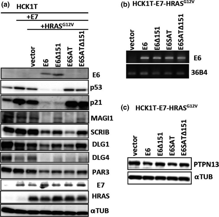
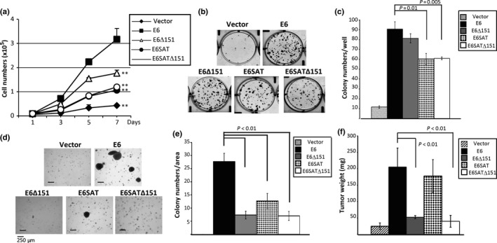
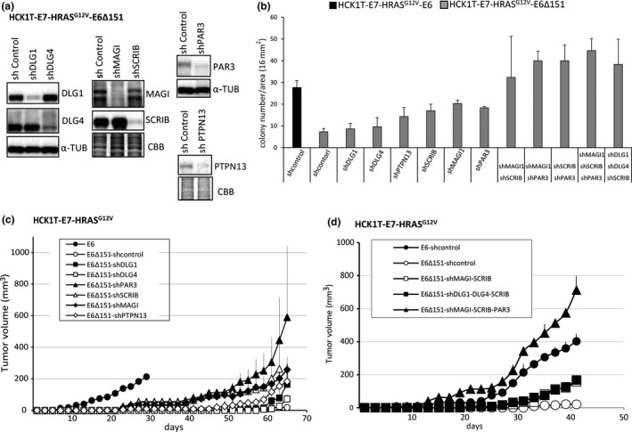
The high‐risk human papillomavirus E6 proteins have been shown to interact with and lead to degradation of PDZ ‐domain‐containing proteins through its carboxy‐terminal motif. This PDZ ‐binding motif plays important roles in transformation of cultured cells and carcinogenesis of E6‐transgenic mice. However, its biological effects on the natural host cells have not been elucidated. We have examined its roles in an in vitro carcinogenesis model for cervical cancer, in which E6 and E7 together with activated HRAS ( HRAS G 12V ) can induce tumorigenic transformation of normal human cervical keratinocytes. In this model, E6Δ151 mutant, which is defective in binding to PDZ domains, almost lost tumorigenic ability, whereas E6 SAT mutant, which is defective in p53 degradation showed activity close to wild‐type E6. Interestingly, we found decreased expression of PAR 3 in E6‐expressing cells independently of E6 AP , which has not been previously recognized. Therefore, we knocked down several PDZ ‐domain containing proteins including PAR 3 in human cervical keratinocytes expressing E7, HRAS G 12V and E6Δ151 to examine whether depletion of these proteins can restore the tumorigenic ability. Single knockdown of SCRIB , MAGI 1 or PAR 3 significantly but partially restored the tumorigenic ability. The combinatorial knockdown of SCRIB and MAGI 1 cooperatively restored the tumorigenic ability, and additional depletion of PAR 3 further enhanced the tumorigenic ability surpassing that induced by wild‐type E6. These data highlight the importance of the carboxy‐terminal motif of the E6 protein and downregulation of PAR 3 in tumorigenic transformation of human cervical keratinocytes.

Overview

The claims and statistics of this paper haven’t been checked yet.

Run the check — free, no account needed. It reads the open-access text and checks the sources it cites.

A few free checks a day · papers already verified open instantly · results are public

Integrity signals

Registry

Crossref · 10.1111/cas.13264

Cites

2 of 2 references verified0 citation errors · 0 not checkable

Statistics

Figures

Figure 1 Establishment of HCKT‐E7‐HRASG 12V expressing the wild type HPV16E6 or its mutants defective for degradation of p53 and/or PDZ domain containing proteins. Expression of HPV16E7, HRAS mutant, HRASG 12V, and HPV16E6 or its mutants was introduced to HCK1T cells by retrovirus mediated transduction as described in Materials and Method. The levels of the wild type 16E6 and its mutant were examined by Western blots (a) and RT‐PCR (b). (a) The mouse monoclonal antibody for HPV16E6 was raised against 16 amino acids of its N‐terminal region and therefore does not react with HPV16E6SAT which contains R8S/P9A/R10T substitution. While the wild type E6 and E6Δ151 which lacks its C‐terminal amino acid induced p53 degradation, E6 mutants with SAT substituions such as E6SAT and E6SATΔ151 did not do so. The levels of PDZ domain containing proteins, such as Scribble (SCRIB), DLG4, MAGI‐1 (MAGI) and PAR3 were decreased in wild‐type E6, but not E6Δ151 or E6SATΔ151 expressing cells. The level of HPV16E7 and HRAS were not affected by the expression of E6. α‐tubulin was detected as a loading control. (b) mRNA levels of the wild type E6 and its mutants were comparable in those cells. 36B4 mRNA was also detected as an internal control. (c) The level of PTPN13 was compared in indicated cells by Western blotting.
Figure 1 Establishment of HCKT‐E7‐HRASG 12V expressing the wild type HPV16E6 or its mutants defective for degradation of p53 and/or PDZ domain containing proteins. Expression of HPV16E7, HRAS mutant, HRASG 12V, and HPV16E6 or its mutants was introduced to HCK1T cells by retrovirus mediated transduction as described in Materials and Method. The levels of the wild type 16E6 and its mutant were examined by Western blots (a) and RT‐PCR (b). (a) The mouse monoclonal antibody for HPV16E6 was raised against 16 amino acids of its N‐terminal region and therefore does not react with HPV16E6SAT which contains R8S/P9A/R10T substitution. While the wild type E6 and E6Δ151 which lacks its C‐terminal amino acid induced p53 degradation, E6 mutants with SAT substituions such as E6SAT and E6SATΔ151 did not do so. The levels of PDZ domain containing proteins, such as Scribble (SCRIB), DLG4, MAGI‐1 (MAGI) and PAR3 were decreased in wild‐type E6, but not E6Δ151 or E6SATΔ151 expressing cells. The level of HPV16E7 and HRAS were not affected by the expression of E6. α‐tubulin was detected as a loading control. (b) mRNA levels of the wild type E6 and its mutants were comparable in those cells. 36B4 mRNA was also detected as an internal control. (c) The level of PTPN13 was compared in indicated cells by Western blotting.CC BY-NC · PubMed Central
Figure 2 Tumorigenic potentials of HCK1T‐E7‐HRASG 12V expressing the wild type or E6 mutants defective for inducing degradation of p53 and/or PDZ domain containing proteins. The ability of E6 mutants to promote cell proliferation was compared to the wild type by examining growth curve (a) and clonogenic potential (b and c). HCK1T‐E7‐HRASG 12V cells transduced with retrovirus containing an empty vector (vector) or expressing the wild type or mutants of E6 were seeded at 2× 10⁴ cells/well in 12 well plates at day 0 and cell number in each well was counted at indicated time points. ** indicates P‐value < 0.05 compared to the wild type E6 expressing cells. (b, c) The cells were seeded on 35‐mm dishes under sparse conditions. After cultivating for 2 weeks, the cells were stained with Giemsa's dye, and number of colonies was counted. The photographs are representative dishes (b), and the graph illustrates means + SDs (c). Anchorage independent growth of HCK1T‐E7‐HRASG 12V expressing wild type or mutants of E6 was examined (d, e). The cells were seeded onto soft agar at 5 × 10⁴ and cultivated for 4 weeks. The representative images are shown (d). Colonies whose size was >50 μm in diameter were counted and the total number of colonies in a 16 mm2 area was compared (e). Tumor promoting potentials of the E6 mutants were compared to the wild type by mouse xenografts. 1 × 10⁶ cells mixed with Matrigel were subcutaneously injected into nude mice. Mice were sacrificed at 40 days after the injection and tumor weights were compared (f). P‐value was evaluated by student's t‐test.
Figure 2 Tumorigenic potentials of HCK1T‐E7‐HRASG 12V expressing the wild type or E6 mutants defective for inducing degradation of p53 and/or PDZ domain containing proteins. The ability of E6 mutants to promote cell proliferation was compared to the wild type by examining growth curve (a) and clonogenic potential (b and c). HCK1T‐E7‐HRASG 12V cells transduced with retrovirus containing an empty vector (vector) or expressing the wild type or mutants of E6 were seeded at 2× 10⁴ cells/well in 12 well plates at day 0 and cell number in each well was counted at indicated time points. ** indicates P‐value < 0.05 compared to the wild type E6 expressing cells. (b, c) The cells were seeded on 35‐mm dishes under sparse conditions. After cultivating for 2 weeks, the cells were stained with Giemsa's dye, and number of colonies was counted. The photographs are representative dishes (b), and the graph illustrates means + SDs (c). Anchorage independent growth of HCK1T‐E7‐HRASG 12V expressing wild type or mutants of E6 was examined (d, e). The cells were seeded onto soft agar at 5 × 10⁴ and cultivated for 4 weeks. The representative images are shown (d). Colonies whose size was >50 μm in diameter were counted and the total number of colonies in a 16 mm2 area was compared (e). Tumor promoting potentials of the E6 mutants were compared to the wild type by mouse xenografts. 1 × 10⁶ cells mixed with Matrigel were subcutaneously injected into nude mice. Mice were sacrificed at 40 days after the injection and tumor weights were compared (f). P‐value was evaluated by student's t‐test.CC BY-NC · PubMed Central
Figure 3 The effect of knock‐down of PDZ domain containing proteins on the tumorigenicity of HCK1T‐E7‐HRASG 12V expressing E6Δ151. Retrovirus expressing shRNA to the indicated PDZ‐ domain containing proteins was infected to HCK1T‐E7‐HRASG 12V expressing E6Δ151 at (a, c). At least two independent shRNA to each PDZ‐containing protein were tested for their efficiency to knockdown target proteins and the most efficient shRNA was used for further analysis. A retrovirus expressing shRNA to luciferase was used as a control. For knockdown of multiple target proteins, three retroviruses each expressing shRNA to either PDZ proteins or luciferases were simultaneously infected to the HCK1T‐E7‐HRASG 12V expressing E6Δ151 at MOI 5 (b, d). (a) Western blot analysis showing knockdown efficiency of shRNA to the indicated target proteins used in the following analysis. α‐tubulin was also detected as a loading control. CBB staining was performed to indicate equal loading. (b) Anchorage independent growth of HCK1T‐E7‐HRASG 12V expressing E6Δ151 with shRNA either to control or PDZ containing proteins was analyzed compared to HCK1T‐E7‐HRASG 12V expressing wild type E6 with control shRNA. (c) Tumor promoting potentials of HCK1T‐E7‐HRASG 12V expressing E6Δ151 with knockdown of single PDZ protein was compared to HCK1T‐E7‐HRASG 12V expressing the wild type E6 by mouse xenograft. The size of tumor was measured at the indicated time points. (d) The effect of knockdown multiple PDZ containing proteins on tumorigenicity of HCK1T‐E7‐HRASG 12V expressing E6Δ151 was also analyzed. Error bar indicates SD. P‐value was evaluated by student's t‐test.
Figure 3 The effect of knock‐down of PDZ domain containing proteins on the tumorigenicity of HCK1T‐E7‐HRASG 12V expressing E6Δ151. Retrovirus expressing shRNA to the indicated PDZ‐ domain containing proteins was infected to HCK1T‐E7‐HRASG 12V expressing E6Δ151 at (a, c). At least two independent shRNA to each PDZ‐containing protein were tested for their efficiency to knockdown target proteins and the most efficient shRNA was used for further analysis. A retrovirus expressing shRNA to luciferase was used as a control. For knockdown of multiple target proteins, three retroviruses each expressing shRNA to either PDZ proteins or luciferases were simultaneously infected to the HCK1T‐E7‐HRASG 12V expressing E6Δ151 at MOI 5 (b, d). (a) Western blot analysis showing knockdown efficiency of shRNA to the indicated target proteins used in the following analysis. α‐tubulin was also detected as a loading control. CBB staining was performed to indicate equal loading. (b) Anchorage independent growth of HCK1T‐E7‐HRASG 12V expressing E6Δ151 with shRNA either to control or PDZ containing proteins was analyzed compared to HCK1T‐E7‐HRASG 12V expressing wild type E6 with control shRNA. (c) Tumor promoting potentials of HCK1T‐E7‐HRASG 12V expressing E6Δ151 with knockdown of single PDZ protein was compared to HCK1T‐E7‐HRASG 12V expressing the wild type E6 by mouse xenograft. The size of tumor was measured at the indicated time points. (d) The effect of knockdown multiple PDZ containing proteins on tumorigenicity of HCK1T‐E7‐HRASG 12V expressing E6Δ151 was also analyzed. Error bar indicates SD. P‐value was evaluated by student's t‐test.CC BY-NC · PubMed Central
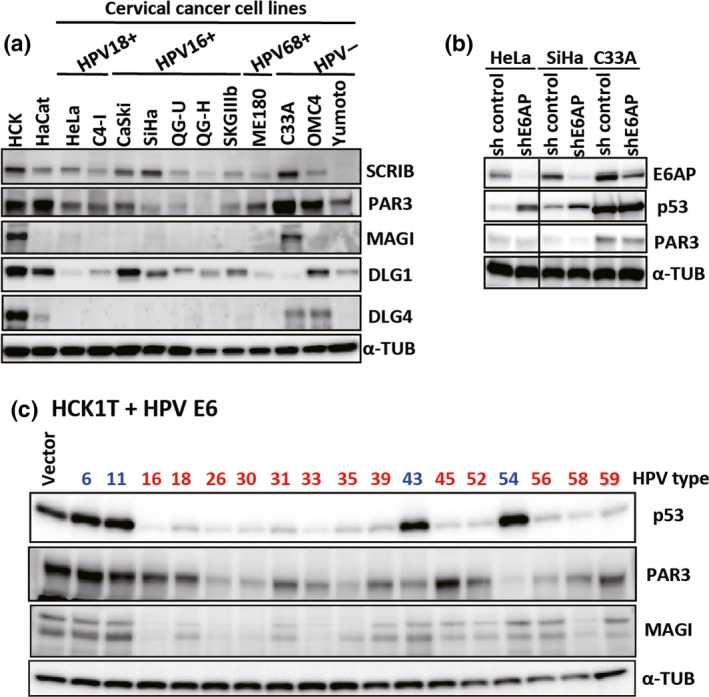
Figure 4 The protein levels of PDZ containing proteins in cervical cancer cell lines. (a) The levels of PDZ containing proteins in HPV positive‐cervical cancer cell lines were compared to that in HPV negative‐cervical cancer cell lines or normal human keratinocytes by Western blot analysis. HaCaT cells are spontaneously immortalized human foreskin keratinocytes. (b) The effect of shRNA to E6AP on the level of PAR3 in cervical cancer cells were examined by Western blot analysis. HeLa, SiHa and C33a cells with E6AP shRNA was generated as described previously. p53 was included as a positive control because an E6 and E6AP complex was known to induce degradation of p53. α‐tubulin was also detected as a loading control. (c) The ability of E6 to induce reduction of PAR3 level was compared between high and low risk HPVs. The expression E6 of high risk HPVs shown in red (HPV16, 18, 26, 30, 31, 33, 35, 39, 45, 52, 56, 58 and 59 or low‐risk HPVs shown in blue (HPV6, 11, 43 and 54) was introduced to HCK1T by retrovirus mediated transduction. A retrovirus carrying empty vector was used as a control (vector). The levels of p53, MAGI‐1 and PAR3 were examined by Western blot analysis.
Figure 4 The protein levels of PDZ containing proteins in cervical cancer cell lines. (a) The levels of PDZ containing proteins in HPV positive‐cervical cancer cell lines were compared to that in HPV negative‐cervical cancer cell lines or normal human keratinocytes by Western blot analysis. HaCaT cells are spontaneously immortalized human foreskin keratinocytes. (b) The effect of shRNA to E6AP on the level of PAR3 in cervical cancer cells were examined by Western blot analysis. HeLa, SiHa and C33a cells with E6AP shRNA was generated as described previously. p53 was included as a positive control because an E6 and E6AP complex was known to induce degradation of p53. α‐tubulin was also detected as a loading control. (c) The ability of E6 to induce reduction of PAR3 level was compared between high and low risk HPVs. The expression E6 of high risk HPVs shown in red (HPV16, 18, 26, 30, 31, 33, 35, 39, 45, 52, 56, 58 and 59 or low‐risk HPVs shown in blue (HPV6, 11, 43 and 54) was introduced to HCK1T by retrovirus mediated transduction. A retrovirus carrying empty vector was used as a control (vector). The levels of p53, MAGI‐1 and PAR3 were examined by Western blot analysis.CC BY-NC · PubMed Central

Verification

Claims the paper makes

The claims and statistics of this paper haven’t been checked yet.

Run the check — free, no account needed. It reads the open-access text and checks the sources it cites.

A few free checks a day · papers already verified open instantly · results are public

Claims other papers make about this one

Each is checked against this paper’s own text: does it say what the citing paper says it does?

Self-check

Percentages, sums and reported differences re-computed from the paper’s own numbers.

The self-check needs the paper’s full text.

Citations

Cites

Research works printed but not linked: 32

  1. 10.1073/pnas.94.21.11612 · DOI · Paper page
  2. 10.1073/pnas.94.13.6670 · DOI · Paper page
  3. 10.1111/j.1349-7006.2006.00315.x · DOI
  4. 10.1128/JVI.01712-06 · DOI
  5. 10.1038/sj.onc.1203906 · DOI · Paper page
  6. 10.1128/JVI.02188-07 · DOI
  7. 10.1128/JVI.77.12.6957-6964.2003 · DOI
  8. 10.1016/j.abb.2007.07.027 · DOI
  9. 10.1073/pnas.97.8.4233 · DOI
  10. 10.1242/jcs.00809 · DOI
  11. 10.1128/JVI.78.22.12366-12377.2004 · DOI · Paper page
  12. 10.1073/pnas.87.20.8170 · DOI
  13. 10.1128/JVI.00233-07 · DOI
  14. 10.1038/onc.2008.365 · DOI
  15. 10.1158/0008-5472.CAN-07-6862 · DOI
  16. 10.1093/carcin/bgs104 · DOI
  17. 10.1016/S0006-291X(02)00661-7 · DOI
  18. 10.1038/sj.onc.1210118 · DOI
  19. 10.1128/MCB.02119-06 · DOI
  20. 10.1016/j.bbrc.2016.07.025 · DOI
  21. 10.1073/pnas.1202684109 · DOI
  22. 10.1074/jbc.M605117200 · DOI · Paper page
  23. 10.1128/JVI.01979-06 · DOI
  24. 10.1016/j.molonc.2014.01.002 · DOI
  25. 10.1128/JVI.01756-10 · DOI
  26. 10.1371/journal.ppat.1004980 · DOI
  27. 10.1002/jcb.20383 · DOI
  28. 10.1016/j.ceb.2009.07.003 · DOI
  29. 10.1126/science.1096096 · DOI
  30. 10.1038/sj.onc.1203374 · DOI
  31. 10.1128/JVI.02545-06 · DOI
  32. 10.1099/vir.0.80035-0 · DOI

Metadata

Authors
Yoshimatsu, Y., Nakahara, T., Tanaka, K., Inagawa, Y., Narisawa‐Saito, M., Yugawa, T., Ohno, S., Fujita, M., Nakagama, H., & Kiyono, T.
Affiliations
  • Division of Carcinogenesis and Cancer Prevention National Cancer Center Research Institute Tokyo Japan
  • Department of Pathology Graduate School of Medicine University of Tokyo Tokyo Japan
Venue
Cancer Science
Publisher
Wiley
Published
2017-06-05
Volume
108
Issue
7
Pages
1303-1309
ISSN
  • 1347-9032
  • 1349-7006
Cited by
44
References
2
Language
en
Funders
Japan Agency for Medical Research and Development (17ck0106178h0003)

About these checks

Registry, retraction, Cites and statistics results are computed, not judged, with one exception: a printed reference that no index lists is read once by a model before it counts as a citation error. Claims are extracted by a model and kept only when their quote appears verbatim in the text; each cited source is then checked, with a model’s help, for whether it says what the claim relies on. Those verdicts can be wrong — read the claim’s quote and the cited source before relying on one.

Statistics and arithmetic are recomputed, not judged.

Confidence bands are shown only for fields calibrated on our annotated test set. None is calibrated yet, so no band is shown.

What counts as a claim ›How we measure confidence ›How we check this ›

Are you an author, or do you see a problem with this page? Request a review