Cited by 221

The DOI matches its Crossref record.

Sec16 Defines Endoplasmic Reticulum Exit Sites and is Required for Secretory Cargo Export in Mammalian Cells

Watson, Peter, Townley, Anna K., Koka, Pratyusha, Palmer, Krysten J., Stephens, David J.·2006·Traffic

Abstract

The selective export of proteins and lipids from the endoplasmic reticulum (ER) is mediated by the coat protein complex II (COPII) that assembles onto the ER membrane. In higher eukaryotes, COPII proteins assemble at discrete sites on the membrane known as ER exit sites (ERES). Here, we identify Sec16 as the protein that defines ERES in mammalian cells. Sec16 localizes to ERES independent of Sec23/24 and Sec13/31. Overexpression, and to a lesser extent, small interfering RNA depletion of Sec16, both inhibit ER‐to‐Golgi transport suggesting that Sec16 is required in stoichiometric amounts. Sar1 activity is required to maintain the localization of Sec16 at discrete locations on the ER membrane, probably through preventing its dissociation. Our data suggest that Sar1‐GTP‐dependent assembly of Sec16 on the ER membrane forms an organized scaffold defining an ERES.

Overview

The claims and statistics of this paper haven’t been checked yet.

Run the check — free, no account needed. It reads the open-access text and checks the sources it cites.

A few free checks a day · papers already verified open instantly · results are public

Integrity signals

Registry

Crossref · 10.1111/j.1600-0854.2006.00493.x

Cites

Statistics

Figures

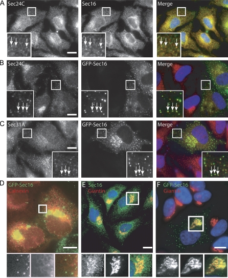
Figure 1 Localization of human Sec16 A) Immunofluorescence labelling of HeLa cells with anti-Sec16 (red in merge) and anti-Sec24C (green) antibodies. B and C) HeLa cells expressing EGFP-Sec16 (green in merge) at low level were methanol fixed and labelled with antibodies specific for B) Sec24C (red) or C) Sec31A (red). D) Venus-Sec16 (green in merge) puncta aligned on filaments of the ER membrane labelled with ECFP-ER (red). E and F) Endogenous Sec16 labelled using E) anti-Sec16 antibodies or F) EGFP-Sec16 in transfected cells (both green in merges) were localized in fixed cells and co-labelled with anti-giantin antibodies to reveal the close apposition of Sec16 puncta and Golgi membranes (red). Bars = 10 μm.
Figure 1 Localization of human Sec16 A) Immunofluorescence labelling of HeLa cells with anti-Sec16 (red in merge) and anti-Sec24C (green) antibodies. B and C) HeLa cells expressing EGFP-Sec16 (green in merge) at low level were methanol fixed and labelled with antibodies specific for B) Sec24C (red) or C) Sec31A (red). D) Venus-Sec16 (green in merge) puncta aligned on filaments of the ER membrane labelled with ECFP-ER (red). E and F) Endogenous Sec16 labelled using E) anti-Sec16 antibodies or F) EGFP-Sec16 in transfected cells (both green in merges) were localized in fixed cells and co-labelled with anti-giantin antibodies to reveal the close apposition of Sec16 puncta and Golgi membranes (red). Bars = 10 μm.CC BY · PubMed Central
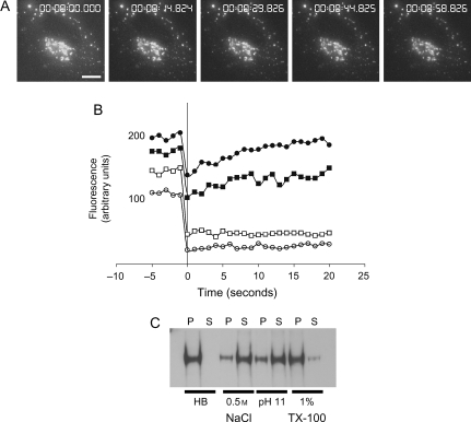
Figure 2 Dynamics and membrane association of Sec16 A) Time-lapse imaging of cells expressing GFP-Sec16; 10 images/second were acquired at 1 μm spacing in z and are displayed as extended focus images. In these movies of whole cell depth at 1 frame/second, we cannot identify any fast-moving punctate structures consistent with what we observe with other EYFP-tagged COPII subunits. Bar = 10 μm. B) Fluorescence recovery after photobleaching of cells expressing low (solid symbols) or high (open symbols) levels of EYFP-Sec16. Individual examples are shown, the half-life for fluorescence recovery was in the range 4.3–26.7 seconds, the mobile fraction was 80–100% (20 structures from 6 cells, two independent experiments, half-lives were calculated using Leica TCS software). Variability in expression level and the size of the mobile fraction probably reflects differences in expression level between cells. C) Ad293 cells were fractionated as described and membranes extracted with buffers [homogenization buffer containing no supplements (HB), 0.5 m NaCl, 0.1 m sodium carbonate, pH 11 or 1% TX-100] as indicated before separation into pellet (P) and supernatant (S) at 25 000 × g.
Figure 2 Dynamics and membrane association of Sec16 A) Time-lapse imaging of cells expressing GFP-Sec16; 10 images/second were acquired at 1 μm spacing in z and are displayed as extended focus images. In these movies of whole cell depth at 1 frame/second, we cannot identify any fast-moving punctate structures consistent with what we observe with other EYFP-tagged COPII subunits. Bar = 10 μm. B) Fluorescence recovery after photobleaching of cells expressing low (solid symbols) or high (open symbols) levels of EYFP-Sec16. Individual examples are shown, the half-life for fluorescence recovery was in the range 4.3–26.7 seconds, the mobile fraction was 80–100% (20 structures from 6 cells, two independent experiments, half-lives were calculated using Leica TCS software). Variability in expression level and the size of the mobile fraction probably reflects differences in expression level between cells. C) Ad293 cells were fractionated as described and membranes extracted with buffers [homogenization buffer containing no supplements (HB), 0.5 m NaCl, 0.1 m sodium carbonate, pH 11 or 1% TX-100] as indicated before separation into pellet (P) and supernatant (S) at 25 000 × g.CC BY · PubMed Central
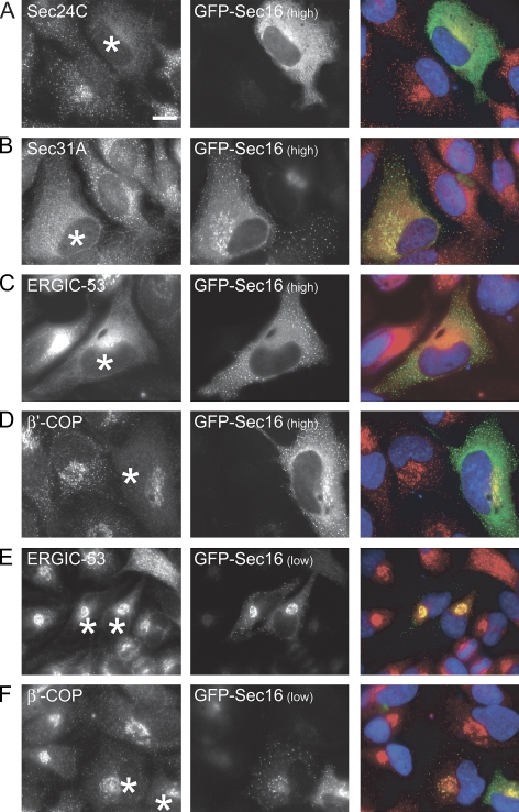
Figure 3 Sec16 is required in stoichiometric amounts for COPII assembly and ER-to-Golgi transport Cells expressing EGFP-Sec16 at high (punctate and cytosolic, A–D) or low levels (exclusively punctate, E and F) were labelled with antibodies directed against A) Sec24C, B) Sec31A, C and E) ERGIC-53 or D and F) β’-COP. Cells expressing EGFP-Sec16 are marked with asterisks. Note loss of Sec24C, Sec31A and β’-COP labelling in cells expressing a high level of EGFP-Sec16; ERGIC-53 is redistributed to the ER in these cells. Bars = 10 μm.
Figure 3 Sec16 is required in stoichiometric amounts for COPII assembly and ER-to-Golgi transport Cells expressing EGFP-Sec16 at high (punctate and cytosolic, A–D) or low levels (exclusively punctate, E and F) were labelled with antibodies directed against A) Sec24C, B) Sec31A, C and E) ERGIC-53 or D and F) β’-COP. Cells expressing EGFP-Sec16 are marked with asterisks. Note loss of Sec24C, Sec31A and β’-COP labelling in cells expressing a high level of EGFP-Sec16; ERGIC-53 is redistributed to the ER in these cells. Bars = 10 μm.CC BY · PubMed Central
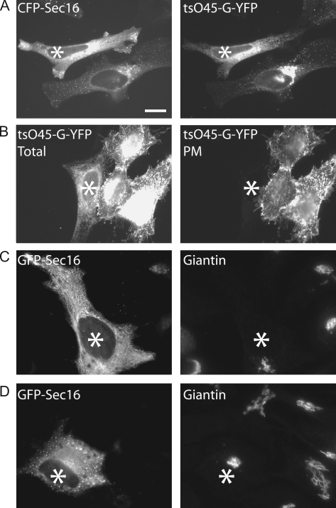
Figure 4 Inhibition of ER-to-Golgi transport following overexpression of CFP-Sec16 Cells expressing high levels of CFP-Sec16 were infected with Adenovirus to express tsO45-G-YFP at 39.5°C. Cells were then shifted to 32°C for A) 30 min or B) 90 min. A) In cells expressing a low level of CFP-Sec16, tsO45-G-YFP accumulates in the Golgi apparatus 30 min following shift to 32°C, while in cells expressing a high level of CFP-Sec16 (asterisk), tsO45-G-YFP remains within the ER. B) Cells incubated at 32°C for 90 min were paraformaldehyde fixed and immunolabelled to detect tsO45-G-YFP that reaches the PM. In cells expressing a high level of CFP-Sec16 (asterisk), tsO45-G-YFP remains within the ER (total) and does not reach the PM. C and D) Cells expressing GFP-Sec16 (asterisks) do not have significantly disrupted Golgi apparatus as determined by giantin localization (right hand panels). Bar (all panels) = 10 μm.
Figure 4 Inhibition of ER-to-Golgi transport following overexpression of CFP-Sec16 Cells expressing high levels of CFP-Sec16 were infected with Adenovirus to express tsO45-G-YFP at 39.5°C. Cells were then shifted to 32°C for A) 30 min or B) 90 min. A) In cells expressing a low level of CFP-Sec16, tsO45-G-YFP accumulates in the Golgi apparatus 30 min following shift to 32°C, while in cells expressing a high level of CFP-Sec16 (asterisk), tsO45-G-YFP remains within the ER. B) Cells incubated at 32°C for 90 min were paraformaldehyde fixed and immunolabelled to detect tsO45-G-YFP that reaches the PM. In cells expressing a high level of CFP-Sec16 (asterisk), tsO45-G-YFP remains within the ER (total) and does not reach the PM. C and D) Cells expressing GFP-Sec16 (asterisks) do not have significantly disrupted Golgi apparatus as determined by giantin localization (right hand panels). Bar (all panels) = 10 μm.CC BY · PubMed Central
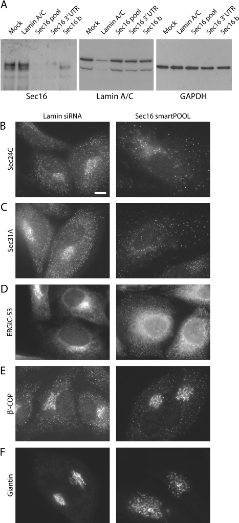
Figure 5 Depletion of Sec16 from human cells disrupts COPII assembly and ER-to-Golgi transport A) Depletion of Sec16 expression using siRNA. HeLa cells were transfected with siRNA duplexes targeting Sec16 [pool of four duplexes (Sec16 pool), lamin A/C, a duplex targeting the 3′ UTR of Sec16 (Sec16 3′ UTR) and a further Sec16 duplex targeting the coding region (Sec16 b)]. Cell lysates were then immunoblotted for Sec16 (250 kD), lamin A/C (84 kD) or GAPDH (as a loading control, 40 kD) as indicated. Note that the duplex termed Sec16b only depletes a subset of splice forms of Sec16. Cells depleted of Sec16 using a pool of four siRNA duplexes were methanol fixed and labelled with antibodies specific for B) Sec24C, C) Sec31A, D) ERGIC-53, E) β’-COP and F) giantin as indicated. Bar (all panels) = 10 μm.
Figure 5 Depletion of Sec16 from human cells disrupts COPII assembly and ER-to-Golgi transport A) Depletion of Sec16 expression using siRNA. HeLa cells were transfected with siRNA duplexes targeting Sec16 [pool of four duplexes (Sec16 pool), lamin A/C, a duplex targeting the 3′ UTR of Sec16 (Sec16 3′ UTR) and a further Sec16 duplex targeting the coding region (Sec16 b)]. Cell lysates were then immunoblotted for Sec16 (250 kD), lamin A/C (84 kD) or GAPDH (as a loading control, 40 kD) as indicated. Note that the duplex termed Sec16b only depletes a subset of splice forms of Sec16. Cells depleted of Sec16 using a pool of four siRNA duplexes were methanol fixed and labelled with antibodies specific for B) Sec24C, C) Sec31A, D) ERGIC-53, E) β’-COP and F) giantin as indicated. Bar (all panels) = 10 μm.CC BY · PubMed Central
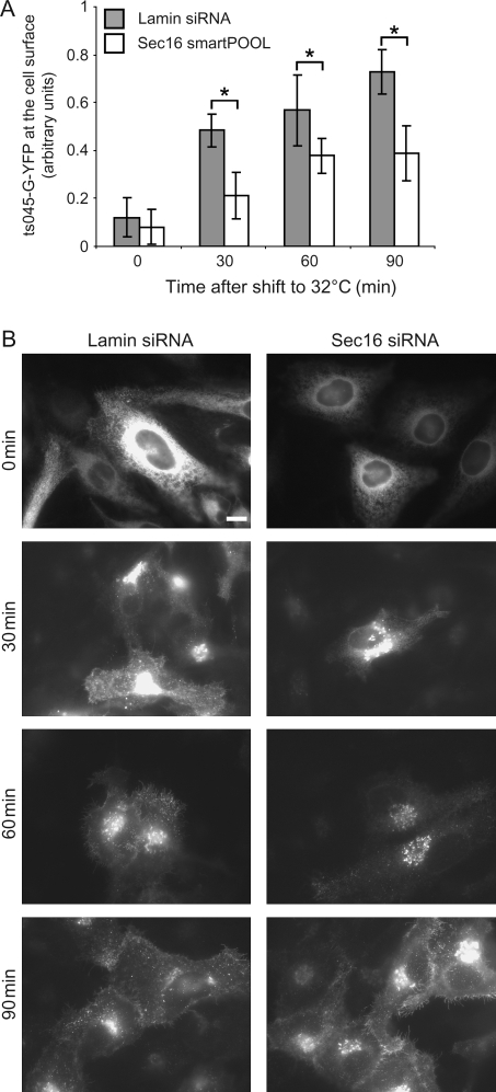
Figure 6 A) Cells depleted of either Lamin A/C or Sec16 were infected with Adenovirus to express tsO45-G-YFP at 39.5°C followed by incubation at 32°C for the times indicated Cells were depleted of either Lamin A/C (filled bars) or Sec16 (using a pool of four duplexes, open bars). The amount of tsO45-G-YFP reaching the cell surface was quantitated for each time-point. tsO45-G-YFP shows a significant (anova, * p = 0.04) delay in reaching the PM in cells depleted of Sec16. B) Seventy-two hours after transfection with siRNA duplexes, cells were infected with Adenovirus to express tsO45-G-YFP at 39°C followed by shift to 32°C for the times indicated and quantification of the amount of tsO45-G-YFP reaching the PM. Bar (all panels) = 10 μm.
Figure 6 A) Cells depleted of either Lamin A/C or Sec16 were infected with Adenovirus to express tsO45-G-YFP at 39.5°C followed by incubation at 32°C for the times indicated Cells were depleted of either Lamin A/C (filled bars) or Sec16 (using a pool of four duplexes, open bars). The amount of tsO45-G-YFP reaching the cell surface was quantitated for each time-point. tsO45-G-YFP shows a significant (anova, * p = 0.04) delay in reaching the PM in cells depleted of Sec16. B) Seventy-two hours after transfection with siRNA duplexes, cells were infected with Adenovirus to express tsO45-G-YFP at 39°C followed by shift to 32°C for the times indicated and quantification of the amount of tsO45-G-YFP reaching the PM. Bar (all panels) = 10 μm.CC BY · PubMed Central
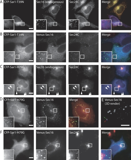
Figure 7 Sar1-GTP mediates assembly of Sec16 at ERES A) Cells expressing ECFP-Sar1-T39N (GDP restricted, blue in merge) were labelled with antibodies to detect endogenous Sec16 (green) and Sec24C (red). The majority of punctate Sec16 localization is lost; those few punctate spots of Sec16 that remain also label with anti-Sec24C antibodies, as they do in non-transfected cells. B) Cells were transfected with both ECFP-Sar1-T39N (blue in merge) and Venus-Sec16 (green) and immunolabelled for endogenous Sec24C (red). C) Cells transfected with ECFP-Sar1-H79G (blue in merge) were labelled for endogenous Sec16 (green) and Sec24C (red). Sec24C is not present on Sec16-positive structures (arrowheads) D) 3D imaging of paraformaldehyde-fixed cells expressing both ECFP-Sar1-H79G (green in merge) and Venus-Sec16 (red) shows that both proteins localize to large curved and round structures. E) 3D rendering of deconvolved stacks from cells expressing both ECFP-Sar1-H79G and Venus-Sec16 reveal that these objects often form near complete spheres. F) Immunolabelling of ECFP-Sar1-H79G (blue in merge) and Venus-Sec16 (green) labelled structures shows that they do not contain Sec24C (red). Boxed regions show threefold enlargements of selected areas. Representative images are shown from four independent experiments in which a total of 400 cells were inspected visually. All bars = 10 μm.
Figure 7 Sar1-GTP mediates assembly of Sec16 at ERES A) Cells expressing ECFP-Sar1-T39N (GDP restricted, blue in merge) were labelled with antibodies to detect endogenous Sec16 (green) and Sec24C (red). The majority of punctate Sec16 localization is lost; those few punctate spots of Sec16 that remain also label with anti-Sec24C antibodies, as they do in non-transfected cells. B) Cells were transfected with both ECFP-Sar1-T39N (blue in merge) and Venus-Sec16 (green) and immunolabelled for endogenous Sec24C (red). C) Cells transfected with ECFP-Sar1-H79G (blue in merge) were labelled for endogenous Sec16 (green) and Sec24C (red). Sec24C is not present on Sec16-positive structures (arrowheads) D) 3D imaging of paraformaldehyde-fixed cells expressing both ECFP-Sar1-H79G (green in merge) and Venus-Sec16 (red) shows that both proteins localize to large curved and round structures. E) 3D rendering of deconvolved stacks from cells expressing both ECFP-Sar1-H79G and Venus-Sec16 reveal that these objects often form near complete spheres. F) Immunolabelling of ECFP-Sar1-H79G (blue in merge) and Venus-Sec16 (green) labelled structures shows that they do not contain Sec24C (red). Boxed regions show threefold enlargements of selected areas. Representative images are shown from four independent experiments in which a total of 400 cells were inspected visually. All bars = 10 μm.CC BY · PubMed Central
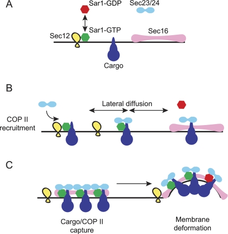
Figure 8 Model to describe Sec16 function in mammalian cells A) The small GTPase Sar1-GDP (red) can be recruited to the ER membrane by Sec12 (its guanine nucleotide exchange factor, yellow). Cargo proteins (blue) diffuse through the ER membrane and Sec16 assemblies (light magenta) are present at ERES. B) Sar1-GTP can bind to some cargo molecules and recruits Sec23/24 (cyan). Complexes of Sar1-GTP-cargo-Sec23/24 can diffuse within the plane of the ER membrane. GTP turnover on Sar1 could lead to its dissociation here while cargo and Sec23/24 remain associated. C) Sec16 assemblies could act to capture and stabilize cargo-Sec23/24 complexes either in the presence or absence of Sar1. Sec16 acts to concentrate Sar1-GTP-cargo-Sec23/24 pre-budding complexes such that membrane deformation occurs. See text for further details.
Figure 8 Model to describe Sec16 function in mammalian cells A) The small GTPase Sar1-GDP (red) can be recruited to the ER membrane by Sec12 (its guanine nucleotide exchange factor, yellow). Cargo proteins (blue) diffuse through the ER membrane and Sec16 assemblies (light magenta) are present at ERES. B) Sar1-GTP can bind to some cargo molecules and recruits Sec23/24 (cyan). Complexes of Sar1-GTP-cargo-Sec23/24 can diffuse within the plane of the ER membrane. GTP turnover on Sar1 could lead to its dissociation here while cargo and Sec23/24 remain associated. C) Sec16 assemblies could act to capture and stabilize cargo-Sec23/24 complexes either in the presence or absence of Sar1. Sec16 acts to concentrate Sar1-GTP-cargo-Sec23/24 pre-budding complexes such that membrane deformation occurs. See text for further details.CC BY · PubMed Central

Verification

Claims the paper makes

The claims and statistics of this paper haven’t been checked yet.

Run the check — free, no account needed. It reads the open-access text and checks the sources it cites.

A few free checks a day · papers already verified open instantly · results are public

Claims other papers make about this one

Each is checked against this paper’s own text: does it say what the citing paper says it does?

Self-check

Percentages, sums and reported differences re-computed from the paper’s own numbers.

The self-check needs the paper’s full text.

Citations

Cites

No references could be resolved yet.

Research works printed but not linked: 40

  1. 10.1016/0092-8674(94)90138-4 · DOI
  2. 10.1016/S0014-5793(02)03068-5 · DOI
  3. 10.1073/pnas.88.19.8611 · DOI
  4. 10.1083/jcb.135.1.19 · DOI
  5. 10.1038/sj.embor.embor736 · DOI
  6. 10.1091/mbc.11.9.3013 · DOI
  7. 10.1242/jcs.113.12.2177 · DOI
  8. 10.1034/j.1600-0854.2001.20704.x · DOI
  9. 10.1083/jcb.125.1.51 · DOI
  10. 10.1091/mbc.7.10.1535 · DOI
  11. 10.1074/jbc.274.12.7833 · DOI
  12. Shugrue CA, 1999, Identification of the putative mammalian orthologue of Sec31P, a component of the COPII coat, J Cell Sci, 112, 4547, 10.1242/jcs.112.24.4547 · DOI
  13. 10.1128/MCB.17.1.256 · DOI
  14. 10.1038/nature04339 · DOI
  15. 10.1091/mbc.7.11.1815 · DOI
  16. 10.1083/jcb.131.2.311 · DOI
  17. 10.1083/jcb.200207053 · DOI
  18. 10.1016/j.cub.2005.06.065 · DOI
  19. 10.1074/jbc.272.41.25413 · DOI
  20. 10.1074/jbc.274.29.20505 · DOI
  21. 10.1016/j.cub.2005.11.076 · DOI
  22. 10.1038/38001 · DOI
  23. 10.1128/jvi.54.2.374-382.1985 · DOI
  24. 10.1093/jb/mvh024 · DOI
  25. 10.1034/j.1600-0854.2002.31108.x · DOI
  26. 10.1242/jcs.01269 · DOI
  27. 10.1038/ncb1371 · DOI
  28. 10.1038/ncb1206 · DOI
  29. 10.1016/j.cell.2005.07.025 · DOI
  30. 10.1083/jcb.200509095 · DOI
  31. 10.1074/jbc.274.7.4389 · DOI
  32. 10.1038/nsmb893 · DOI
  33. 10.1038/sj.emboj.7600428 · DOI
  34. 10.1083/jcb.131.2.325 · DOI
  35. 10.1038/35078500 · DOI
  36. 10.1046/j.1365-2443.2000.00391.x · DOI
  37. 10.1242/jcs.02999 · DOI
  38. 10.1093/dnares/4.2.141 · DOI
  39. 10.1038/nbt0102-87 · DOI
  40. 10.1038/35055042 · DOI · Paper page

Cited by

Sorted by citations

Metadata

Authors
Watson, P., Townley, A. K., Koka, P., Palmer, K. J., & Stephens, D. J.
Venue
Traffic
Publisher
Wiley
Published
2006-10-02
Volume
7
Issue
12
Pages
1678-1687
ISSN
  • 1398-9219
  • 1600-0854
Cited by
221
References
0
Language
en

About these checks

Registry, retraction, Cites and statistics results are computed, not judged, with one exception: a printed reference that no index lists is read once by a model before it counts as a citation error. Claims are extracted by a model and kept only when their quote appears verbatim in the text; each cited source is then checked, with a model’s help, for whether it says what the claim relies on. Those verdicts can be wrong — read the claim’s quote and the cited source before relying on one.

Statistics and arithmetic are recomputed, not judged.

Confidence bands are shown only for fields calibrated on our annotated test set. None is calibrated yet, so no band is shown.

What counts as a claim ›How we measure confidence ›How we check this ›

Are you an author, or do you see a problem with this page? Request a review