

The DOI matches its Crossref record; 27 of the 27 references it cites were found in a registry or an index.
Differentiation and risk stratification of basal cell carcinoma with deep learning on histopathologic images and measuring nuclei and tumor microenvironment features
Open accessAbstract
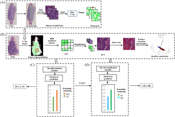
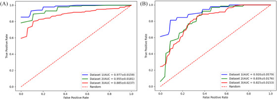
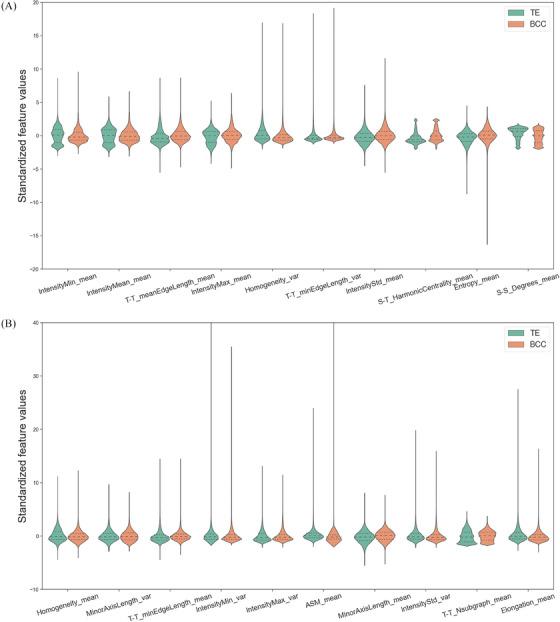
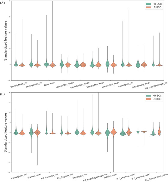
BACKGROUND: Nuclear pleomorphism and tumor microenvironment (TME) play a critical role in cancer development and progression. Identifying most predictive nuclei and TME features of basal cell carcinoma (BCC) may provide insights into which characteristics pathologists can use to distinguish and stratify this entity. OBJECTIVES: To develop an automated workflow based on nuclei and TME features from basaloid cell tumor regions to differentiate BCC from trichoepithelioma (TE) and stratify BCC into high-risk (HR) and low-risk (LR) subtypes, and to identify the nuclear and TME characteristics profile of different basaloid cell tumors. METHODS: The deep learning systems were trained on 161 H&E -stained sections which contained 51 sections of HR-BCC, 50 sections of LR-BCC and 60 sections of TE from one institution (D1), and externally and independently validated on D2 (46 sections) and D3 (76 sections), from 2015 to 2022. 60%, 20% and 20% of D1 data were randomly splitted for training, validation and testing, respectively. The framework comprised four stages: tumor regions identification by multi-head self-attention (MSA) U-Net, nuclei segmentation by HoVer-Net, quantitative feature by handcrafted extraction, and differentiation and risk stratification classifier construction. Pixel accuracy, precision, recall, dice score, intersection over union (IoU) and area under the curve (AUC) were used to evaluate the performance of tumor segmentation model and classifiers. RESULTS: MSA-U-Net model detected tumor regions with 0.910 precision, 0.869 recall, 0.889 dice score and 0.800 IoU. The differentiation classifier achieved 0.977 ± 0.0159, 0.955 ± 0.0181, 0.885 ± 0.0237 AUC in D1, D2 and D3, respectively. The most discriminative features between BCC and TE contained Homogeneity, Elongation, T-T_meanEdgeLength, T-T_Nsubgraph, S-T_HarmonicCentrality, S-S_Degrees. The risk stratification model can well predict HR-BCC and LR-BCC with 0.920 ± 0.0579, 0.839 ± 0.0176, 0.825 ± 0.0153 AUC in D1, D2 and D3, respectively. The most discriminative features between HR-BCC and LR-BCC comprised IntensityMin, Solidity, T-T_minEdgeLength, T-T_Coreness, T-T_Degrees, T-T_Betweenness, S-T_Degrees. CONCLUSIONS: This framework hold potential for future use as a second opinion helping inform diagnosis of BCC, and identify nuclei and TME features related with malignancy and tumor risk stratification.
Overview
The claims and statistics of this paper haven’t been checked yet.
Run the check — free, no account needed. It reads the open-access text and checks the sources it cites.
Integrity signals
Registry
Cites
Statistics
Figures
Verification
Claims the paper makes
The claims and statistics of this paper haven’t been checked yet.
Run the check — free, no account needed. It reads the open-access text and checks the sources it cites.
Claims other papers make about this one
Each is checked against this paper’s own text: does it say what the citing paper says it does?
The cited source says what the claim attributes to it.
In the citing paper
“Using a multi-stage deep learning pipeline to segment tumor and nuclei, Lan et al. extracted interpretable morphologic and microenvironment features to distinguish BCC from trichoepithelioma (TE) and stratify BCC into high- vs. low-risk subtypes, achieving high AUCs across internal and external cohorts [23].”
From Artificial Intelligence in Dermatopathology: An Update and Review of the Current Literature (2026) · cites this paper as [23]
Self-check
Percentages, sums and reported differences re-computed from the paper’s own numbers.
The self-check needs the paper’s full text.
Citations
Cites
Peris, K., Fargnoli, M. C., Garbe, C., Kaufmann, R., Bastholt, L., Basset‐Séguin, N., Bataille, V. A., del Mármol, V., Dummer, R., Harwood, C. A., et al. (2019) · European Journal of Cancer · doi:10.1016/j.ejca.2019.06.003 · Cited by 600
Automatic Nuclei Segmentation in H&E Stained Breast Cancer Histopathology Images
Veta, M., van Diest, P. J., Kornegoor, R., Huisman, A., Viergever, M. A., & Pluim, J. P. W. (2013) · PLoS ONE · doi:10.1371/journal.pone.0070221 · Cited by 370
Bilal, M., Raza, S. E. A., Azam, A., Graham, S., Ilyas, M., Cree, I. A., Snead, D., Minhas, F., & Rajpoot, N. M. (2021) · The Lancet Digital Health · doi:10.1016/s2589-7500(21)00180-1 · Cited by 299
Basal Cell Carcinoma: A Comprehensive Review
Dika, E., Scarfì, F., Ferracin, M., Broseghini, E., Marcelli, E., Bortolani, B., Campione, E., Riefolo, M., Ricci, C., & Lambertini, M. (2020) · International Journal of Molecular Sciences · doi:10.3390/ijms21155572 · Cited by 205
Thomas, S. M., Lefevre, J., Baxter, G. D., & Hamilton, N. (2020) · Medical Image Analysis · doi:10.1016/j.media.2020.101915 · Cited by 170
SnapShot: Cancer-Associated Fibroblasts
Park, D., Sahai, E. A., & Rullan, A. (2020) · Cell · doi:10.1016/j.cell.2020.03.013 · Cited by 162
Diagnosis and Management of Basal Cell Carcinoma
Tanese, K. (2019) · Current Treatment Options in Oncology · doi:10.1007/s11864-019-0610-0 · Cited by 161
Histologic Mimics of Basal Cell Carcinoma
Stanoszek, L. M., Wang, G., & Harms, P. W. (2017) · Archives of Pathology & Laboratory Medicine · doi:10.5858/arpa.2017-0222-ra · Cited by 124
Niculeț, E., Crăescu, M., Rebegea, L. F., Bobeică, C., Năstase, F., Lupăşteanu, G., Stan, D. L., Chioncel, V., Anghel, L., Lungu, M., et al. (2021) · Experimental and Therapeutic Medicine · doi:10.3892/etm.2021.10982 · Cited by 93
Romo‐Bucheli, D., Janowczyk, A. R., Gilmore, H. L., et al. (2017) · Cytometry Part A · doi:10.1002/cyto.a.23065 · Cited by 74
Cited by
Sorted by citations
Artificial Intelligence in Dermatopathology: An Update and Review of the Current Literature
Abu-Dayeh, A., Cazzato, G., & Giubellino, A. (2026) · Diagnostics · doi:10.3390/diagnostics16111702 · Cited by 1
Metadata
- Authors
- Lan, X., Guo, G., Wang, X., Yan, Q., Xue, R., Li, Y., Zhu, J., Dong, Z., Wang, F., Li, G., Wang, X., Xu, J., & Jiang, Y.
- Affiliations
- Chinese Academy of Medical Sciences & Peking Union Medical College (CN)
- Nanjing University of Information Science and Technology (CN)
- Zhongda Hospital Southeast University (CN)
- Southern Medical University (CN)
- Corresponding author
- Wang, Xiangxue
- Xu, Jun
- Jiang, Yiqun
- ORCID
- Venue
- Skin Research and Technology
- Publisher
- Wiley
- Published
- 2024-01
- Volume
- 30
- Issue
- 1
- Pages
- e13571-e13571
- ISSN
- 0909-752X (ISSN-L)
- 1600-0846
- PMC
- PMC10776893
- OpenAlex
- W4390699781
- Cited by
- 11
- References
- 27
- Citations by year
- 2026: 3 · 2025: 4 · 2024: 4
- FWCI
- 1.62
- Type
- journal article
- Language
- en
- Open access
- hybrid
- doi.org
- Licence
- Locations
- Skin Research and Technology (publishedVersion, cc-by-nc-nd)
- PubMed Central (publishedVersion, cc-by-nc-nd)
- PubMed (publishedVersion)
- Funders
- National Natural Science Foundation of China (62171230, 92159301, 91959207, 62301265)
- Topics
- Physical Sciences › Computer Science › Artificial Intelligence › AI in cancer detection
- Health Sciences › Medicine › Dermatology › Nonmelanoma Skin Cancer Studies
- Physical Sciences › Computer Science › Computer Vision and Pattern Recognition › Medical Image Segmentation Techniques
- Keywords
- basal cell carcinoma, trichoepithelioma, tumor differentiation, risk stratification, tumor microenvironment, deep learning, nuclei segmentation, H&E histopathology, automated histopathology, high-risk basal cell carcinoma, tumor segmentation
- MeSH terms
- Deep Learning, Carcinoma, Basal Cell, Basal Cell Carcinoma, Humans, Skin Neoplasms, Risk Assessment, Tumor Microenvironment
About these checks
Registry, retraction, Cites and statistics results are computed, not judged, with one exception: a printed reference that no index lists is read once by a model before it counts as a citation error. Claims are extracted by a model and kept only when their quote appears verbatim in the text; each cited source is then checked, with a model’s help, for whether it says what the claim relies on. Those verdicts can be wrong — read the claim’s quote and the cited source before relying on one.
Statistics and arithmetic are recomputed, not judged.
Confidence bands are shown only for fields calibrated on our annotated test set. None is calibrated yet, so no band is shown.
What counts as a claim ›How we measure confidence ›How we check this ›
Are you an author, or do you see a problem with this page? Request a review
