

The DOI matches its Crossref record; 68 of the 68 references it cites were found in a registry or an index.
Microbial hauberks: composition and function of surface layer proteins in gammaproteobacterial methanotrophs
Open accessPublished version of a preprint
Abstract
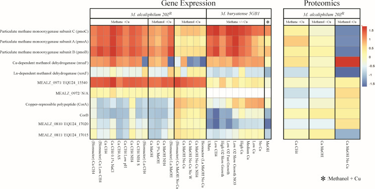
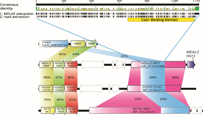
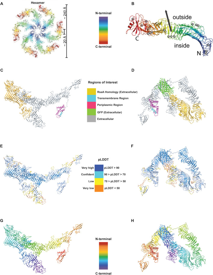
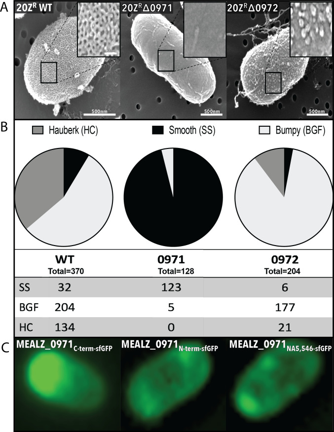
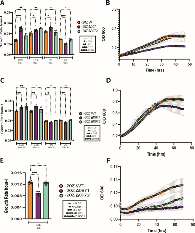
Many species of proteobacterial methane-consuming bacteria (methanotrophs) form a hauberk-like envelope represented by a surface (S-) layer protein (SLP) matrix. While several proteins were predicted to be associated with the cell surface, the composition and function of the hauberk matrix remained elusive. Here, we report the identification of the genes encoding the hauberk-forming proteins in two gamma-proteobacterial (Type I) methanotrophs, Methylotuvimicrobium buryatense 5GB1 (EQU24_15540) and Methylotuvimicrobium alcaliphilum 20Z R (MEALZ_0971 and MEALZ_0972). The proteins share 40% amino acid (AA) sequence identity with each other and are distantly related to the RsaA proteins from Caulobacter crescentus (20% AA sequence identity). Deletion of these genes resulted in loss of the characteristic hauberk pattern on the cell surface. A set of transcriptional fusions between the MEALZ_0971 and a superfolder green fluorescent protein (sfGFP) further confirmed its surface localization. The functional roles of the hauberk and cell-surface-associated proteins, including MEALZ_0971, MEALZ_0972, EQU24_15540, and a copper-induced CorA protein, were further investigated via gene expression studies and phenotypic tests. The hauberk core protein of M. alcaliphilum 20Z R showed constitutive expression across 18 growth conditions with reduced growth at high salinity, high methanol, and metal-limited conditions, suggesting a role in cell-envelope stability and metal scavenging. Overall, understanding the genetics, composition, and cellular functions of S-layers contributes to our knowledge of methanotroph adaptation to environmental perturbations and opens a promising prospect for (nano)biotechnology applications. IMPORTANCE: Understanding the genetics, composition, and cellular functions of the cell envelope proteins, such as S-layers, contributes to our knowledge of microbial cell biology and stress responses and molecular adaptations to environmental perturbations. In addition, this study opens a promising prospect for (nano)biotechnology applications of methane-derived self-assembling protein materials.
Overview
The claims and statistics of this paper haven’t been checked yet.
Run the check — free, no account needed. It reads the open-access text and checks the sources it cites.
Integrity signals
Registry
Cites
Statistics
Figures
Verification
Claims the paper makes
The claims and statistics of this paper haven’t been checked yet.
Run the check — free, no account needed. It reads the open-access text and checks the sources it cites.
Self-check
Percentages, sums and reported differences re-computed from the paper’s own numbers.
The self-check needs the paper’s full text.
Citations
Cites
Moderated estimation of fold change and dispersion for RNA-seq data with DESeq2
Love, M. I., Huber, W., & Anders, S. (2014) · Genome Biology · doi:10.1186/s13059-014-0550-8 · Cited by 95,106
MAFFT Multiple Sequence Alignment Software Version 7: Improvements in Performance and Usability
Katoh, K., & Standley, D. M. (2013) · Molecular Biology and Evolution · doi:10.1093/molbev/mst010 · Cited by 46,209
Highly accurate protein structure prediction with AlphaFold
Jumper, J., Evans, R., Pritzel, A., Green, T., Figurnov, M., Ronneberger, O., Tunyasuvunakool, K., Bates, R., Žídek, A., Potapenko, A., et al. (2021) · Nature · doi:10.1038/s41586-021-03819-2 · Cited by 43,764
fastp: an ultra-fast all-in-one FASTQ preprocessor
Chen, S., Zhou, Y., Chen, Y., & Gu, J. (2018) · Bioinformatics · doi:10.1093/bioinformatics/bty560 · Cited by 29,721
Kearse, M., Moir, R., Wilson, A., Stones-Havas, S., Cheung, M., Sturrock, S., Buxton, S., Cooper, A., Markowitz, S., Duran, C., et al. (2012) · Bioinformatics · doi:10.1093/bioinformatics/bts199 · Cited by 20,027
IQ-TREE 2: New Models and Efficient Methods for Phylogenetic Inference in the Genomic Era
Minh, B. Q., Schmidt, H. A., Chernomor, O., Schrempf, D., Woodhams, M. D., von Haeseler, A., & Lanfear, R. (2020) · Molecular Biology and Evolution · doi:10.1093/molbev/msaa015 · Cited by 16,873
trimAl: a tool for automated alignment trimming in large-scale phylogenetic analyses
Capella-Gutiérrez, S., Silla-Martínez, J. M., & Gabaldón, T. (2009) · Bioinformatics · doi:10.1093/bioinformatics/btp348 · Cited by 12,751
Salmon provides fast and bias-aware quantification of transcript expression
Patro, R., Duggal, G., Love, M. I., Irizarry, R. A., & Kingsford, C. (2017) · Nature Methods · doi:10.1038/nmeth.4197 · Cited by 12,700
ColabFold - Making protein folding accessible to all
Mirdita, M., Schütze, K., Moriwaki, Y., Heo, L., Ovchinnikov, S., & Steinegger, M. (2021) · doi:10.1101/2021.08.15.456425 · Cited by 9,391
MAFFT online service: multiple sequence alignment, interactive sequence choice and visualization
Katoh, K., Rozewicki, J., & Yamada, K. D. (2019) · Briefings in Bioinformatics · doi:10.1093/bib/bbx108 · Cited by 8,909
UniProt: the Universal Protein knowledgebase
Apweiler, R., Bairoch, A., Wu, C. H., Barker, W. C., Boeckmann, B., Ferro, S., Gasteiger, E., Huang, H., Lopez, R., Magrane, M., et al. (2003) · Nucleic Acids Research · doi:10.1093/nar/gkh131 · Cited by 7,622
UniProt: the Universal Protein Knowledgebase in 2023
The UniProt Consortium, Bateman, A., Martin, M.-J., Orchard, S., Magrane, M., Ahmad, S., Alpi, E., Bowler-Barnett, E. H., Britto, R., Bye-A-Jee, H., et al. (2023) · Nucleic Acids Research · doi:10.1093/nar/gkac1052 · Cited by 7,210
Letunic, I., & Bork, P. (2024) · Nucleic Acids Research · doi:10.1093/nar/gkae268 · Cited by 4,497
Cui, P., Zhang, S., Ding, F., Ali, S., & Xiong, L. (2014) · Genome Biology · doi:10.1186/gb-2014-15-1-r1 · Cited by 2,294
Outer-membrane vesicles from Gram-negative bacteria: biogenesis and functions
Schwechheimer, C., & Kuehn, M. J. (2015) · Nature Reviews Microbiology · doi:10.1038/nrmicro3525 · Cited by 1,922
DeepTMHMM predicts alpha and beta transmembrane proteins using deep neural networks
Hallgren, J., Tsirigos, K. D., Pedersen, M. D., Almagro Armenteros, J. J., Marcatili, P., Nielsen, H., Krogh, A., & Winther, O. (2022) · bioRxiv (Cold Spring Harbor Laboratory) · doi:10.1101/2022.04.08.487609 · Cited by 1,499
Mol* Viewer: modern web app for 3D visualization and analysis of large biomolecular structures
Sehnal, D., Bittrich, S., Deshpande, M., Svobodová, R., Berka, K., Bazgier, V., Velankar, S., Burley, S. K., Koča, J., & Rose, A. S. (2021) · Nucleic Acids Research · doi:10.1093/nar/gkab314 · Cited by 1,154
Kuraku, S., Zmasek, C. M., Nishimura, O., & Katoh, K. (2013) · Nucleic Acids Research · doi:10.1093/nar/gkt389 · Cited by 962
Sára, M., & Sleytr, U. B. (2000) · Journal of Bacteriology · doi:10.1128/jb.182.4.859-868.2000 · Cited by 766
Outer Membrane Vesicles (OMVs) of Gram-negative Bacteria: A Perspective Update
Jan, A. T. (2017) · Frontiers in Microbiology · doi:10.3389/fmicb.2017.01053 · Cited by 643
S-layers: principles and applications
Sleytr, U. B., Schuster, B., Egelseer, E.-M., & Pum, D. (2014) · FEMS Microbiology Reviews · doi:10.1111/1574-6976.12063 · Cited by 408
Biogenesis and functions of bacterial S-layers
Fagan, R. P., & Fairweather, N. F. (2014) · Nature Reviews Microbiology · doi:10.1038/nrmicro3213 · Cited by 387
Protein secretion by Gram-negative bacterial ABC exporters – a review
Binet, R., Létoffé, S., Ghigo, J., Delepelaire, P., & Wandersman, C. (1997) · Gene · doi:10.1016/s0378-1119(96)00829-3 · Cited by 211
Development of a broad-host-range sacB-based vector for unmarked allelic exchange
Marx, C. J. (2008) · BMC Research Notes · doi:10.1186/1756-0500-1-1 · Cited by 204
Qin, W., Heal, K. R., Ramdasi, R., Kobelt, J. N., Martens‐Habbena, W., Bertagnolli, A. D., Amin, S. A., Walker, C. B., Urakawa, H., Könneke, M., et al. (2017) · INTERNATIONAL JOURNAL OF SYSTEMATIC AND EVOLUTIONARY MICROBIOLOGY · doi:10.1099/ijsem.0.002416 · Cited by 201
Genetic Tools for the Industrially Promising Methanotroph Methylomicrobium buryatense
Puri, A. W., Owen, S., Chu, F., Chavkin, T., Beck, D. A. C., Kalyuzhnaya, M., & Lidstrom, M. E. (2014) · Applied and Environmental Microbiology · doi:10.1128/aem.03795-14 · Cited by 196
Self-citationOsmoadaptation in halophilic and alkaliphilic methanotrophs
Khmelenina, V. N., Kalyuzhnaya, M., SAKHAROVSKY, V. G., Сузина, Н. Е., Trotsenko, Y. A., & Gottschalk, G. (1999) · Archives of Microbiology · doi:10.1007/s002030050786 · Cited by 162
Self-citation3D Inkjet Printing of Complex, Cell-Laden Hydrogel Structures
Negro, A., Cherbuin, T., & Lütolf, M. P. (2018) · Scientific Reports · doi:10.1038/s41598-018-35504-2 · Cited by 158
Torre, A. D. L., Metivier, A., Chu, F., Laurens, L. M. L., Beck, D. A. C., Pienkos, P. T., Lidstrom, M. E., & Kalyuzhnaya, M. (2015) · Microbial Cell Factories · doi:10.1186/s12934-015-0377-3 · Cited by 156
Self-citationKaluzhnaya, M., Khmelenina, V. N., Eshinimaev, B. T., Сузина, Н. Е., Nikitin, D., Solonin, A. S., Lin, J., McDonald, I., Murrell, C. J., & Trotsenko, Y. A. (2001) · Systematic and Applied Microbiology · doi:10.1078/0723-2020-00028 · Cited by 155
Nielsen, A. K., Gerdes, K., & Murrell, J. C. (1997) · Molecular Microbiology · doi:10.1046/j.1365-2958.1997.4801846.x · Cited by 150
Beveridge, T. J., Pouwels, P. H., Sára, M., Kotiranta, A. K., Lounatmaa, K., Kari, K., Kerosuo, E., Haapasalo, M., Egelseer, E. M., Schocher, I., et al. (1997) · FEMS Microbiology Reviews · doi:10.1111/j.1574-6976.1997.tb00305.x · Cited by 150
Methane utilization in Methylomicrobium alcaliphilum 20ZR: a systems approach
Akberdin, I. R., Thompson, M., Hamilton, R., Desai, N., Alexander, D. C., Henard, C. A., Guarnieri, M. T., & Kalyuzhnaya, M. (2018) · Scientific Reports · doi:10.1038/s41598-018-20574-z · Cited by 130
Self-citationStructure of the hexagonal surface layer on Caulobacter crescentus cells
Bharat, T. A. M., Kureisaite-Ciziene, D., Hardy, G. G., Yu, E. W., Devant, J. M., Hagen, W. J. H., Brun, Y. V., Briggs, J. A. G., & Löwe, J. (2017) · Nature Microbiology · doi:10.1038/nmicrobiol.2017.59 · Cited by 111
In Situ Structure of an Intact Lipopolysaccharide-Bound Bacterial Surface Layer
von Kügelgen, A., Tang, H., Hardy, G. G., Kureisaite-Ciziene, D., Brun, Y. V., Stansfeld, P. J., Robinson, C. V., & Bharat, T. A. M. (2019) · Cell · doi:10.1016/j.cell.2019.12.006 · Cited by 111
Electroporation-Based Genetic Manipulation in Type I Methanotrophs
Yan, X., Chu, F., Puri, A. W., Fu, Y., & Lidstrom, M. E. (2016) · Applied and Environmental Microbiology · doi:10.1128/aem.03724-15 · Cited by 106
Awram, P., & Smit, J. (1998) · Journal of Bacteriology · doi:10.1128/jb.180.12.3062-3069.1998 · Cited by 97
The Structure and Binding Behavior of the Bacterial Cell Surface Layer Protein SbsC
Pavkov‐Keller, T., Egelseer, E. M., Tesarz, M., Svergun, D. I., Sleytr, U. B., & Keller, W. (2008) · Structure · doi:10.1016/j.str.2008.05.012 · Cited by 91
Wong, L. L., Lü, Y., Ho, J. C. S., Mugunthan, S., Law, Y., Conway, P. L., Kjelleberg, S. J., & Seviour, T. W. (2023) · The ISME Journal · doi:10.1038/s41396-023-01388-y · Cited by 91
Molecular Logic of Prokaryotic Surface Layer Structures
Bharat, T. A. M., von Kügelgen, A., & Alva, V. (2020) · Trends in Microbiology · doi:10.1016/j.tim.2020.09.009 · Cited by 82
Zytnick, A. M., Gutenthaler, S. M., Aron, A. T., Reitz, Z. L., Phi, M. T., Good, N. M., Petras, D., Daumann, L. J., & Martinez-Gomez, N. C. (2024) · Proceedings of the National Academy of Sciences · doi:10.1073/pnas.2322096121 · Cited by 68
Konrad, Z., & Eichler, J. (2002) · Biochemical Journal · doi:10.1042/bj20020757 · Cited by 63
Hemmann, J. L., Keller, P., Hemmerle, L., Vonderach, T., Ochsner, A. M., Bortfeld‐Miller, M., Günther, D., & Vorholt, J. A. (2023) · Journal of Biological Chemistry · doi:10.1016/j.jbc.2023.102940 · Cited by 62
S-layer proteins of Lactobacillus acidophilus inhibits JUNV infection
Martínez, M. G., Acosta, M., Candurra, N. A., & Ruzal, S. M. (2012) · Biochemical and Biophysical Research Communications · doi:10.1016/j.bbrc.2012.05.031 · Cited by 61
Juma, P., Fujitani, Y., Alessa, O., Oyama, T., Yurimoto, H., Sakai, Y., & Tani, A. (2022) · Frontiers in Microbiology · doi:10.3389/fmicb.2022.921635 · Cited by 57
Campylobacter fetus Surface Layer Proteins Are Transported by a Type I Secretion System
Thompson, S. A., Shedd, O. L., Ray, K. C., Beins, M. H., Jorgensen, J. P., & Blaser, M. J. (1998) · Journal of Bacteriology · doi:10.1128/jb.180.24.6450-6458.1998 · Cited by 56
Lipid Anchoring of Archaeosortase Substrates and Midcell Growth in Haloarchaea
Halim, M. F. A., Schulze, S., DiLucido, A., Pfeiffer, F., Filho, A. W. B., & Pohlschröder, M. (2020) · mBio · doi:10.1128/mbio.00349-20 · Cited by 55
Caulobacter crescentus: model system extraordinaire
Govers, S. K., & Jacobs, C. (2020) · Current Biology · doi:10.1016/j.cub.2020.07.033 · Cited by 54
Complete atomic structure of a native archaeal cell surface
von Kügelgen, A., Alva, V., & Bharat, T. A. M. (2021) · Cell Reports · doi:10.1016/j.celrep.2021.110052 · Cited by 53
Rare Earth Elements Alter Redox Balance in Methylomicrobium alcaliphilum 20ZR
Akberdin, I. R., Collins, D. A., Hamilton, R., Oshchepkov, D. Y., Shukla, A. K., Nicora, C., Nakayasu, E. S., Adkins, J., & Kalyuzhnaya, M. (2018) · Frontiers in Microbiology · doi:10.3389/fmicb.2018.02735 · Cited by 50
Self-citationGómez Guzmán, O. M., Álvarez Acevedo, L. C., Muñoz Gómez, J. F., Misas Rivas, E., Jiménez Alzate, M. D. P., Arango Arteaga, M., McEwen Ochoa, J. G., Hernández Ruiz, O., Clay, O., & Gallo, J. (2017) · Institutional Repository University of Antioquia (University of Antioquia) · doi:10.1128/genomea · Cited by 45
Analysis of high-level S-layer protein secretion in Caulobacter crescentus.
Lau, J., Nomellini, J. F., & Smit, J. (2010) · Canadian Journal of Microbiology (print) · doi:10.1139/w10-036 · Cited by 40
Structure of the two-component S-layer of the archaeon Sulfolobus acidocaldarius
Gambelli, L., McLaren, M., Conners, R., Sanders, K., Gaines, M. C., Clark, L., Gold, V. A. M., Kattnig, D. R., Sikora, M., Hanus, C., et al. (2024) · eLife · doi:10.7554/elife.84617 · Cited by 37
Structural and functional features of methanotrophs from hypersaline and alkaline lakes
Khmelenina, V., Shchukin, V., Reshetnikov, A., Mustakhimov, I., Suzina, N., Eshinimaev, B., & Trotsenko, Y. (2010) · Microbiology · doi:10.1134/s0026261710040090 · Cited by 36
von Kügelgen, A., van Dorst, S., Yamashita, K., Sexton, D. L., Tocheva, E. I., Murshudov, G., Alva, V., & Bharat, T. A. M. (2023) · Proceedings of the National Academy of Sciences · doi:10.1073/pnas.2215808120 · Cited by 35
A bacterial surface layer protein exploits multistep crystallization for rapid self-assembly
Herrmann, J. R., Li, P.-N., Jabbarpour, F., Chan, A., Rajković, I., Matsui, T., Shapiro, L., Smit, J., Weiss, T. M., Murphy, M. E. P., et al. (2019) · Proceedings of the National Academy of Sciences · doi:10.1073/pnas.1909798116 · Cited by 33
Berson, O., & Lidstrom, M. E. (1997) · FEMS Microbiology Letters · doi:10.1016/s0378-1097(97)00029-3 · Cited by 31
Cobos, I. M., García, M. I. I., Moreno, F. D. L. P., Melero, L. P. S., Luque-Almagro, V., Rodríguez, F. C., Ruiz, M. D. R., Jiménez, M. A. P., & Vivián, C. M. (2015) · Microbial Cell Factories · doi:10.1186/s12934-015-0267-8 · Cited by 27
Glukhova, L. B., Frank, Y. A., Danilova, E. V., Avakyan, M. R., Banks, D. A., Tuovinen, O. H., & Karnachuk, O. V. (2018) · Microbial Ecology · doi:10.1007/s00248-018-1186-0 · Cited by 27
Johnson, K. A., Ve, T., Larsen, Ø., Pedersen, R. B., Lillehaug, J. R., Jensen, H. B., Helland, R., & Karlsen, O. A. (2014) · PLoS ONE · doi:10.1371/journal.pone.0087750 · Cited by 21
Lifecycle of a predatory bacterium vampirizing its prey through the cell envelope and S-layer
Santin, Y. G., Sogues, A., Bourigault, Y., Remaut, H. K., & Laloux, G. (2024) · Nature Communications · doi:10.1038/s41467-024-48042-5 · Cited by 21
Surface layers of methanotrophic bacteria
Khmelenina, V. N., Сузина, Н. Е., & Trotsenko, Y. A. (2013) · Microbiology · doi:10.1134/s0026261713050068 · Cited by 19
Shchukin, V. N., Khmelenina, V. N., Eshinimaev, B. T., Сузина, Н. Е., & Trotsenko, Y. A. (2011) · Microbiology · doi:10.1134/s0026261711050122 · Cited by 19
Archaeal Host Cell Recognition and Viral Binding of HFTV1 to Its Haloferax Host
Schwarzer, S., Hackl, T., Oksanen, H. M., & Quax, T. E. F. (2023) · mBio · doi:10.1128/mbio.01833-22 · Cited by 17
Identification of a bacterial di-haem cytochrome c peroxidase from Methylomicrobium album BG8
Karlsen, O. A., Larsen, Ø., & Jensen, H. B. (2010) · Microbiology · doi:10.1099/mic.0.037119-0 · Cited by 14
Electron microscopy of the cell wall complex ofMethylomonas albus
Jeffries, P., & Wilkinson, J. F. (1978) · Archives of Microbiology · doi:10.1007/bf00964278 · Cited by 12
Liu, C., Mesman, R., Pol, A., Angius, F., & den Camp, H. J. M. O. (2023) · Antonie van Leeuwenhoek · doi:10.1007/s10482-023-01879-0 · Cited by 3
Khmelenina, V., Beck, D. A., Munk, C., Davenport, K., Daligault, H., Erkkila, T. H., Goodwin, L., Gu, W., Lo, C.-C., Scholz, M., et al. (2013) · Genome Announcements · doi:10.1128/genomea.00053-13 · Cited by 2
Self-citation
Research works printed but not linked: 6
- Tyurin V, Gorskaya L, Kaftanova A, Logi nova TM, Mikhailov AM. 1985. Some features of the ultrastructure of Methylococcus capsulatus grown under different conditions. Prikl Biokhim Mikrobiol 21:770–775.
- 10.1111/j.1574-6968.1997.tb10284.x · DOI
- 10.1038/s41592-022-01488-1 · DOI · Paper page
- Vital AV. 2023. Surface (S)-layers from methanotrophic bacteria: composition and function. San Diego State University.
- Ismail SS. 2019. Fundamentals of protein biosynthesis in Methylomicrobium alcaliphilum 20Z R, a model methanotroph. San Diego State University.
- Collins DAI. 2021. Dissecting the cellular biology of the model methanotroph Methylotuvimicrobium alcaliphilum 20Z R through multi-omics, mutagenesis, and microscopy. University of California, San Diego.
Metadata
- Authors
- Hamilton, R., Gebbie, W., Bowman, C., Mantanona, A., & Kalyuzhnaya, M. G.
- Affiliations
- San Diego State University (US)
- Corresponding author
- Kalyuzhnaya, Marina G.
- Venue
- Applied and Environmental Microbiology
- Publisher
- American Society for Microbiology
- Published
- 2025-01-31
- Volume
- 91
- Issue
- 1
- Pages
- e0136424-e0136424
- ISSN
- 0099-2240 (ISSN-L)
- 1098-5336
- PMC
- PMC11784148
- OpenAlex
- W4405944986
- Cited by
- 1
- References
- 68
- Citations by year
- 2026: 1
- FWCI
- 0.14
- Type
- journal article
- Language
- en
- Open access
- hybrid
- doi.org
- Licence
- cc-by
- creativecommons.org/licenses/by/4.0/ (vor)
- journals.asm.org/non-commercial-tdm-license (tdm)
- Locations
- Applied and Environmental Microbiology (publishedVersion, cc-by)
- bioRxiv (Cold Spring Harbor Laboratory) (acceptedVersion)
- PubMed Central (publishedVersion, cc-by)
- PubMed (publishedVersion)
- DOAJ (DOAJ: Directory of Open Access Journals) (submittedVersion, cc-by-sa)
- Funders
- DOD | ARPA | Defense Sciences Office, DARPA (DE-AR000350)
- Topics
- Life Sciences › Biochemistry, Genetics and Molecular Biology › Molecular Biology › Microbial metabolism and enzyme function
- Life Sciences › Biochemistry, Genetics and Molecular Biology › Genetics › Bacterial Genetics and Biotechnology
- Physical Sciences › Environmental Science › Environmental Chemistry › Methane Hydrates and Related Phenomena
- Keywords
- surface layer protein, methanotrophic bacteria, Gammaproteobacteria, cell envelope, gene deletion, gene expression, metal scavenging, salt tolerance, methanol tolerance
- MeSH terms
- Bacterial Proteins, Membrane Glycoproteins, Methane, Methylococcaceae, Gammaproteobacteria
About these checks
Registry, retraction, Cites and statistics results are computed, not judged, with one exception: a printed reference that no index lists is read once by a model before it counts as a citation error. Claims are extracted by a model and kept only when their quote appears verbatim in the text; each cited source is then checked, with a model’s help, for whether it says what the claim relies on. Those verdicts can be wrong — read the claim’s quote and the cited source before relying on one.
Statistics and arithmetic are recomputed, not judged.
Confidence bands are shown only for fields calibrated on our annotated test set. None is calibrated yet, so no band is shown.
What counts as a claim ›How we measure confidence ›How we check this ›
Are you an author, or do you see a problem with this page? Request a review
Data sources
Bibliographic data: Semantic Scholar, OpenAlex, Crossref.
Contains information from the Semantic Scholar Academic Graph Datasets, made available under the ODC Attribution License (ODC-BY 1.0).
Semantic Scholar’s data is described in: Kinney, R., et al. (2023). The Semantic Scholar Open Data Platform. arXiv:2301.10140.
