

The DOI matches its Crossref record; 52 of the 52 references it cites were found in a registry or an index.
Type I partition-related proteins enhance conjugative transfer through transcriptional activation and oriT region binding
Open accessPublished version of a preprint
Abstract
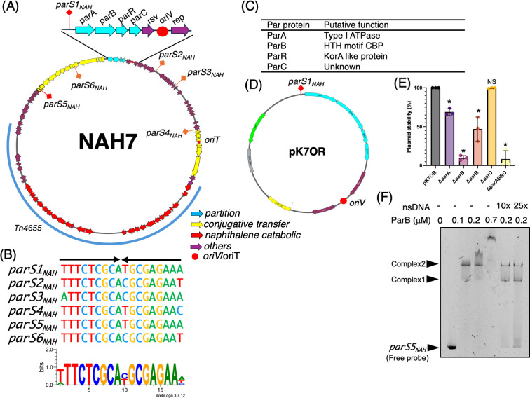
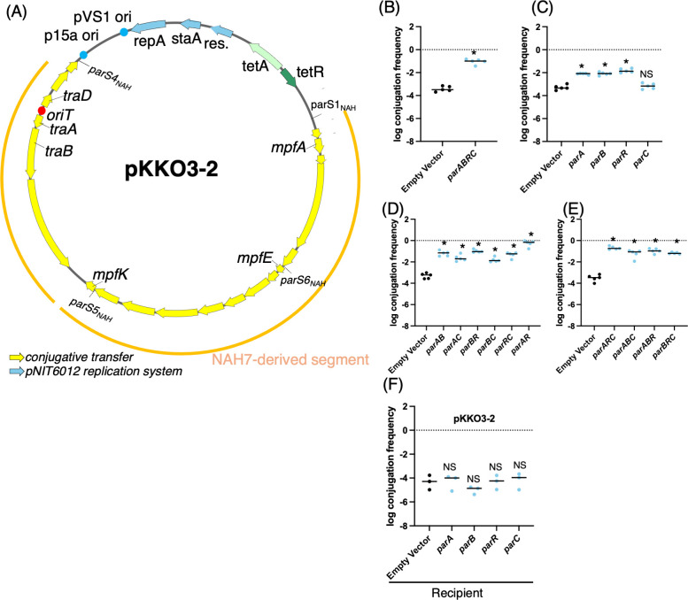
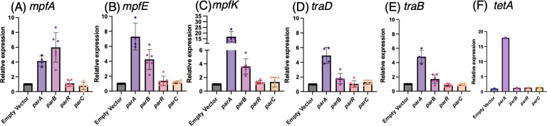
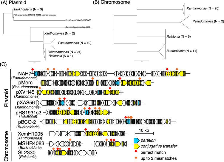
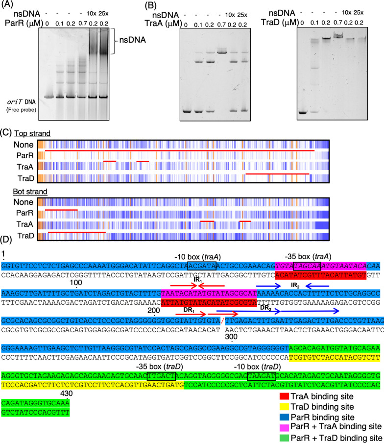
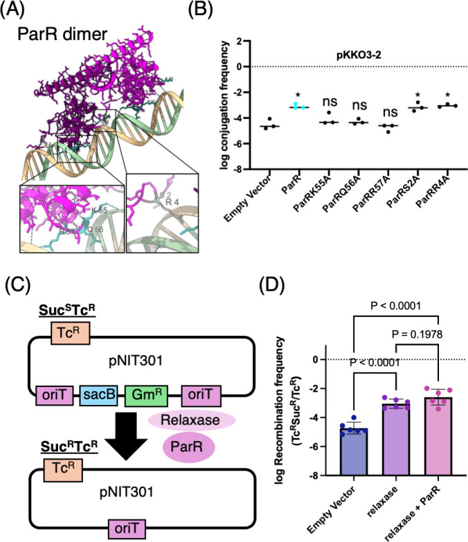
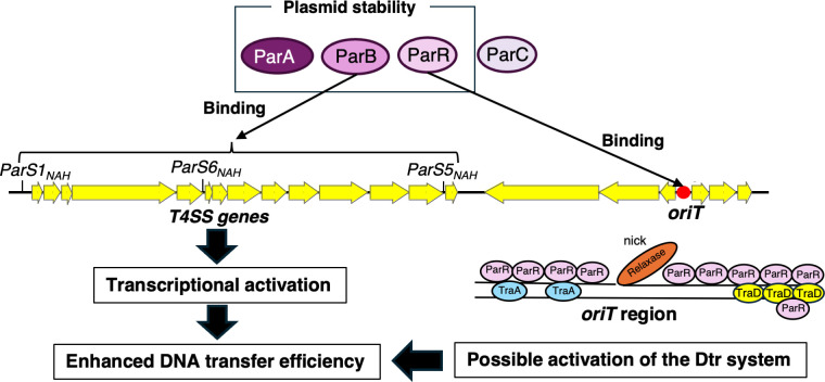
Plasmid partitioning and bacterial conjugation are critical processes ensuring plasmid maintenance and dissemination, respectively, within bacterial populations. Although traditionally regarded as distinct phenomena, these two processes are increasingly recognized as interconnected. While partitioning ensures plasmid inheritance during cell division, its potential influence on conjugative transfer remains poorly understood. A major impediment to understanding their interplay is that partition systems are often essential for plasmid stability, making it difficult to distinguish their direct effects on conjugation. In this study, we addressed this challenge using a mini-conjugative plasmid derived from the Pseudomonas putida NAH7 plasmid. This engineered plasmid, containing all conjugation-related genes, was cloned into an Escherichia coli -compatible vector. Additionally, the par genes from NAH7 were expressed from a separate plasmid to investigate their roles in conjugative transfer. Our results revealed that the par gene cluster plays a significant role in enhancing the conjugative transfer of the mini-conjugative plasmid. Specifically, ParB, a centromere-binding protein, functions as a transcriptional activator of conjugation-related genes by binding the parS NAH site. In contrast, ParR, a KorA homolog, was not found to enhance transcription directly but binds extensively to the oriT region. This binding probably facilitates the recruitment or stabilization of the relaxosome, thereby enhancing conjugation efficiency. Together, these findings unveil a previously unappreciated role for partition proteins in stimulating bacterial conjugation, providing new insights into how plasmids coordinate vertical and horizontal dissemination, highlighting that these processes can occur simultaneously within bacterial communities. IMPORTANCE: Plasmid partition systems are classified into three types. While some systems have been reported to influence conjugative transfer, this study uncovers a novel mechanism utilized by a Type I system to enhance DNA transfer. Strikingly, repeat sequences perfectly matching the parS NAH site—bound by ParB to activate downstream conjugative transfer genes—were identified on both plasmids and chromosomes across diverse proteobacterial taxa. Furthermore, many of these repeat sequences were localized near genes involved in conjugative transfer and partitioning, suggesting the presence of a conserved regulatory mechanism mediated by these repeats. This study provides important insights into how plasmid partition systems coordinate both vertical and horizontal dissemination. Such knowledge is essential for understanding and mitigating the spread of antibiotic resistance and other plasmid-encoded traits, and it offers a foundation for developing strategies to manage plasmid-associated genetic exchange.
Overview
The claims and statistics of this paper haven’t been checked yet.
Run the check — free, no account needed. It reads the open-access text and checks the sources it cites.
Integrity signals
Registry
Cites
Statistics
Figures
Verification
Claims the paper makes
The claims and statistics of this paper haven’t been checked yet.
Run the check — free, no account needed. It reads the open-access text and checks the sources it cites.
Self-check
Percentages, sums and reported differences re-computed from the paper’s own numbers.
The self-check needs the paper’s full text.
Citations
Cites
Molecular Cloning. A Laboratory Manual
Wood, E. (1983) · Biochemical Education · doi:10.1016/0307-4412(83)90068-7 · Cited by 29,533
Accurate structure prediction of biomolecular interactions with AlphaFold 3
Abramson, J., Adler, J., Dunger, J., Evans, R., Green, T., Pritzel, A., Ronneberger, O., Willmore, L., Ballard, A. J., Bambrick, J., et al. (2024) · Nature · doi:10.1038/s41586-024-07487-w · Cited by 15,861
WebLogo: A Sequence Logo Generator: Figure 1
Crooks, G. E., Hon, G., Chandonia, J.-M., & Brenner, S. E. (2004) · Genome Research · doi:10.1101/gr.849004 · Cited by 12,386
MAFFT online service: multiple sequence alignment, interactive sequence choice and visualization
Katoh, K., Rozewicki, J., & Yamada, K. D. (2019) · Briefings in Bioinformatics · doi:10.1093/bib/bbx108 · Cited by 8,909
Use of bacteriophage T7 RNA polymerase to direct selective high-level expression of cloned genes
Studier, F. W., & Moffatt, B. A. (1986) · Journal of Molecular Biology · doi:10.1016/0022-2836(86)90385-2 · Cited by 6,405
Letunic, I., & Bork, P. (2024) · Nucleic Acids Research · doi:10.1093/nar/gkae268 · Cited by 4,497
UCSF ChimeraX: Tools for structure building and analysis
Meng, E. C., Goddard, T. D., Pettersen, E. F., Couch, G. S., Pearson, Z. J., Morris, J. H., & Ferrin, T. E. (2023) · Protein Science · doi:10.1002/pro.4792 · Cited by 4,442
Construction of improved M13 vectors using oligodeoxynucleotide-directed mutagenesis
Norrander, J. M., Kempe, T., & Messing, J. W. (1983) · Gene · doi:10.1016/0378-1119(83)90040-9 · Cited by 2,441
Varadi, M., Bertoni, D., Magana, P., Paramval, U., Pidruchna, I., Radhakrishnan, M., Tsenkov, M., Nair, S., Mirdita, M., Yeo, J., et al. (2024) · Nucleic Acids Research · doi:10.1093/nar/gkad1011 · Cited by 2,052 · cited 1×
“To obtain predicted structures of these Par proteins, we utilized the AlphaFold Database (44) and AlphaFold Server (45) for structure prediction.”
Improved gfp and inaZ Broad-Host-Range Promoter-Probe Vectors
Miller, W. G., Leveau, J. H. J., & Lindow, S. E. (2000) · Molecular Plant-Microbe Interactions® · doi:10.1094/mpmi.2000.13.11.1243 · Cited by 557
Metadata
- Authors
- Kishida, K., Kudo, K., Kumagai, R., Ijima, S., Deng, W., Stari, L., Ogawa-Kishida, N., Ohtsubo, Y., Nagata, Y., & Tsuda, M.
- Affiliations
- Tohoku University (JP)
- Department of Molecular and Chemical Life Sciences, Graduate School of Life Sciences, Tohoku University
- Corresponding author
- Kishida, Kouhei
- Venue
- mBio
- Publisher
- American Society for Microbiology
- Published
- 2025-09-10
- Volume
- 16
- Issue
- 9
- Pages
- e0160025-e0160025
- ISSN
- 2150-7511 (ISSN-L)
- PMC
- PMC12421886
- OpenAlex
- W4412782267
- Cited by
- 1
- References
- 52
- Citations by year
- 2026: 1
- FWCI
- 0.59
- Type
- journal article
- Language
- en
- Open access
- gold
- doi.org
- Licence
- cc-by
- creativecommons.org/licenses/by/4.0/ (vor)
- journals.asm.org/non-commercial-tdm-license (tdm)
- Locations
- mBio (publishedVersion, cc-by)
- bioRxiv (Cold Spring Harbor Laboratory) (acceptedVersion)
- Europe PMC (PubMed Central) (publishedVersion, cc-by)
- PubMed (publishedVersion)
- PubMed Central (publishedVersion, cc-by)
- DOAJ (DOAJ: Directory of Open Access Journals) (submittedVersion, cc-by-sa)
- Funders
- Japan Society for the Promotion of Science (19K15725)
- Institute for Fermentation, Osaka (Y-2024-1-007)
- Topics
- Life Sciences › Biochemistry, Genetics and Molecular Biology › Genetics › Bacterial Genetics and Biotechnology
- Health Sciences › Medicine › Microbiology (medical) › Antibiotic Resistance in Bacteria
- Life Sciences › Immunology and Microbiology › Virology › Bacteriophages and microbial interactions
- Keywords
- plasmid partitioning, bacterial conjugation, ParB, parr, transcriptional activation, relaxosome, plasmid maintenance, plasmid dissemination, horizontal gene transfer
- MeSH terms
- Bacterial Proteins, Conjugation, Genetic, Escherichia coli, Multigene Family, Plasmids, Protein Binding, Transcriptional Activation, Gene Expression Regulation, Bacterial, Pseudomonas putida, Replication Origin
About these checks
Registry, retraction, Cites and statistics results are computed, not judged, with one exception: a printed reference that no index lists is read once by a model before it counts as a citation error. Claims are extracted by a model and kept only when their quote appears verbatim in the text; each cited source is then checked, with a model’s help, for whether it says what the claim relies on. Those verdicts can be wrong — read the claim’s quote and the cited source before relying on one.
Statistics and arithmetic are recomputed, not judged.
Confidence bands are shown only for fields calibrated on our annotated test set. None is calibrated yet, so no band is shown.
What counts as a claim ›How we measure confidence ›How we check this ›
Are you an author, or do you see a problem with this page? Request a review
How to cite
Cite CoChat for the checks on this page, and the paper for its own findings.
- This page
- CoChat. (2026). Citation check: Type I partition-related proteins enhance conjugative transfer through transcriptional activation and oriT region binding. https://app.cochat.ai/papers/10.1128/mbio.01600-25
- The paper
- Kishida, K., Kudo, K., Kumagai, R., Ijima, S., Deng, W., Stari, L., Ogawa-Kishida, N., Ohtsubo, Y., Nagata, Y., & Tsuda, M. (2025). Type I partition-related proteins enhance conjugative transfer through transcriptional activation and oriT region binding. mBio. https://doi.org/10.1128/mbio.01600-25
Data sources
Bibliographic data: Semantic Scholar, OpenAlex, Crossref.
Contains information from the Semantic Scholar Academic Graph Datasets, made available under the ODC Attribution License (ODC-BY 1.0).
Semantic Scholar’s data is described in: Kinney, R., et al. (2023). The Semantic Scholar Open Data Platform. arXiv:2301.10140.
