

An algorithmic approach to structural imaging in dementia
Open accessThe DOI matches its Crossref record; 75 of the 75 references it cites were found in a registry or an index.
Abstract
Accurate and timely diagnosis of dementia is important to guide management and provide appropriate information and support to patients and families. Currently, with the exception of individuals with genetic mutations, postmortem examination of brain tissue remains the only definitive means of establishing diagnosis in most cases, however, structural neuroimaging, in combination with clinical assessment, has value in improving diagnostic accuracy during life. Beyond the exclusion of surgical pathology, signal change and cerebral atrophy visible on structural MRI can be used to identify diagnostically relevant imaging features, which provide support for clinical diagnosis of neurodegenerative dementias. While no structural imaging feature has perfect sensitivity and specificity for a given diagnosis, there are a number of imaging characteristics which provide positive predictive value and help to narrow the differential diagnosis. While neuroradiological expertise is invaluable in accurate scan interpretation, there is much that a non-radiologist can gain from a focused and structured approach to scan analysis. In this article we describe the characteristic MRI findings of the various dementias and provide a structured algorithm with the aim of providing clinicians with a practical guide to assessing scans.
Overview
The claims and statistics of this paper haven’t been checked yet.
Free: it reads the open-access text and checks the first 5 claims that cite a source we can check. Create a free account to check every claim.
Integrity signals
Registry
Cites
Statistics
Figures



Verification
Claims the paper makes
The claims and statistics of this paper haven’t been checked yet.
Free: it reads the open-access text and checks the first 5 claims that cite a source we can check. Create a free account to check every claim.
Claims other papers make about this one
Each is checked against this paper’s own text: does it say what the citing paper says it does?
The source backs part of the claim, not all of it — or backs it for a different population, condition or comparison.
In the citing paper
“Structural imaging can exclude conditions such as space-occupying lesions, stroke, normal-pressure hydrocephalus, as well as many other pathologies”
From The use of neuroimaging techniques in the early and differential diagnosis of dementia (2023) · cites this paper as [10]
Experimental — accuracy not yet measuredThe cited source says what the claim attributes to it.
In the citing paper
“and can also help with the differential diagnosis of dementia based on characteristic patterns of atrophy, white matter changes and the presence or absence of cerebrovascular disease [10].”
From The use of neuroimaging techniques in the early and differential diagnosis of dementia (2023) · cites this paper as [10]
Self-check
Percentages, sums and reported differences re-computed from the paper’s own numbers.
The self-check needs the paper’s full text.
Citations
Cites
McKhann, G. M., Knopman, D. S., Chertkow, H., Hyman, B. T., Jack, C. R., Kawas, C. H., Klunk, W. E., Koroshetz, W. J., Manly, J. J., Mayeux, R., et al. (2011) · Alzheimer's & Dementia · doi:10.1016/j.jalz.2011.03.005 · Cited by 18,657
Self-citationDiagnostic criteria for multiple sclerosis: 2010 revisions to the McDonald criteria
Polman, C. H., Reingold, S. C., Banwell, B., Clanet, M., Cohen, J. A., Filippi, M., Fujihara, K., Havrdova, E., Hutchinson, M., Kappos, L., et al. (2011) · Annals of Neurology · doi:10.1002/ana.22366 · Cited by 9,802 · cited 1×
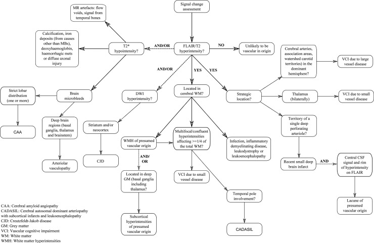
“33 Less often, multifocal/con fl uent regions of hyperintensity in a patient with suspected dementia may result from a number of other conditions, including infections, in fl ammatory demyelinating diseases, leukodystrophies or leukoencephalopathies.”
BackgroundWardlaw, J. M., Smith, E. E., Biessels, G. J., Cordonnier, C., Fazekas, F., Frayne, R., Lindley, R. I., O'Brien, J. T., Barkhof, F., Benavente, O. R., et al. (2013) · The Lancet Neurology · doi:10.1016/s1474-4422(13)70124-8 · Cited by 5,544
Self-citationMR signal abnormalities at 1.5 T in Alzheimer's dementia and normal aging
Fazekas, F., Chawluk, J., Alavi, A., Hurtig, H., & Zimmerman, R. (1987) · American Journal of Roentgenology · doi:10.2214/ajr.149.2.351 · Cited by 5,334 · cited 1×
“29 Con fl uence of hyperintensities in at least two regions, and the beginning of con fl uence of hyperintensities in a further two regions, is considered to represent the involvement of at least a quarter of the total white matter and is suf fi cient to assume SVD is the cause of VCI or VaD.”
BackgroundSensitivity of revised diagnostic criteria for the behavioural variant of frontotemporal dementia
Rascovsky, K., Hodges, J. R., Knopman, D., Mendez, M. F., Kramer, J. H., Neuhaus, J., van Swieten, J. C., Seelaar, H., Dopper, E. G. P., Onyike, C. U., et al. (2011) · Brain · doi:10.1093/brain/awr179 · Cited by 5,221
Self-citationClassification of primary progressive aphasia and its variants
Gorno-Tempini, M. L., Hillis, A. E., Weintraub, S., Kertesz, A., Mendez, M., Cappa, S. F., Ogar, J. M., Rohrer, J. D., Black, S., Boeve, B. F., et al. (2011) · Neurology · doi:10.1212/wnl.0b013e31821103e6 · Cited by 5,141
Diagnosis and management of dementia with Lewy bodies: Third report of the DLB consortium
McKeith, I. G., Dickson, D. W., Lowe, J., Emre, M., O'Brien, J. T., Feldman, H., Cummings, J., Duda, J. E., Lippa, C., Perry, E. K., et al. (2005) · Neurology · doi:10.1212/01.wnl.0000187889.17253.b1 · Cited by 5,069 · cited 1×
“…suggested as a means of distinguishing DLB from AD at a group level, 65 however this may not be true of older patients or patients with advanced DLB and may not be reliable in individual cases 66 (see fi gure 4), particularly as many patients with DLB pathology also have AD pathology at postmortem.”
BackgroundRomán, G. C., Tatemichi, T. K., Erkinjuntti, T., Cummings, J. L., Masdeu, J. C., Garcia, J. H., Amaducci, L., Orgogozo, J.-M., Brun, A., Hofman, A., et al. (1993) · Neurology · doi:10.1212/wnl.43.2.250 · Cited by 4,921 · cited 1×
“23 24 In the absence of evidence of vascular damage on MRI a vascular cause for cognitive impairment is very unlikely; conversely, extensive vascular changes are very likely to produce signi fi cant cognitive de fi cits.”
BackgroundResearch criteria for the diagnosis of Alzheimer's disease: revising the NINCDS–ADRDA criteria
Dubois, B., Feldman, H. H., Jacova, C., DeKosky, S. T., Barberger-Gateau, P., Cummings, J., Delacourte, A., Galasko, D., Gauthier, S., Jicha, G., et al. (2007) · The Lancet Neurology · doi:10.1016/s1474-4422(07)70178-3 · Cited by 4,411
Self-citationGorelick, P. B., Scuteri, A., Black, S. E., DeCarli, C., Greenberg, S. M., Iadecola, C., Launer, L. J., Laurent, S., Lopez, O. L., Nyenhuis, D., et al. (2011) · Stroke · doi:10.1161/str.0b013e3182299496 · Cited by 3,811
Cited by
Dementia prevention, intervention, and care
Livingston, G., Sommerlad, A., Orgeta, V., Costafreda, S. G., Huntley, J., Ames, D., Ballard, C., Banerjee, S., Burns, A., Cohen-Mansfield, J., et al. (2017) · The Lancet · doi:10.1016/s0140-6736(17)31363-6 · Cited by 6,072
Self-citationDiagnosis of Early Alzheimer's Disease: Clinical Practice in 2021
Porsteinsson, A. P., Isaacson, R. S., Knox, S., Sabbagh, M. N., & Rubino, I. (2021) · The Journal of Prevention of Alzheimer's Disease · doi:10.14283/jpad.2021.23 · Cited by 744
“Structural imaging, such as MRI, provides clinically useful information when investigating causes of cognitive impairment (69) (Figure 2).”
Qiu, S., Joshi, P. S., Miller, M. I., Xue, C., Zhou, X., Karjadi, C., Chang, G. H., Joshi, A. S., Dwyer, B., Zhu, S., et al. (2020) · Brain · doi:10.1093/brain/awaa137 · Cited by 472
Cognition and dementia in older patients with epilepsy
Sen, A., Capelli, V., & Husain, M. (2018) · Brain · doi:10.1093/brain/awy022 · Cited by 280
“there are biomarkers suggesting the presence of both types of pathology (Harper et al., 2014).”
BackgroundMRI visual rating scales in the diagnosis of dementia: evaluation in 184 post-mortem confirmed cases
Harper, L., Fumagalli, G. G., Barkhof, F., Scheltens, P., O’Brien, J. T., Bouwman, F., Burton, E. J., Rohrer, J. D., Fox, N. C., Ridgway, G. R., et al. (2016) · Brain · doi:10.1093/brain/aww005 · Cited by 264
“The high resolution and excellent tissue contrast afforded by MRI in particular allows for global and regional cerebral atrophy to be assessed, offering positive predictive value for underlying disease pathology (Harper et al., 2014).”
BackgroundSelf-citation
Metadata
- Authors
- Harper, L., Barkhof, F., Scheltens, P., Schott, J. M., & Fox, N. C.
- Affiliations
- UCL Queen Square Institute of Neurology (GB)
- Amsterdam UMC Location Vrije Universiteit Amsterdam (NL)
- Corresponding author
- Fox, N. C.
- Venue
- Journal of Neurology, Neurosurgery & Psychiatry
- Publisher
- BMJ
- Published
- 2013-10-16
- Volume
- 85
- Issue
- 6
- Pages
- 692-698
- ISSN
- 0022-3050 (ISSN-L)
- PMC
- PMC4033032
- OpenAlex
- W2141470005
- Cited by
- 137
- References
- 75
- Citations by year
- 2026: 4 · 2025: 11 · 2024: 13 · 2023: 11 · 2022: 17 · 2021: 12 · 2020: 18 · 2019: 16 · 2018: 9 · 2017: 6 · 2016: 10 · 2015: 5 · 2014: 4
- FWCI
- 0.86
- Type
- journal article
- Language
- en
- Open access
- hybrid
- doi.org
- Licence
- cc-by-nc
- Locations
- Journal of Neurology Neurosurgery & Psychiatry (publishedVersion, cc-by-nc)
- Europe PMC (PubMed Central) (publishedVersion, cc-by-nc)
- PubMed Central (publishedVersion, cc-by-nc)
- PubMed (publishedVersion)
- Pure Amsterdam UMC (publishedVersion)
- Amsterdam UMC (VU Amsterdam) - Institutional Repository (publishedVersion)
- UCL Discovery (University College London) (submittedVersion)
- HighWire Press Open Archive (submittedVersion)
- Data Archiving and Networked Services (DANS) (submittedVersion, other-oa)
- Data Archiving and Networked Services (DANS) (submittedVersion)
- Funders
- National Institute for Health and Care Research (NF-SI-0513-10134, NF-SI-0508-10123)
- Topics
- Health Sciences › Medicine › Psychiatry and Mental health › Dementia and Cognitive Impairment Research
- Health Sciences › Medicine › Radiology, Nuclear Medicine and Imaging › Radiology practices and education
- Health Sciences › Medicine › Radiology, Nuclear Medicine and Imaging › Medical Imaging Techniques and Applications
- Keywords
- dementia, structural MRI, neurodegenerative dementia, diagnostic algorithm, brain atrophy, differential diagnosis, neuroradiology, diagnostic accuracy
- MeSH terms
- Aged, Aged, 80 and over, Algorithms, Alzheimer Disease, Atrophy, Brain, Dementia, Diagnosis, Differential, Europe, Female, United Kingdom, Hippocampus, Humans, Magnetic Resonance Imaging, Male, Middle Aged, Predictive Value of Tests, Supranuclear Palsy, Progressive, United States, Practice Guidelines as Topic, Neuroimaging
About these checks
Registry, retraction, Cites and statistics results are computed, not judged, with one exception: a printed reference that no index lists is read once by a model before it counts as a citation error. Claims are extracted by a model and kept only when their quote appears verbatim in the text; each cited source is then checked, with a model’s help, for whether it says what the claim relies on. Those verdicts can be wrong — read the claim’s quote and the cited source before relying on one.
Statistics and arithmetic are recomputed, not judged.
Confidence bands are shown only for fields calibrated on our annotated test set. None is calibrated yet, so no band is shown.
What counts as a claim ›How we measure confidence ›How we check this ›
Are you an author, or do you see a problem with this page? Request a review
How to cite
Cite CoChat for the checks on this page, and the paper for its own findings.
- This page
- CoChat. (2026). Citation check: An algorithmic approach to structural imaging in dementia. https://app.cochat.ai/papers/10.1136/jnnp-2013-306285
- The paper
- Harper, L., Barkhof, F., Scheltens, P., Schott, J. M., & Fox, N. C. (2014). An algorithmic approach to structural imaging in dementia. Journal of Neurology, Neurosurgery & Psychiatry. https://doi.org/10.1136/jnnp-2013-306285
Data sources
Bibliographic data: Semantic Scholar, OpenAlex, Crossref.
Contains information from the Semantic Scholar Academic Graph Datasets, made available under the ODC Attribution License (ODC-BY 1.0).
Semantic Scholar’s data is described in: Kinney, R., et al. (2023). The Semantic Scholar Open Data Platform. arXiv:2301.10140.
