

Modulation of Vascular Cell Function by Bim Expression
Open accessThe DOI matches its Crossref record; 29 of the 29 references it cites were found in a registry or an index.
Abstract
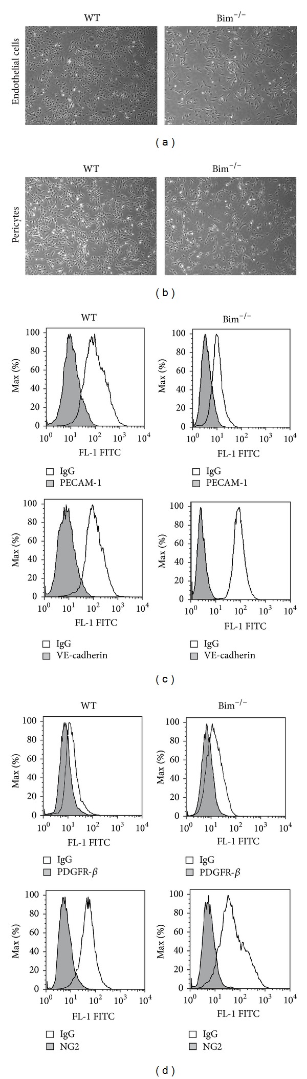
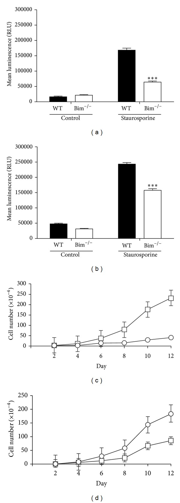
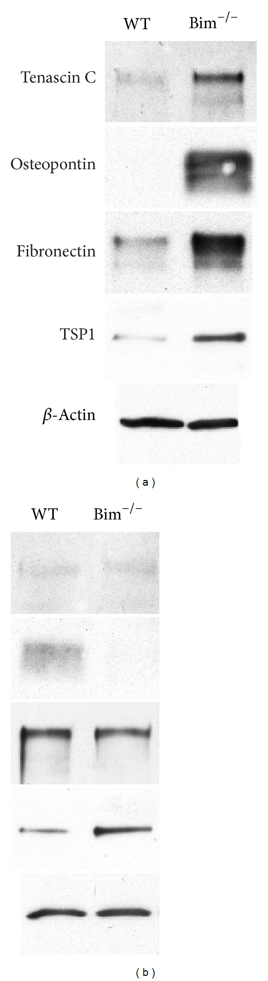
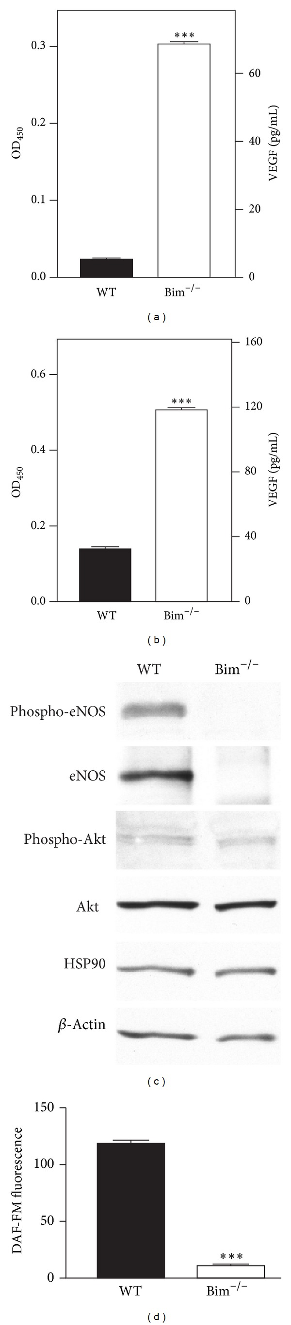
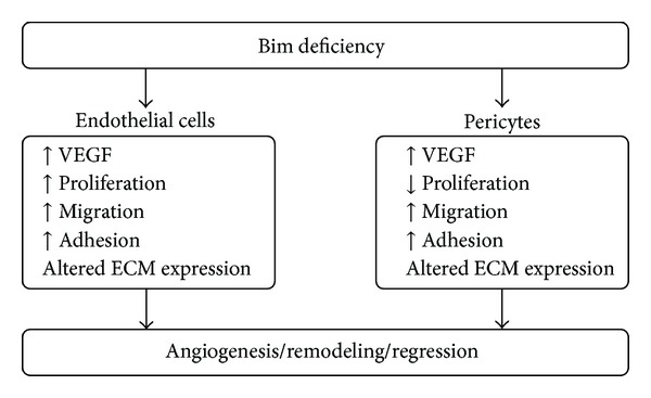
Apoptosis of vascular cells, including pericytes and endothelial cells, contributes to disease pathogenesis in which vascular rarefaction plays a central role. Bim is a proapoptotic protein that modulates not only apoptosis but also cellular functions such as migration and extracellular matrix (ECM) protein expression. Endothelial cells and pericytes each make a unique contribution to vascular formation and function although the details require further delineation. Here we set out to determine the cell autonomous impact of Bim expression on retinal endothelial cell and pericyte function using cells prepared from Bim deficient (Bim−/−) mice. Bim−/−endothelial cells displayed an increased production of ECM proteins, proliferation, migration, adhesion, and VEGF expression but, a decreased eNOS expression and nitric oxide production. In contrast, pericyte proliferation decreased in the absence of Bim while migration, adhesion, and VEGF expression were increased. In addition, we demonstrated that the coculturing of either wild-type or Bim−/−endothelial cells with Bim−/−pericytes diminished their capillary morphogenesis. Thus, our data further emphasizes the importance of vascular cell autonomous regulatory mechanisms in modulation of vascular function.
Overview
The claims and statistics of this paper haven’t been checked yet.
Free: it reads the open-access text and checks the first 5 claims that cite a source we can check. Create a free account to check every claim.
Integrity signals
Registry
Cites
Statistics
Figures




Verification
Claims the paper makes
The claims and statistics of this paper haven’t been checked yet.
Free: it reads the open-access text and checks the first 5 claims that cite a source we can check. Create a free account to check every claim.
Claims other papers make about this one
Each is checked against this paper’s own text: does it say what the citing paper says it does?
The source backs part of the claim, not all of it — or backs it for a different population, condition or comparison.
In the citing paper
“The impact BIM has both on apoptosis and the extracellular matrix milieu is integral to its role during vascular remodeling, regression and resolution of neovascularization [32, 41].”
From Thrombospondin-1, BIM and CFH polymorphisms and response to anti-VEGF treatment in neovascular age- related macular degeneration patients (2024) · cites this paper as [41]
Experimental — accuracy not yet measured
Self-check
Percentages, sums and reported differences re-computed from the paper’s own numbers.
The self-check needs the paper’s full text.
Citations
Cites
The BCL-2 protein family: opposing activities that mediate cell death
Youle, R. J., & Strasser, A. (2008) · Nature Reviews Molecular Cell Biology · doi:10.1038/nrm2308 · Cited by 4,679 · cited 1×
“Hindawi Publishing Corporation International Journal of Cell Biology Volume 2013, Article ID 297537, 15 pages http://dx.doi.org/10.1155/2013/297537”
BackgroundInfluentialAngiogenesis in life, disease and medicine
Carmeliet, P. (2005) · Nature · doi:10.1038/nature04478 · Cited by 3,482
Role of platelet-derived growth factors in physiology and medicine
Andrae, J., Gallini, R., & Betsholtz, C. (2008) · Genes & Development · doi:10.1101/gad.1653708 · Cited by 2,438
Bouillet, P., Metcalf, D., Huang, D. C. S., Tarlinton, D. M., Kay, T. W. H., Köntgen, F., Adams, J. M., & Strasser, A. (1999) · Science · doi:10.1126/science.286.5445.1735 · Cited by 1,504 · cited 1×
“Hindawi Publishing Corporation International Journal of Cell Biology Volume 2013, Article ID 297537, 15 pages http://dx.doi.org/10.1155/2013/297537”
BackgroundInfluentialFukumura, D., Gohongi, T., Kadambi, A., Izumi, Y., Ang, J., Yun, C.-O., Buerk, D. G., Huang, P. L., & Jain, R. K. (2001) · Proceedings of the National Academy of Sciences · doi:10.1073/pnas.041359198 · Cited by 1,020
A role for VEGF as a negative regulator of pericyte function and vessel maturation
Greenberg, J. I., Shields, D. J., Barillas, S. G., Acevedo, L. M., Murphy, E., Huang, J., Scheppke, L., Stockmann, C., Johnson, R. S., Angle, N., et al. (2008) · Nature · doi:10.1038/nature07424 · Cited by 629
Development of the retinal vasculature
Fruttiger, M. (2007) · Angiogenesis · doi:10.1007/s10456-007-9065-1 · Cited by 505 · cited 1×
“Remodeling and pruning of the retinal vasculature continue until P42 [7, 8].”
BackgroundDevelopment and pathology of the hyaloid, choroidal and retinal vasculature
Saint-Geniez, M., & D'amore, P. A. (2004) · The International Journal of Developmental Biology · doi:10.1387/ijdb.041895ms · Cited by 416
Regulatory phosphorylation of Bim: sorting out the ERK from the JNK
Ley, R., Ewings, K. E., Hadfield, K., & Cook, S. J. (2005) · Cell Death & Differentiation · doi:10.1038/sj.cdd.4401688 · Cited by 313
Degenerative Disorders Caused by Bcl-2 Deficiency Prevented by Loss of Its BH3-Only Antagonist Bim
Bouillet, P., Cory, S., Zhang, L.-C., Strasser, A., & Adams, J. M. (2001) · Developmental Cell · doi:10.1016/s1534-5807(01)00083-1 · Cited by 274
Cited by
The role of RNA uptake in platelet heterogeneity
Beaulieu, L. M., Tanrıverdi, K., Freedman, J. E., & Clancy, L. (2017) · Thrombosis and Haemostasis · doi:10.1160/th16-11-0873 · Cited by 98
“Both BIM and BAX have previously been linked to platelet clearance and vascular cell apoptosis, though the extent of the role they play is not fully understood (35-39).”
BackgroundFerro, A., Marinato, G. M., Mulargiu, C., Marino, M., Pasello, G., Guarneri, V., & Bonanno, L. (2024) · Critical Reviews in Oncology/Hematology · doi:10.1016/j.critrevonc.2024.104295 · Cited by 82
Jamali, N., Wang, S., Darjatmoko, S. R., Sorenson, C. M., & Sheibani, N. (2017) · PLoS ONE · doi:10.1371/journal.pone.0190131 · Cited by 47
Self-citationWang, S., Zaitoun, I. S., Johnson, R. P., Jamali, N., Gürel, Z., Wintheiser, C. M., Strasser, A., Lindner, V., Sheibani, N., & Sorenson, C. M. (2017) · PLoS ONE · doi:10.1371/journal.pone.0178198 · Cited by 26
Self-citationNegative feedback between TAp63 and Mir-133b mediates colorectal cancer suppression
Dai, J., Wu, H., Zhang, Y., Gao, K., Hu, G. M., Guo, Y., Lin, C., & Li, X. (2016) · Oncotarget · doi:10.18632/oncotarget.13515 · Cited by 19
“In our previous experiments, cells were counted on consecutive days for 1 week using a hematocytometer [36], and this culture time is too long to maintain stable gene overexpression.”
Method
Metadata
- Authors
- Morrison, M. E., Palenski, T. L., Jamali, N., Sheibani, N., & Sorenson, C. M.
- Affiliations
- University of Wisconsin–Madison (US)
- Corresponding author
- Sorenson, Christine M.
- Venue
- International Journal of Cell Biology
- Publisher
- Wiley
- Published
- 2013
- Volume
- 2013
- Pages
- 1-15
- ISSN
- 1687-8876 (ISSN-L)
- 1687-8884
- PMC
- PMC3833316
- OpenAlex
- W1979620853
- Cited by
- 12
- References
- 28
- Citations by year
- 2024: 3 · 2023: 1 · 2022: 2 · 2021: 1 · 2020: 1 · 2017: 3 · 2016: 1
- FWCI
- 0.15
- Type
- journal article
- Language
- en
- Open access
- gold
- doi.org
- Locations
- International Journal of Cell Biology (publishedVersion, cc-by)
- Europe PMC (PubMed Central) (publishedVersion, cc-by)
- PubMed Central (publishedVersion, cc-by)
- PubMed (publishedVersion)
- DOAJ (DOAJ: Directory of Open Access Journals) (submittedVersion, cc-by-sa)
- Funders
- National Institutes of Health (EY 023024, EY016995, EY018179, RC4 EY 021357, P30 CA014520, T32 ES007015, 1-10-BS-160, P30 EY016665)
- American Diabetes Association (EY 023024, EY016995, EY018179, RC4 EY 021357, P30 CA014520, T32 ES007015, 1-10-BS-160, P30 EY016665)
- Retina Research Foundation (EY 023024, EY016995, EY018179, RC4 EY 021357, P30 CA014520, T32 ES007015, 1-10-BS-160, P30 EY016665)
- Carbone Cancer Center (EY 023024, EY016995, EY018179, RC4 EY 021357, P30 CA014520, T32 ES007015, 1-10-BS-160, P30 EY016665)
- Topics
- Life Sciences › Biochemistry, Genetics and Molecular Biology › Molecular Biology › Angiogenesis and VEGF in Cancer
- Life Sciences › Neuroscience › Neurology › Barrier Structure and Function Studies
- Life Sciences › Biochemistry, Genetics and Molecular Biology › Molecular Biology › Cell death mechanisms and regulation
- Keywords
- building information modeling, apoptosis, retinal endothelial cells, pericytes, vascular rarefaction, extracellular matrix proteins, cell migration, cell adhesion, VEGF expression, endothelial nitric oxide synthase, nitric oxide, endothelial cell proliferation, capillary morphogenesis, cell-autonomous regulation
About these checks
Registry, retraction, Cites and statistics results are computed, not judged, with one exception: a printed reference that no index lists is read once by a model before it counts as a citation error. Claims are extracted by a model and kept only when their quote appears verbatim in the text; each cited source is then checked, with a model’s help, for whether it says what the claim relies on. Those verdicts can be wrong — read the claim’s quote and the cited source before relying on one.
Statistics and arithmetic are recomputed, not judged.
Confidence bands are shown only for fields calibrated on our annotated test set. None is calibrated yet, so no band is shown.
What counts as a claim ›How we measure confidence ›How we check this ›
Are you an author, or do you see a problem with this page? Request a review
How to cite
Cite CoChat for the checks on this page, and the paper for its own findings.
- This page
- CoChat. (2026). Citation check: Modulation of Vascular Cell Function by Bim Expression. https://app.cochat.ai/papers/10.1155/2013/297537
- The paper
- Morrison, M. E., Palenski, T. L., Jamali, N., Sheibani, N., & Sorenson, C. M. (2013). Modulation of Vascular Cell Function by Bim Expression. International Journal of Cell Biology. https://doi.org/10.1155/2013/297537
Data sources
Bibliographic data: Semantic Scholar, OpenAlex, Crossref.
Contains information from the Semantic Scholar Academic Graph Datasets, made available under the ODC Attribution License (ODC-BY 1.0).
Semantic Scholar’s data is described in: Kinney, R., et al. (2023). The Semantic Scholar Open Data Platform. arXiv:2301.10140.
