

The DOI matches its Crossref record; 45 of the 48 references it cites were found in a registry or an index.
The Role of Intrinsic Pathway in Apoptosis Activation and Progression in Peyronie’s Disease
Open accessAbstract
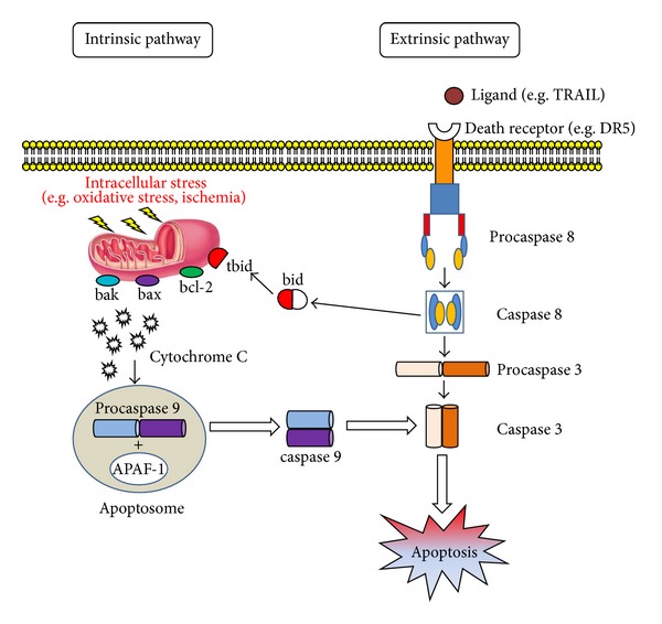
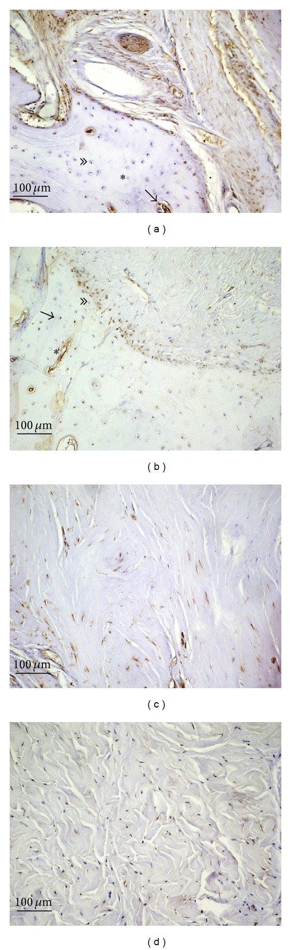
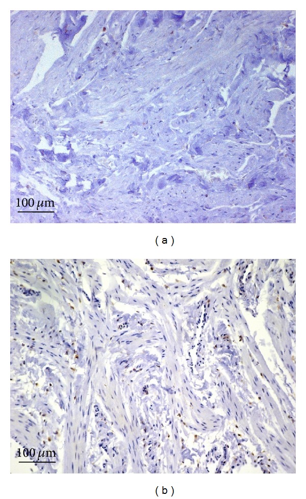
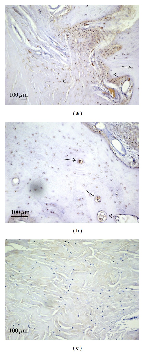
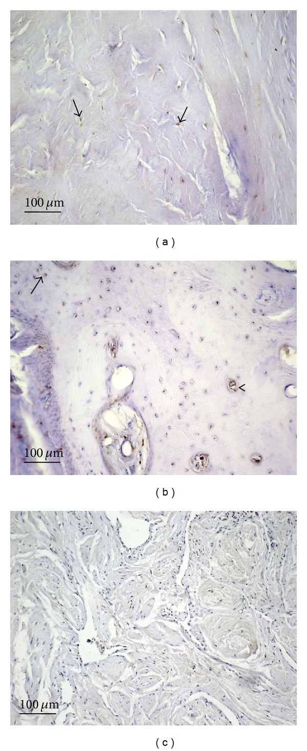
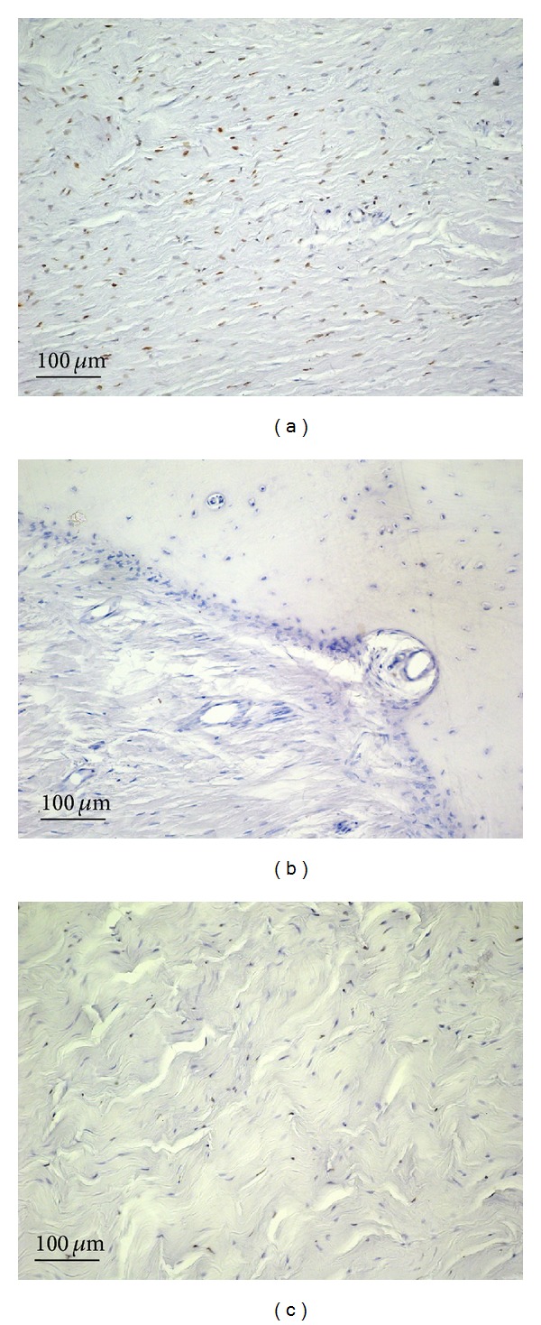
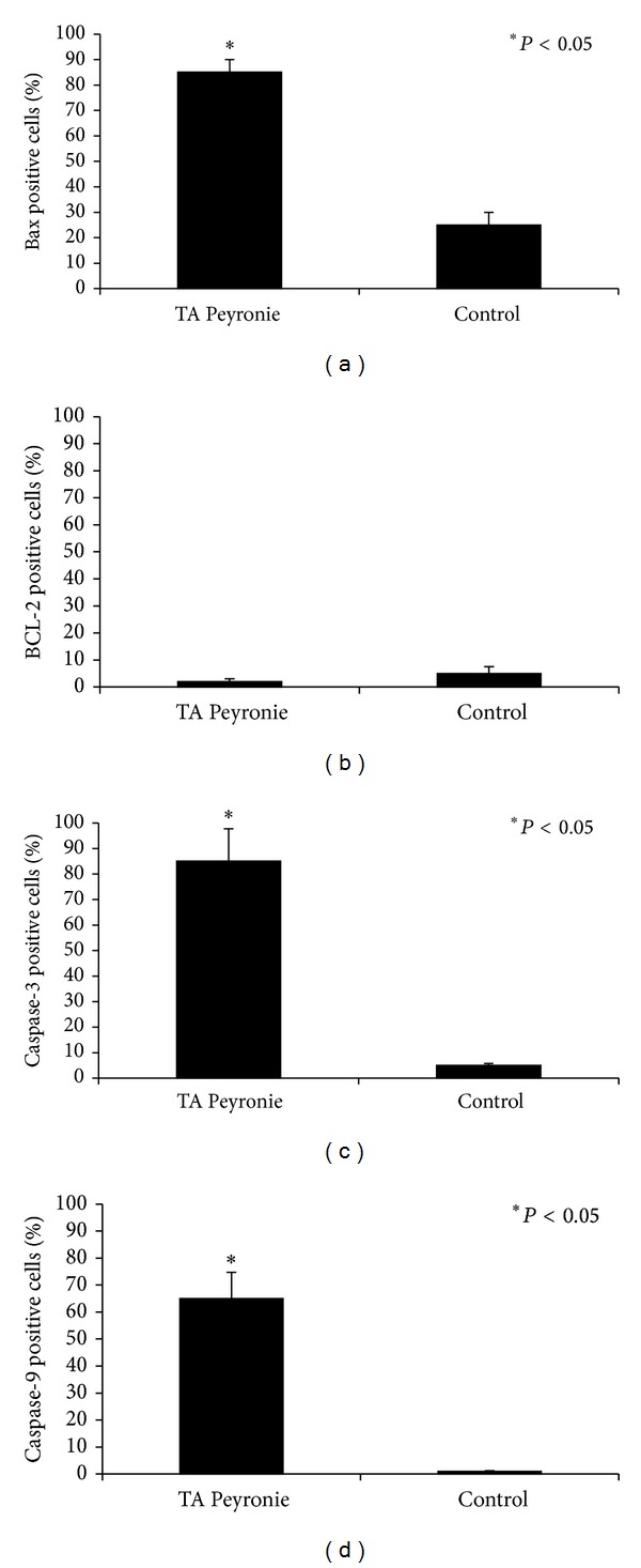
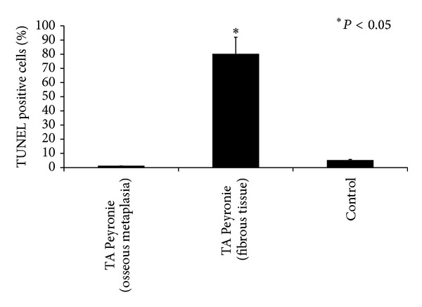
Peyronie’s disease (PD) is characterized with formation of fibrous plaques which result in penile deformity, pain, and erectile dysfunction. The aim of this study was to investigate the activation of the intrinsic apoptotic pathway in plaques from PD patients. Tunica albuginea from either PD or control patients was assessed for the expression of bax, bcl-2 and caspases 9 and 3 using immunohistochemistry and by measurement of apoptotic cells using TUNEL assay. Bax overexpression was observed in metaplastic bone tissue, in fibroblasts, and in myofibroblast of plaques from PD patients. Little or no bcl-2 immunostaining was detected in samples from either patients or controls. Caspase 3 immunostaining was very strong in fibrous tissue, in metaplasic bone osteocytes, and in primary ossification center osteoblasts. Moderate caspase 9 immunostaining was seen in fibrous cells plaques and in osteocytes and osteoblasts of primary ossification centers from PD patients. Control samples were negative for caspase 9 immunostaining. In PD patients the TUNEL immunoassay showed intense immunostaining of fibroblasts and myofibroblasts, the absence of apoptotic cells in metaplasic bone tissue and on the border between fibrous and metaplastic bone tissue. Apoptosis occurs in stabilized PD plaques and is partly induced by the intrinsic pathway.
Overview
The claims and statistics of this paper haven’t been checked yet.
Run the check — free, no account needed. It reads the open-access text and checks the sources it cites.
Integrity signals
Registry
Cites
Statistics
Figures
Verification
Claims the paper makes
The claims and statistics of this paper haven’t been checked yet.
Run the check — free, no account needed. It reads the open-access text and checks the sources it cites.
Claims other papers make about this one
Each is checked against this paper’s own text: does it say what the citing paper says it does?
The source backs part of the claim, not all of it — or backs it for a different population, condition or comparison.
In the citing paper
“These pathways often interact and crosstalk with each other, amplifying fibrotic processes and tissue remodeling in the penile tissue.”
From Research advances in Peyronie's disease: a comprehensive review on genomics, pathways, phenotypic manifestation, and therapeutic targets. (2024) · cites this paper as [79]
Experimental — accuracy not yet measuredThe cited source says what the claim attributes to it.
In the citing paper
“Elevated levels of Bcl-2 prevent the release of cytochrome c from mitochondria, a crucial step in initiating apoptosis.”
From Research advances in Peyronie's disease: a comprehensive review on genomics, pathways, phenotypic manifestation, and therapeutic targets. (2024) · cites this paper as [79]
Self-check
Percentages, sums and reported differences re-computed from the paper’s own numbers.
The self-check needs the paper’s full text.
Citations
Cites
Bcl-2 heterodimerizes in vivo with a conserved homolog, Bax, that accelerates programed cell death
Oltval, Z. N., Milliman, C. L., & Korsmeyer, S. J. (1993) · Cell · doi:10.1016/0092-8674(93)90509-o · Cited by 6,003
Bcl-2 gene promotes haemopoietic cell survival and cooperates with c-myc to immortalize pre-B cells
Vaux, D., Cory, S., & Adams, J. (1988) · Nature · doi:10.1038/335440a0 · Cited by 3,355
Yin, X.-M., Oltvai, Z., & Korsmeyer, S. (1994) · Nature · doi:10.1038/369321a0 · Cited by 1,383
Bcl-2 initiates a new category of oncogenes: regulators of cell death
Korsmeyer, S. J. (1992) · Blood · doi:10.1182/blood.v80.4.879.bloodjournal804879 · Cited by 1,115
(2005) · doi:10.1016/j.ucl.2005.08.007 · Cited by 759 · cited 1×
“Fibrosis, its major pathological manifestation, arises from fibroblast proliferation and accumulation of extracellular matrix; PD progresses with formation of plaques or even ectopic calcification having the appearance of scar tissue, which prevents TA expansion during erections [6, 7].”
BackgroundCONGENITAL CURVATURE OF THE PHALLUS: REPORT OF THREE CASES WITH DESCRIPTION OF CORRECTIVE OPERATION.
Nesbit, R. M. (1965) · Transactions of the American Association of Genito-Urinary Surgeons · doi:10.1016/s0022-5347(17)63751-0 · Cited by 471 · cited 1×
“Control samples were specimens from 4 patients (mean age 23 ± 3 years; range 21–27) with congenital penile curvature who underwent Nesbit’s corrective procedure [22].”
BackgroundProposal: Trauma as the Cause of the Peyronie's Lesion
Devine, C. J. J., Somers, K. D., Jordan, G. H., & Schlossberg, S. M. (1997) · Journal of Urology · doi:10.1016/s0022-5347(01)65361-8 · Cited by 381
The apoptosis cascade — morphological and immunohistochemical methods for its visualization
Huppertz, B., Frank, H., & Kaufmann, P. (1999) · Anatomy and Embryology · doi:10.1007/s004290050254 · Cited by 325 · cited 1×
“These complex events lead to apoptotic death, with collapse of the nucleus and of the cell itself and DNA fragmentation [31].”
BackgroundInsights into Programmed Cell Death through Structural Biology
Fesik, S. W. (2000) · Cell · doi:10.1016/s0092-8674(00)00119-7 · Cited by 317
Surgery of the Penis and Urethra
Jordan, G., & Mccammon, K. (2012) · doi:10.1016/b978-1-4160-6911-9.00036-0 · Cited by 216
Research works printed but not linked: 7
- 10.1016/S0094-0143(05)70146-4 · DOI
- 2012, Advances in Clinical and Experimental Medicine, 21, 607
- 2012, Sinaling pathway that regulate life and cell death: evolution of apoptosis in the contex of self-defence, 124
- 10.1038/aja.2012.9 · DOI
- 1992, Blood, 80, 879, 10.1182/blood.V80.4.879.879 · DOI
- 10.1016/S0022-5347(01)65360-6 · DOI
- 10.1095/biolreprod64.3.974 · DOI
Cited by
Sorted by citations
Items 101–200 of 238
Cheng, Q., Lou, G., Huang, W.-H., & Li, X.-D. (2017) · Environmental science and pollution research international · doi:10.1007/s11356-017-9059-5 · Cited by 24
Adaptive rhythm sequencing: A method for dynamic rhythm classification during CPR.
Kwok, H., Coult, J., Drton, M., Rea, T., & Sherman, L. (2015) · Resuscitation · doi:10.1016/j.resuscitation.2015.02.031 · Cited by 24
Khodamoradi, K., Amini-Khoei, H., Khosravizadeh, Z., Hosseini, S. R., Dehpour, A. R., & Hassanzadeh, G. (2019) · Reproductive Biology · doi:10.1016/j.repbio.2019.10.003 · Cited by 26
Gupta, R., Pandey, P., Singh, S., Singh, D., Saxena, A., Luqman, S., Bawankule, D., & Banerjee, S. (2016) · Protoplasma · doi:10.1007/s00709-015-0875-5 · Cited by 23
Emojevwe, V. O., Nwangwa, E. K., Naiho, A. O., Oyovwi, M. O., & Ben‐Azu, B. (2022) · Middle East Fertility Society Journal · doi:10.1186/s43043-022-00096-5 · Cited by 23
Chaudhry, G., Jan, R., Zafar, M., Mohammad, H., & Muhammad, T. (2019) · Asian Pacific Journal of Cancer Prevention · doi:10.31557/apjcp.2019.20.12.3555 · Cited by 22
“While caspase-8 plays a major role in the extrinsic pathway of apoptosis (Loreto et al., 2014).”
BackgroundShah, N., & Patel, B. M. (2016) · Biomedicine & pharmacotherapy = Biomedecine & pharmacotherapie · doi:10.1016/j.biopha.2016.07.041 · Cited by 22
Insights on aspects of apoptosis in neurodegenerative disorders: a comprehensive review
Goyal, R., Wilson, K., Saharan, A., Gautam, R., Chopra, H., Gupta, S., & Kamal, M. A. (2024) · Exploration of Medicine · doi:10.37349/emed.2024.00208 · Cited by 21
The recent colonization of south Brazil by the Azores chromis Chromis limbata
Anderson, A. B., Salas, E., Rocha, L. A., & Floeter, S. R. (2017) · Journal of Fish Biology · doi:10.1111/jfb.13363 · Cited by 24
Cytochrome c: Using Biological Insight toward Engineering an Optimized Anticancer Biodrug
Delinois, L. J., León-Vélez, O. D., Vázquez-Medina, A., Vélez-Cabrera, A., Marrero-Sánchez, A., Nieves-Escobar, C., Alfonso-Cano, D., Caraballo-Rodríguez, D., Rodriguez-Ortiz, J., Acosta‐Mercado, J., et al. (2021) · Inorganics · doi:10.3390/inorganics9110083 · Cited by 22
“Apoptosis is a genetically directed process of cell self-destruction that occurs in multicellular organisms, which can take place either through the extrinsic or the intrinsic pathway (Figure 9) [46].”
BackgroundIodine-catalyzed C3-formylation of indoles using hexamethylenetetramine and air
Wang, Q.-D., Yang, J.-M., Fang, D., Ren, J.-M., & Zeng, B.-B. (2017) · doi:10.1016/j.tetlet.2017.05.050 · Cited by 20
Boukraa, S., Grandiere, M. A. D. L., Bawin, T., Raharimalala, F. N., Zimmer, J., Haubruge, E., Thiry, E., & Francis, F. (2016) · Preventive Veterinary Medicine · doi:10.1016/j.prevetmed.2015.12.013 · Cited by 20
Plant-produced viral bovine vaccines: what happened during the last 10 years?
Ruiz, V., Mozgovoj, M., Santos, M. D. D., & Wigdorovitz, A. (2015) · Plant Biotechnology Journal · doi:10.1111/pbi.12440 · Cited by 20
Antimicrobial Food Additives and Disinfectants
Nair, M. S., Upadhyaya, I., Amalaradjou, M. A. R., & Venkitanarayanan, K. (2016) · Wiley eBooks · doi:10.1002/9781119139188.ch12 · Cited by 29
Vitamin E Mitigates Polystyrene-Nanoplastic-Induced Visual Dysfunction in Zebrafish Larvae
Saputra, F., Pramata, A. D., Soegianto, A., & Hu, S. (2025) · International Journal of Molecular Sciences · doi:10.3390/ijms26031216 · Cited by 23
“The activation of BCL-2 family members bax and bak , along with caspase-3, is a crucial step in the intrinsic apoptosis pathway [56].”
Peptide-functionalized nanoparticles for the selective induction of apoptosis in target cells.
Sibuyi, N. R., Thovhogi, N., Gabuza, K., Meyer, M., Drah, M., Onani, M., Skepu, A., Madiehe, A., & Meyer, M. (2017) · Nanomedicine · doi:10.2217/nnm-2017-0085 · Cited by 19
Biomaterials to suppress cancer stem cells and disrupt their tumoral niche.
Garcia-Mazas, C., Csaba, N., & Garcia-Fuentes, M. (2017) · International journal of pharmaceutics · doi:10.1016/j.ijpharm.2016.12.013 · Cited by 19
Maddaloni, E., D’Onofrio, L., & Pozzilli, P. (2015) · Diabetes/Metabolism Research and Reviews · doi:10.1002/dmrr.2762 · Cited by 19
Hou, X.-J., Du, Q., Li, R., Zhou, M., Wang, H., Wang, L., Guo, C., Chen, T.-B., & Shaw, C. (2015) · Chemical Biology and Drug Design · doi:10.1111/cbdd.12396 · Cited by 19
Candidiasis, Bacterial Vaginosis, Trichomoniasis and Other Vaginal Conditions Affecting the Vulva
Vieira‐Baptista, P., & Bornstein, J. (2019) · Springer eBooks · doi:10.1007/978-3-319-61621-6_24 · Cited by 18
Nano-TRAIL: a promising path to cancer therapy
Gampa, S. C., Garimella, S., & Pandrangi, S. (2023) · Cancer Drug Resistance · doi:10.20517/cdr.2022.82 · Cited by 17
Interplay Between Non-Coding RNAs and Programmed Cell Death Proteins
Ghafouri‐Fard, S., Hussen, B. M., Mohaqiq, M., Shoorei, H., Baniahmad, A., Taheri, M. N., & Jamali, E. (2022) · Frontiers in Oncology · doi:10.3389/fonc.2022.808475 · Cited by 18
“Consequently, the initiator caspase-9 can cleave and upregulate the executioner caspases-3/6/7, leading to cell apoptosis (42, 43).”
BackgroundYuan, C.-H., Ma, Y.-L., Shih, P.-C., Chen, C., Cheng, S.-Y., Pan, C.-Y., Jean, Y., Chu, Y.-M., Lin, S., Lai, Y.-C., et al. (2020) · Biochemical Pharmacology · doi:10.1016/j.bcp.2020.114064 · Cited by 17
Amara, I., Timoumi, R., Annabi, E., Salem, I. B., & Abid-Essefi, S. (2019) · Cell stress & chaperones (Print) · doi:10.1007/s12192-019-01060-5 · Cited by 17
Keeran, N. S., Ganesan, G., & Parida, A. K. (2016) · Transgenic Research · doi:10.1007/s11248-016-0002-1 · Cited by 24
Park, J. Y., Park, W., Song, G., Jung, S.-J., Kim, B., Choi, M.-J., Kim, S. H., Park, J., Kwak, H., Ahn, K., et al. (2023) · Frontiers in Pharmacology · doi:10.3389/fphar.2023.1039622 · Cited by 16
“In particular, the intrinsic apoptotic pathway is activated by various microenvironmental changes such as reduction of growth factors, DNA damage, ER stress, active oxygen overload, and DNA replication stress (Loreto et al., 2014).”
BackgroundMicrobial Interactions and Perspectives for Bioremediation of Pesticides in the Soils
Prabha, R., Singh, D. P., & Verma, M. (2017) · Springer eBooks · doi:10.1007/978-981-10-6593-4_27 · Cited by 16
Lerat, Y., Cornish, M. L., & Critchley, A. T. (2018) · Blue Biotechnology · doi:10.1002/9783527801718.ch5 · Cited by 16
Fenning, R. M., Steinberg-Epstein, R., Butter, E. M., Chan, J. G. M., McKinnon-Bermingham, K., Hammersmith, K. J., Moffitt, J., Shui, A. M., Parker, R. A., Coury, D. L., et al. (2020) · Journal of Autism and Developmental Disorders · doi:10.1007/s10803-020-04420-8 · Cited by 20
Melo, Â., Veríssimo, R., Farinha, M., Martins, N. N., & Martins, F. N. (2018) · Journal of Obstetrics and Gynaecology Research · doi:10.1080/01443615.2018.1457632 · Cited by 15
Bortolini, C., Pivato, A., Bogialli, S., & Pastore, P. (2015) · Analytical and Bioanalytical Chemistry · doi:10.1007/s00216-015-8801-4 · Cited by 15
Data Reliability and Quality in Body Area Networks for Diabetes Monitoring
Huzooree, G., Khedo, K. K., & Joonas, N. (2018) · EAI/Springer Innovations in Communication and Computing · doi:10.1007/978-3-030-00865-9_4 · Cited by 13
Chi, J., Bi, W., Lou, K., Ma, J., Wu, J., & Cui, Y. (2024) · Sexual Medicine Reviews · doi:10.1093/sxmrev/qeae006 · Cited by 14
Hassan, O., Mahmood, A. A. R., Hamzah, A. H., & Tahtamouni, L. (2022) · ChemistrySelect · doi:10.1002/slct.202203726 · Cited by 14
Ross, A., & Searle, M. S. (2019) · International Journal of Community Well-Being · doi:10.1007/s42413-019-00018-w · Cited by 15
Dash, T., & Konkimalla, V. (2017) · Pharmaceutical Research · doi:10.1007/s11095-017-2182-7 · Cited by 14
Amalia, E., Diantini, A., Prabandari, E. E., Waluyo, D., & Subarnas, A. (2022) · Journal of Experimental Pharmacology · doi:10.2147/jep.s365159 · Cited by 13
Lenis-Rojas, O. A., Cabral, R., Carvalho, B., Friães, S., Roma-Rodrigues, C., Fernández, J. A. A., Vila, S. F., Sánchez, L., Gomes, C. S. B., Fernandes, A. R., et al. (2021) · Inorganic Chemistry · doi:10.1021/acs.inorgchem.1c00527 · Cited by 13
Teoh, S., & Yap, M. K. K. (2020) · Toxin Reviews · doi:10.1080/15569543.2020.1799408 · Cited by 13
Martin, G., Landrock, D., Dangott, L., McIntosh, A., Kier, A., & Schroeder, F. (2018) · Lipids · doi:10.1002/lipd.12008 · Cited by 13
Doan, N. Q. H., Nguyen, N. T. K., Nguyen, N., Tran, T. T., Tran, Q. N., & Truong, T. (2023) · Biochimica et Biophysica Acta - General Subjects · doi:10.1016/j.bbagen.2023.130414 · Cited by 12
Martínez-Aguilar, K., Pérez-Legaspi, I. A., Ramírez-Fuentes, E., Trujillo-Tapia, M. N., & Ortega-Clemente, L. A. (2018) · Journal of Environmental Science and Health. Part B - Pesticides, Food Contaminants, and Agricultural Wastes · doi:10.1080/03601234.2018.1505070 · Cited by 12
Hyperleptinemia positively associated with central arterial stiffness in hemodialysis patients
Kuo, C.-H., Lin, Y.-L., Lee, C.-J., Wang, C.-H., Lai, Y.-H., Liou, H., & Hsu, B. (2018) · PLoS ONE · doi:10.1371/journal.pone.0190694 · Cited by 12
Shichkin, V., Gorbach, O., Zuieva, O. A., Grabchenko, N., Aksyonova, I. A., & Todurov, B. (2017) · Cryobiology · doi:10.1016/j.cryobiol.2017.06.010 · Cited by 12
Recombinant Production of 12 Catalytically Active Human CA Isoforms
Mickevičiūtė, A., Juozapaitienė, V., Michailovienė, V., Jachno, J., Matulienė, J., & Matulis, D. (2019) · Carbonic Anhydrase as Drug Target · doi:10.1007/978-3-030-12780-0_2 · Cited by 12
Kumari, R., Ray, A. G., Mukherjee, D., Chander, V., Kar, D., Kumar, U. S., Deepak Bharadwaj, P. V. P., Banerjee, S. K., Konar, A., & Bandyopadhyay, A. (2022) · Frontiers in Cardiovascular Medicine · doi:10.3389/fcvm.2022.798639 · Cited by 14
“Since Caspase 9 is one of the key components of the intrinsic apoptotic pathway (37, 38), its down regulation implies possible interference in the basic apoptotic signaling induced by the deficiency of PPAR α as similar results were observed in both the cardiac hypertrophy models.”
ResultZhang, Y., Tu, L., Lu, L.-L., Li, Y.-X., Song, L.-J., Qi, Q.-R., Song, H., Li, Z.-C., & Huang, W. (2020) · Analytical Methods · doi:10.1039/d0ay00173b · Cited by 11
Nguyen, C., Nguyen, K. V. A., Eom, S., Choi, Y., Kang, L.-J., Lee, J.-E., Kim, C., Lee, S., Lee, S., & Lee, J.-H. H. (2019) · Phytomedicine · doi:10.1016/j.phymed.2019.153096 · Cited by 11
Simultaneous study of mechanobiology and calcium dynamics on hESC‐derived cardiomyocytes clusters
Caluori, G., Přibyl, J., Čmiel, V., Pešl, M., Potocnak, T., Provazník, I., Skládal, P., & Rotrekl, V. (2018) · Journal of Molecular Recognition · doi:10.1002/jmr.2760 · Cited by 14
Kiptiyah, K., Widodo, W., Ciptadi, G., Aulanni'am, A., Widodo, M., & Sumitro, S. (2017) · Journal of Taibah University Medical Sciences · doi:10.1016/j.jtumed.2017.05.012 · Cited by 11
Selim, M. S., Mounier, M. M., Abdelhamid, S., Hamed, A. A., Elsoud, M. A. A., & Mohamed, S. (2024) · BMC Biotechnology · doi:10.1186/s12896-024-00829-6 · Cited by 10
“These proteins act as pro-or anti-apoptotic regulatory proteins [79].”
BackgroundHuang, Y., Hsia, T., Yeh, C.-A., Ma, Y., Hsu, S.-Y., Liu, Y., Lyu, P. C., Lai, K., Peng, S., Lien, J., et al. (2023) · Oxidative Medicine and Cellular Longevity · doi:10.1155/2023/3479688 · Cited by 9
“) proteins regulate intrinsic/mitochondrial pathways [34, 35].”
Sakellakis, M., & Flores, L. (2022) · Current Urology · doi:10.1097/cu9.0000000000000120 · Cited by 10
Guardian of genome on the tract: Wild type p53-mdm2 complex inhibition in healing the breast cancer
Janani, S. K., Dhanabal, S. P., Sureshkumar, R., Surya, N. U. S., & Karthika, C. (2021) · Gene · doi:10.1016/j.gene.2021.145616 · Cited by 13
Fieldhouse, A., & O'Leary, T. (2020) · BMJ Military Health · doi:10.1136/bmjmilitary-2020-001500 · Cited by 10
Toxicology of natural and synthetic aphrodisiacs
Hess, C., & Boehmer, A. (2020) · Rechtsmedizin · doi:10.1007/s00194-019-00368-x · Cited by 10
Angarita-Fonseca, A., Trask, C., Shah, T., & Bath, B. (2019) · BMC Public Health · doi:10.1186/s12889-019-7395-8 · Cited by 10
Pirnat, A., DeRoo, L., Skjærven, R., & Morken, N. (2018) · BMJ Open · doi:10.1136/bmjopen-2017-021188 · Cited by 10
Major Environmental Issues and Problems
Dowlath, M. J. H., Karuppannan, S. K., Rajadesingu, S., & Arunachalam, K. (2020) · Springer eBooks · doi:10.1007/978-3-319-58538-3_197-1 · Cited by 10
Ranjithkumar, R., Saravanan, K., Balaji, B., Hima, S., Sreeja, S., Timane, S. R., Kumar, M., Kabilan, S., & Ramanathan, M. (2021) · Journal of chemotherapy · doi:10.1080/1120009x.2021.1924935 · Cited by 9
Terhuerne, J., van Diepen, M., Kramann, R., Erpenbeck, J., Dekker, F., Marx, N., Floege, J., Becker, M., & Schlieper, G. (2021) · Clinical Kidney Journal · doi:10.1093/ckj/sfaa161 · Cited by 9
Puja, A. M., Xu, X.-Y., Wang, R.-B., Kim, H., & Kim, Y. (2022) · Cancer nanotechnology · doi:10.1186/s12645-022-00133-y · Cited by 8
Toxicity of rhizomes of the invasive Hedychium coronarium (Zingiberaceae) on aquatic species
Costa, R. O., Vieira, B. H., Espíndola, E. L. G., Ribeiro, A. I., Ferro, J. L. R., Fernandes, J. B., & Matos, D. M. S. (2021) · Biological Invasions · doi:10.1007/s10530-021-02501-y · Cited by 11
Ukwade, C. E., Ebuehi, O. A. T., Adisa, R. A., Singh, S. K., & Singh, R. (2020) · Journal of Ovarian Research · doi:10.1186/s13048-020-00679-8 · Cited by 7
Kotawong, K., Chaijaroenkul, W., Roytrakul, S., Phaonakrop, N., & Na-Bangchang, K. (2020) · Asian Pacific Journal of Cancer Prevention · doi:10.31557/apjcp.2020.21.3.621 · Cited by 8
“Finally, the supernatant was transferred into a new tube and protein concentration was measured the concentration by Lowry method (Lowry et al., 1951).”
MethodPropolis prevents inhibition of apoptosis by potassium bromate in CCD 841 human colon cell
Memmedov, H., Oktay, L. M., Durmaz, B., Günel, N., Yıldırım, H., & Sözmen, E. (2020) · Cell Biochemistry and Function · doi:10.1002/cbf.3475 · Cited by 8
“The intrinsic pathway is regulated by the mitochondria, where the proapoptotic proteins such as cytochrome-c are released into the cytosol and activate the multimeric complex (apoptosome), which is composed of apoptosis protease activating factor-1 (APAF-1), and procaspase-9 leads to caspase-9 activation followed by c…”
BackgroundGhatebi, M., Zavareh, S., Lashkarbolouki, T., & Salmani, M. E. (2019) · The journal of obstetrics and gynaecology research · doi:10.1111/jog.14007 · Cited by 8
Unusual complication of a tattoo in an immunosuppressed patient
Wilson, W., O'Boyle, M., & Leach, W. (2018) · BMJ Case Reports · doi:10.1136/bcr-2018-224968 · Cited by 8
Coêlho, A. G., Lima, J. F. D. S., Moura, A. K. S., de Sousa, T. O., Morais, I. C. P. D. S., Carvalho, G. D., Cunha, F. V. M., de Medeiros, M. D. G. F., Vasconcelos, E. A., de Oliveira, A. P., et al. (2015) · doi:10.1016/j.indcrop.2015.10.033 · Cited by 8
Cyanobacterial Degradation of Organophosphorus Pesticides
Nimisha Vijayan, P., & Abdulhameed, S. (2020) · Applied environmental science and engineering for a sustainable future · doi:10.1007/978-3-030-39137-9_12 · Cited by 8
Razack, S. A., Surendhiran, D., Kozani, P. S., Kozani, P. S., Hamidi, M., Cabrera‐Barjas, G., & Delattre, C. (2021) · Algal Research · doi:10.1016/j.algal.2021.102556 · Cited by 14
Kotawong, K., Chajaroenkul, W., Roytrakul, S., Phaonakrop, N., & Na‐Bangchang, K. (2021) · Asian Pacific Journal of Cancer Prevention · doi:10.31557/apjcp.2021.22.3.909 · Cited by 9
“Various signaling pathways are activated through the p53, followed by bcl2, bax, cytochrome c (Cytc), caspase 9, caspase 3, and apoptosis (Loreto et al., 2014).”
BackgroundFriedl, K. E., & Gifford, R. M. (2020) · BMJ Military Health · doi:10.1136/bmjmilitary-2020-001568 · Cited by 9
Wang, X.-J., Chen, X., Yang, Z., Dou, H.-B., Lu, L., Bi, J., Zou, L., Yu, J., & Bao, L. (2018) · Genes, Chromosomes and Cancer · doi:10.1002/gcc.22540 · Cited by 7
Faverjon, C., Leblond, A., Lecollinet, S., Bødker, R., de Koeijer, A. D., & Fischer, E. (2017) · Transboundary and Emerging Diseases · doi:10.1111/tbed.12577 · Cited by 7
Effects of donor cells' sex on nuclear transfer efficiency and telomere lengths of cloned goats.
Liu, H.-J., Peng, H., Hu, C.-C. T., Li, X.-Y., Zhang, J.-L., Zheng, Z., & Zhang, W.-C. (2016) · Reproduction in domestic animals = Zuchthygiene · doi:10.1111/rda.12752 · Cited by 7
Sayed, M. M., Nabil, Z., El-Shenawy, N., Al-Eisa, R. A., & Nafie, M. S. (2023) · Pharmaceuticals · doi:10.3390/ph16111524 · Cited by 6
“Apoptotic protease activating factor (APAF-1) is then recruited by this multimeric complex [32,33].”
Broggi, G., Angelico, G., Filetti, V., Ledda, C., Lombardo, C., Vitale, E., Rapisarda, V., Loreto, C., & Caltabiano, R. (2021) · International Journal of Environmental Research and Public Health · doi:10.3390/ijerph18126249 · Cited by 6
Self-citationTavallaei, O., Heidarian, M., Marzbany, M., & Aliabadi, A. (2021) · Iranian Journal of Basic Medical Sciences · doi:10.22038/ijbms.2021.53845.12103 · Cited by 6
Review—Aptamer-Based Electrochemical Sensing Strategies for Breast Cancer
Sunday, C. E., & Chowdhury, M. (2021) · doi:10.1149/1945-7111/abe34d · Cited by 6
Anticancer activity of a 1,4-dihydropyridine in DMBA-induced mouse skin tumor model
Manna, D., Akhtar, S., Maiti, P., Mondal, S., Mandal, T. K., & Ghosh, R. (2020) · Anti-Cancer Drugs · doi:10.1097/cad.0000000000000887 · Cited by 6
Karaszewski, J., Zaręba, I., Guszczyn, T., Darewicz, J., & Palka, J. A. (2019) · Molecular and Cellular Probes · doi:10.1016/j.mcp.2019.101488 · Cited by 6
The study of Zr adsorption on nanodispersed hydroxyapatite: X-ray photoelectron study
Teterin, Y., Kazakov, A. G., Teterin, A., Severin, A., Dvorak, S. V., Maslakov, K., & Ivanov, K. (2019) · Journal of Radioanalytical and Nuclear Chemistry · doi:10.1007/s10967-019-06586-7 · Cited by 6
Coffee consumption in hemodialysis patients: how many?
Caetano, C. G., Valente, A., Oliveira, T. S., & Garagarza, C. A. (2018) · European Journal of Clinical Nutrition · doi:10.1038/s41430-018-0328-2 · Cited by 7
Shroff, G. (2018) · The Neuroradiology Journal · doi:10.1177/1971400917742470 · Cited by 6
Recombinant Therapeutic Molecules Produced in Plants
Chen, Q. (2018) · Advances in botanical research · doi:10.1016/bs.abr.2017.11.006 · Cited by 6
Al-Ali, A., Al-Tamimi, S. Q., Al-Maliki, S., & Abdullah, M. A. (2022) · Applied Nanoscience · doi:10.1007/s13204-021-02248-x · Cited by 5
“Intrinsic apoptotic pathway involves intracellular signals which act within the cell as a result of the non-receptor-mediated stimuli, and are mitochondrialbased events (Elmore 2007; Loreto et al. 2014).”
BackgroundSmirnova, I., Drăghici, G., Kazakova, O., Vlaia, L., Avram, Ș., Mioc, A., Mioc, M., Macașoi, I., Dehelean, C., Voicu, A., et al. (2021) · Bioorganic chemistry (Print) · doi:10.1016/j.bioorg.2021.105535 · Cited by 5
Adeowo, F. Y., Lawal, M. M., & Kumalo, H. M. (2020) · ChemistrySelect · doi:10.1002/slct.202003573 · Cited by 6
Ibáñez, Á. G. S., Estrada, C. S. M. L., Favier, G., Vega, A., Stagnitta, P. V., Mattar, M. A., Zolezzi, G., Carbonari, C., Miliwebsky, E., Cortiñas, T. I., et al. (2018) · Journal of food safety · doi:10.1111/jfs.12488 · Cited by 5
Tsuboi, C., Kawasaki, Y., Yoshitome, K., Yagi, K., Miura, T., Esumi, S., Miyazaki, I., Asanuma, M., Kitamura, Y., & Sendo, T. (2016) · Environmental science and pollution research international · doi:10.1007/s11356-016-6332-y · Cited by 5
Castorina, A., Loreto, C., Vespasiani, G., Giunta, S., Musumeci, G., Castorina, S., Bašić, D., & Sansalone, S. (2016) · Histology and Histopathology · doi:10.14670/hh-11-755 · Cited by 5
“…fibrin, plasminogen activator inhibitor-1, tissue inhibitors of metalloproteinases, etc) and antifibrotic (metalloproteases, etc.) processes (Somers and Dawson, 1997; Hellstrom and Bivalacqua, 2000; Gelbard, 2008; Gholami and Lue, 2011; Zorba et al., 2012; Gokce et al., 2014; Loreto et al., 2014).”
BackgroundSelf-citationInterface between Periodontal Tissues and Dental Materials: Dynamic Changes and Challenges
Isola, G. (2021) · Coatings · doi:10.3390/coatings11050485 · Cited by 4
Lazalde-Ramos, B., Zamora-Pérez, A., Ortega-Guerrero, A. I., Quintero-Fraire, S. Z., Palacios-Lara, O., Quirarte-Báez, S. M., Galaviz-Hernández, C., Sosa-Macías, M., Ortiz-García, Y. M., & Morales-Velazquez, G. (2020) · Journal of Medicinal Food · doi:10.1089/jmf.2019.0199 · Cited by 4
“13,14 The genomic instability can be evaluated through comet test (single-cell gel electrophoresis assay), FISH (fluores-cence in situ hybridization), CGH (comparative genomic hybridization), TUNEL (terminal deoxynucleotidyl transferase [TdT] dUTP nick-end labeling) assay, micronucleus (MN) assay,…”
Gibby, C. L. K., Palacios, C. S., Campos, M., Lim, E., & Banna, J. C. (2018) · BMC Obesity · doi:10.1186/s40608-018-0219-z · Cited by 3
Asgari, R., Bidmeshkipour, A., Mansouri, K., Bakhtiari, M., Mozafari, H., & Abdolmaleki, A. (2022) · The Indian journal of medical research · doi:10.4103/ijmr.ijmr_2058_19 · Cited by 3
Kadir, N.-A., Acosta, A., Sarmiento, M., & Norazmi, M. (2020) · Pathogens · doi:10.3390/pathogens9121000 · Cited by 3
Pasquali, C., Deletis, V., & Sala, F. (2020) · Child's Nervous System · doi:10.1007/s00381-020-04746-9 · Cited by 3
Efficacy of Combining PRP and MMP Inhibitors in Treating Moderately Damaged Tendons Ex Vivo
Jafari, L., Hassanisaber, H., Savard, M., Gobeil, F., & Langelier, È. (2019) · Journal of Orthopaedic Research · doi:10.1002/jor.24319 · Cited by 3
Metadata
- Authors
- Loreto, C., La Rocca, G., Anzalone, R., Caltabiano, R., Vespasiani, G., Castorina, S., Ralph, D. J., Cellek, S., Musumeci, G., Giunta, S., Djinovic, R., Basic, D., & Sansalone, S.
- Affiliations
- University of Catania (IT)
- Department of Experimental Biomedicine and Clinical Neuroscience, University of Palermo, 90100 Palermo, Italy
- University of Palermo (IT)
- University of Rome Tor Vergata (IT)
- University College Hospital (GB)
- Cranfield University (GB)
- Sava Perovic Foundation, Center for Genito-Urinary Reconstructive Surgery, 11000 Belgrade, Serbia
- Clinic of Urology, Clinical Center Nis, 18000 Nis, Serbia
- Corresponding author
- Loreto, Carla
- ORCID
- Venue
- BioMed Research International
- Publisher
- Wiley
- Published
- 2014
- Volume
- 2014
- Pages
- 1-10
- ISSN
- 1110-7243 (ISSN-L)
- 2314-6133
- 2314-6141
- PMC
- PMC4147380
- OpenAlex
- W1973666710
- Cited by
- 108
- References
- 48
- Citations by year
- 2026: 1 · 2025: 6 · 2024: 4 · 2023: 14 · 2022: 20 · 2021: 22 · 2020: 15 · 2019: 12 · 2018: 4 · 2017: 3 · 2016: 5 · 2015: 2
- FWCI
- 2.77
- Type
- journal article
- Language
- en
- Open access
- hybrid
- doi.org
- Locations
- BioMed Research International (publishedVersion, cc-by)
- Europe PMC (PubMed Central) (publishedVersion, cc-by)
- PubMed Central (publishedVersion, cc-by)
- PubMed (publishedVersion)
- Cineca Institutional Research Information System (Tor Vergata University) (submittedVersion)
- DOAJ (DOAJ: Directory of Open Access Journals) (submittedVersion, cc-by-sa)
- Nova Science Publishers (Nova Science Publishers, Inc.) (submittedVersion, other-oa)
- Topics
- Health Sciences › Medicine › Rheumatology › Dupuytren's Contracture and Treatments
- Health Sciences › Medicine › Urology › Urological Disorders and Treatments
- Health Sciences › Medicine › Psychiatry and Mental health › Sexual function and dysfunction studies
- Keywords
- Peyronie's disease, mitochondrial apoptosis, apoptosis, tunica albuginea, fibrous plaque, Bax, Bcl-2, caspase-3, caspase-9, myofibroblasts, fibroblasts, ossification centers, penile deformity
- MeSH terms
- Adult, Aged, Humans, Male, Middle Aged, Penile Induration, Staining and Labeling, Signal Transduction, Apoptosis, Disease Progression, In Situ Nick-End Labeling, bcl-2-Associated X Protein, Caspase 3, Caspase 9, Young Adult
About these checks
Registry, retraction, Cites and statistics results are computed, not judged, with one exception: a printed reference that no index lists is read once by a model before it counts as a citation error. Claims are extracted by a model and kept only when their quote appears verbatim in the text; each cited source is then checked, with a model’s help, for whether it says what the claim relies on. Those verdicts can be wrong — read the claim’s quote and the cited source before relying on one.
Statistics and arithmetic are recomputed, not judged.
Confidence bands are shown only for fields calibrated on our annotated test set. None is calibrated yet, so no band is shown.
What counts as a claim ›How we measure confidence ›How we check this ›
Are you an author, or do you see a problem with this page? Request a review
