Cited by 13

The DOI matches its Crossref record.

Biochemical and Biophysical Characterization of the Enolase from Helicobacter pylori

López-López, María de J., Rodríguez-Luna, Isabel C., Lara-Ramírez, Edgar E., López-Hidalgo, Marisol, Benítez-Cardoza, Claudia G., Guo, Xianwu·2018·BioMed Research International

Abstract

Enolase, which catalyses the conversion of 2-phospho-D-glycerate to phosphoenolpyruvate, is an important enzyme in the classic glycolysis pathway in cells. Enolase is highly conserved in organisms from bacteria to humans, indicating its importance in cells. Thus, enolase is a good target for developing new drugs. In the last decade, new functions of this enzyme have been found.Helicobacter pylori is a common human pathogen that causes gastric diseases and even gastric cancer. In this study, the sequence of H. pylori enolase (HpEno) was analysed; the conservation (at least partial) of binding sites for cofactor, plasminogen, and host extracellular RNA, as well as catalytic site, indicates that HpEno should be capable of performing the functions. Recombinant HpEno was overexpressed and purified from E. coli. Compared to the enolases from other species, HpEno had similar characteristics for its secondary structure. The temperature-induced profiles indicate that HpEno is quite stable to temperature, compared to other homologs. Regarding the kinetics of the unfolding reaction, we found that the activation enthalpy associated with the thermal unfolding reaction is equivalent to the reported activation enthalpy for yeast enolase, indicating a similar scaffold and kinetic stability. Although a wide range of experimental conditions were assayed, it was not possible to detect any enzymatic activity of HpEno. To prove the lack of activity, still a much wider range of experiments should be carried out.

Overview

The claims and statistics of this paper haven’t been checked yet.

Run the check — free, no account needed. It reads the open-access text and checks the sources it cites.

A few free checks a day · papers already verified open instantly · results are public

Integrity signals

Registry

Crossref · 10.1155/2018/9538193

Cites

Statistics

Figures

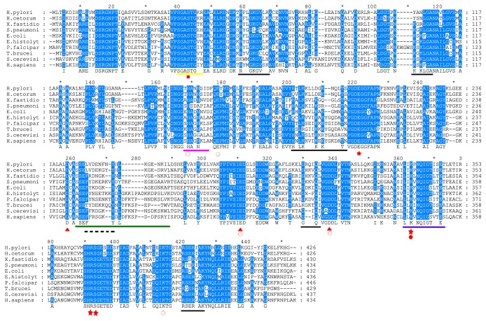
Figure 1 Alignment of the amino acid sequences of HpEno with homologs from different biological species. The species include H. pylori 26695 (P48285), H. cetorum (I0ELQ0), X. fastidiosa (Q9PDT8), S. pneumoniae (Q97QS2), E.coli (P0A6P9)and E. histolytica (N9UZM6), P. falciparum (Q27727), T. brucei (Q38BV6), S. cerevisiae (P00924), and human (α-enolase, P06733). The red signals under the letters represent rectangle, metal binding site reported only for humanα-enolase; open circle, substrate binding sites; star, active site residues; triangle, metal binding sites; close circle, subtract binding site in inhibited form. The black lines indicate the sites for RNA-binding and the dashed lines regions for plasminogen binding in S. pneumonia. The yellow line corresponds to loop L1, pink line to loop L2, and green line to loop L3, blue line corresponds to signature of enolase.
Figure 1 Alignment of the amino acid sequences of HpEno with homologs from different biological species. The species include H. pylori 26695 (P48285), H. cetorum (I0ELQ0), X. fastidiosa (Q9PDT8), S. pneumoniae (Q97QS2), E.coli (P0A6P9)and E. histolytica (N9UZM6), P. falciparum (Q27727), T. brucei (Q38BV6), S. cerevisiae (P00924), and human (α-enolase, P06733). The red signals under the letters represent rectangle, metal binding site reported only for humanα-enolase; open circle, substrate binding sites; star, active site residues; triangle, metal binding sites; close circle, subtract binding site in inhibited form. The black lines indicate the sites for RNA-binding and the dashed lines regions for plasminogen binding in S. pneumonia. The yellow line corresponds to loop L1, pink line to loop L2, and green line to loop L3, blue line corresponds to signature of enolase.CC BY · PubMed Central
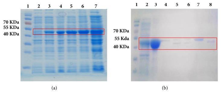
Figure 2 SDS-PAGE electrophoresis of expression and purification of HpEno. (a) Overexpression of HpEno in E.coli strain Rosetta-gami, lane 1: prestained protein marker (7-175 kDa), lane 2: induction with IPTG (To), lane 3: induction for 1 hour (T1), lane 4: induction for 2 hours (T2), lane 5: induction for 3 hours (T3), lane 6: induction for 4 hours (T4), and lane 7: induction overnight (TO∖N). (b) Affinity chromatography and His-tag removal, lane 1: prestained protein marker (7-175 kDa), lane 2: overexpression of HpEno, lane 3: solubilization of inclusion bodies, lane 4: flow through, lane 5: his-tag cut with PSP after 18 hr, lane 6: his-tag cut with PSP after 24 hr, lane 7: enolase no-cleaved, abd lane 8: HpEno cleaved.
Figure 2 SDS-PAGE electrophoresis of expression and purification of HpEno. (a) Overexpression of HpEno in E.coli strain Rosetta-gami, lane 1: prestained protein marker (7-175 kDa), lane 2: induction with IPTG (To), lane 3: induction for 1 hour (T1), lane 4: induction for 2 hours (T2), lane 5: induction for 3 hours (T3), lane 6: induction for 4 hours (T4), and lane 7: induction overnight (TO∖N). (b) Affinity chromatography and His-tag removal, lane 1: prestained protein marker (7-175 kDa), lane 2: overexpression of HpEno, lane 3: solubilization of inclusion bodies, lane 4: flow through, lane 5: his-tag cut with PSP after 18 hr, lane 6: his-tag cut with PSP after 24 hr, lane 7: enolase no-cleaved, abd lane 8: HpEno cleaved.CC BY · PubMed Central
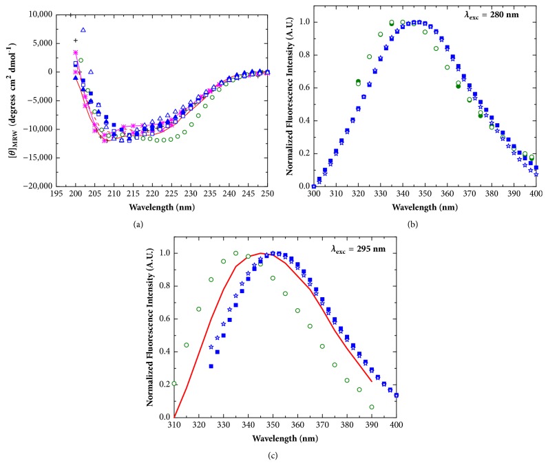
Figure 3 Secondary and tertiary structure of enolases. (a) Far-UV circular dichroism spectra; black cross: Streptococcal surface enolase; red line: P. falciparum enolase (dimer); red dashed line: P. falciparum enolase (monomer);pink line with asterisks: S. pyogenes enolase (TME buffer); pink asterisks: S. pyogenes enolase (Glycine buffer); green circles: S. cerevisiae enolase; blue closed squares: apo-HpEno (Tris-HCl); blue open squares: holo-HpEno (Tris-HCl); blue closed triangles: apo-HpEno (potassium phosphate); blue open triangles: holo-HpEno (potassium phosphate). (b) Fluorescence emission spectra for the excitation wavelength of 280 nm; Green closed circles: S. cerevisiae enolase (Tris-acetate); green open circles: S. cerevisiae enolase (Tris-HCl); blue closed squares: apo-HpEno(Tris-HCl); blue stars: apo-HpEno (Tris-acetate). (c) Fluorescence emission spectra for the excitation wavelength of 295 nm; red line: P. falciparum enolase; green open circle: S. cerevisiae enolase (Tris-HCl); blue closed squares: apo-HpEno(Tris-HCl); blue stars: apo-HpEno (Tris-acetate).
Figure 3 Secondary and tertiary structure of enolases. (a) Far-UV circular dichroism spectra; black cross: Streptococcal surface enolase; red line: P. falciparum enolase (dimer); red dashed line: P. falciparum enolase (monomer);pink line with asterisks: S. pyogenes enolase (TME buffer); pink asterisks: S. pyogenes enolase (Glycine buffer); green circles: S. cerevisiae enolase; blue closed squares: apo-HpEno (Tris-HCl); blue open squares: holo-HpEno (Tris-HCl); blue closed triangles: apo-HpEno (potassium phosphate); blue open triangles: holo-HpEno (potassium phosphate). (b) Fluorescence emission spectra for the excitation wavelength of 280 nm; Green closed circles: S. cerevisiae enolase (Tris-acetate); green open circles: S. cerevisiae enolase (Tris-HCl); blue closed squares: apo-HpEno(Tris-HCl); blue stars: apo-HpEno (Tris-acetate). (c) Fluorescence emission spectra for the excitation wavelength of 295 nm; red line: P. falciparum enolase; green open circle: S. cerevisiae enolase (Tris-HCl); blue closed squares: apo-HpEno(Tris-HCl); blue stars: apo-HpEno (Tris-acetate).CC BY · PubMed Central
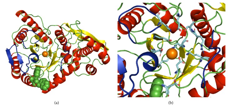
Figure 4 Ribbon representation of the structure HpEnomonomer. β-Strands are coloured in yellow, and α-helices are red. Tryptophans (W294 and W297) are shown as green spheres. The orange sphere represents a Mg2+ ion in the model that was obtained by SWISS-MODEL and rendered using PyMOL. Loops of the active site (L1, L2, and L3) are coloured in dark blue. In panel (b), the side chains of catalytic residues are shown.
Figure 4 Ribbon representation of the structure HpEnomonomer. β-Strands are coloured in yellow, and α-helices are red. Tryptophans (W294 and W297) are shown as green spheres. The orange sphere represents a Mg2+ ion in the model that was obtained by SWISS-MODEL and rendered using PyMOL. Loops of the active site (L1, L2, and L3) are coloured in dark blue. In panel (b), the side chains of catalytic residues are shown.CC BY · PubMed Central
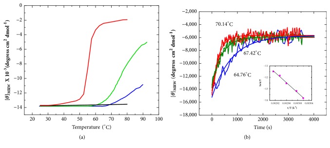
Figure 5 Thermal unfolding of H. pylori enolase. (a) Unfolding transitions of apo- and holoenolase from H. pylori monitored by CD spectroscopy at 220 nm; the experiments were carried out in 50 mMTris-HCl buffer at pH 7.4. Blue line: apo-HpEno, green line: holo-enolase (Mg+ 2 mM), black line: apo-ScEno, and red line: holo-ScEno (Mg+ 2 mM). (b) Kinetics of the thermal denaturation curves of holo-HpEno at different heating rates. The denaturation process was monitored by recording the ellipticity at 220 nm at a constant temperature. The insets correspond to the plots ofln(k1/T) versus 1/T, which represents the Eyring equation.
Figure 5 Thermal unfolding of H. pylori enolase. (a) Unfolding transitions of apo- and holoenolase from H. pylori monitored by CD spectroscopy at 220 nm; the experiments were carried out in 50 mMTris-HCl buffer at pH 7.4. Blue line: apo-HpEno, green line: holo-enolase (Mg+ 2 mM), black line: apo-ScEno, and red line: holo-ScEno (Mg+ 2 mM). (b) Kinetics of the thermal denaturation curves of holo-HpEno at different heating rates. The denaturation process was monitored by recording the ellipticity at 220 nm at a constant temperature. The insets correspond to the plots ofln(k1/T) versus 1/T, which represents the Eyring equation.CC BY · PubMed Central
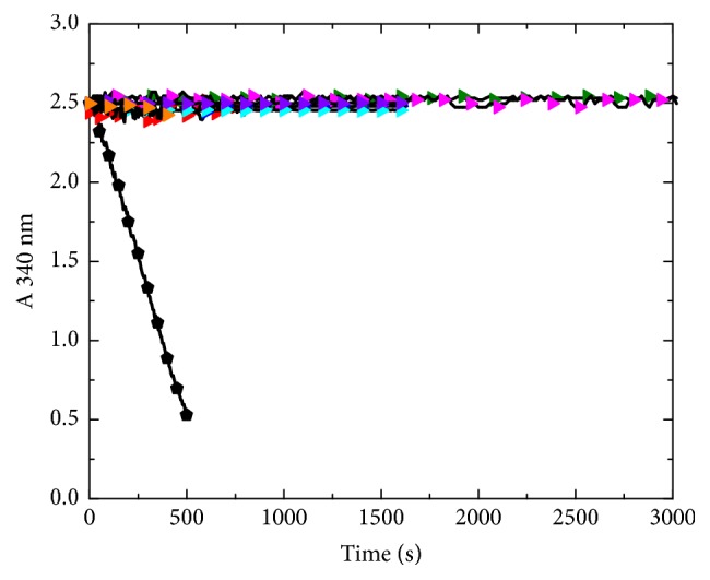
Figure 6 Enzymatic Activity of HpEno. Black circle: ScEno TEA buffer with MgSO4 (25 mM); red triangle: HpEno in tris-acetate buffer with MgSO4 (5 mM); green triangle: HpEno in TEA buffer with MgSO4 (5 mM); blue triangle: HpEno in HEPES buffer with MgSO4 (0.5 mM); cyan triangle: HpEno in TEA buffer with MgSO4 (0.5 mM); yellow triangle: HpEno with the His-tag in TEA buffer with MgSO4 (25 mM); pink triangle: crude extract from inclusion bodies in TEA buffer with MgSO4 (5 mM); orange triangle: HpEno in TEA buffer with MgSO4 (25 mM).
Figure 6 Enzymatic Activity of HpEno. Black circle: ScEno TEA buffer with MgSO4 (25 mM); red triangle: HpEno in tris-acetate buffer with MgSO4 (5 mM); green triangle: HpEno in TEA buffer with MgSO4 (5 mM); blue triangle: HpEno in HEPES buffer with MgSO4 (0.5 mM); cyan triangle: HpEno in TEA buffer with MgSO4 (0.5 mM); yellow triangle: HpEno with the His-tag in TEA buffer with MgSO4 (25 mM); pink triangle: crude extract from inclusion bodies in TEA buffer with MgSO4 (5 mM); orange triangle: HpEno in TEA buffer with MgSO4 (25 mM).CC BY · PubMed Central

Verification

Claims the paper makes

The claims and statistics of this paper haven’t been checked yet.

Run the check — free, no account needed. It reads the open-access text and checks the sources it cites.

A few free checks a day · papers already verified open instantly · results are public

Claims other papers make about this one

Each is checked against this paper’s own text: does it say what the citing paper says it does?

Self-check

Percentages, sums and reported differences re-computed from the paper’s own numbers.

The self-check needs the paper’s full text.

Citations

Cites

No references could be resolved yet.

Research works printed but not linked: 57

  1. 10.1007/PL00000910 · DOI
  2. 10.1016/0014-5793(85)81143-1 · DOI
  3. 10.1007/BF00556365 · DOI
  4. 10.1155/2012/156795 · DOI
  5. 10.4061/2011/932549 · DOI
  6. 2009, Malaria Journal, 8
  7. 10.1016/j.bbagen.2007.01.015 · DOI
  8. 10.1111/j.1742-4658.2010.07819.x · DOI
  9. 10.1016/j.bbapap.2008.03.017 · DOI
  10. 10.1046/j.1365-2958.2001.02448.x · DOI
  11. 10.1016/j.bbrc.2007.10.098 · DOI
  12. 2009, RNA Biology, 6
  13. 10.1002/pro.5560040209 · DOI
  14. 10.1023/A:1020790604887 · DOI
  15. 10.1093/jb/mvg104 · DOI · Paper page
  16. 10.1006/jmbi.2001.5065 · DOI
  17. 10.1111/j.1742-4658.2007.06027.x · DOI
  18. 10.1016/j.jmb.2004.08.088 · DOI
  19. 1989, The Journal of Biological Chemistry, 264, 3685, 10.1016/S0021-9258(19)84906-2 · DOI
  20. 10.1016/S1286-4579(03)00111-4 · DOI
  21. 10.1155/2012/723183 · DOI
  22. 10.1016/j.micres.2017.04.008 · DOI
  23. 10.1016/j.sbi.2005.05.008 · DOI
  24. 10.1046/j.1432-1033.2003.03692.x · DOI
  25. 10.1016/j.ijbiomac.2011.07.021 · DOI
  26. 10.1111/j.1742-4658.2007.05738.x · DOI
  27. 10.1016/0076-6879(86)31045-0 · DOI
  28. 10.1038/nprot.2006.204 · DOI
  29. 10.1021/bi010528w · DOI
  30. 1996, Computer Applications in the Biosciences, 12, 543
  31. 10.1038/84513 · DOI
  32. 10.1002/pmic.200600875 · DOI
  33. 2008, Acta Crystallographica Section D, 64, 651, 10.1107/S0907444908008561 · DOI
  34. 10.1107/S0907444911016544 · DOI
  35. 10.1021/bi00225a012 · DOI
  36. 10.1021/bi952859c · DOI
  37. 10.1021/bi00636a023 · DOI
  38. 10.1016/S0167-4838(99)00233-2 · DOI
  39. 10.1021/bi0346345 · DOI
  40. 10.1186/s12917-014-0223-6 · DOI
  41. 10.1016/j.micres.2012.10.001 · DOI
  42. 10.1128/IAI.72.1.94-105.2004 · DOI
  43. 10.1074/jbc.M109.004317 · DOI
  44. 10.1099/mic.0.028795-0 · DOI
  45. 2016, Scientific Reports, 6, 37758, 10.1038/srep37758 · DOI
  46. 10.1016/j.pep.2008.02.015 · DOI
  47. 10.1021/bi00197a038 · DOI
  48. 10.1107/S1399004715018830 · DOI
  49. 10.1038/nprot.2008.197 · DOI
  50. 10.1002/prot.24333 · DOI
  51. 10.1016/j.abb.2009.02.012 · DOI
  52. 10.2174/1568026611212220009 · DOI
  53. 10.1590/S1415-47572003000200015 · DOI
  54. 10.1007/s10930-009-9215-y · DOI
  55. 10.1128/9781555818005 · DOI
  56. 10.1099/13500872-140-8-2085 · DOI
  57. 10.1128/IAI.71.5.2920-2923.2003 · DOI

Cited by

Sorted by citations

See all 13

Metadata

Authors
López-López, M. D. J., Rodríguez-Luna, I. C., Lara-Ramírez, E. E., López-Hidalgo, M., Benítez-Cardoza, C. G., & Guo, X.
Venue
BioMed Research International
Publisher
Wiley
Published
2018-12-17
Volume
2018
Pages
1-12
ISSN
  • 2314-6133
  • 2314-6141
Cited by
13
References
0
Language
en
Funders
  • Consejo Nacional de Ciencia y Tecnología (168541, SIP20120202, SIP20140076)
  • Secretaría de Investigación y Posgrado, Instituto Politécnico Nacional (168541, SIP20120202, SIP20140076)

About these checks

Registry, retraction, Cites and statistics results are computed, not judged, with one exception: a printed reference that no index lists is read once by a model before it counts as a citation error. Claims are extracted by a model and kept only when their quote appears verbatim in the text; each cited source is then checked, with a model’s help, for whether it says what the claim relies on. Those verdicts can be wrong — read the claim’s quote and the cited source before relying on one.

Statistics and arithmetic are recomputed, not judged.

Confidence bands are shown only for fields calibrated on our annotated test set. None is calibrated yet, so no band is shown.

What counts as a claim ›How we measure confidence ›How we check this ›

Are you an author, or do you see a problem with this page? Request a review

How to cite

Cite CoChat for the checks on this page, and the paper for its own findings.

This page
CoChat. (2026). Citation check: Biochemical and Biophysical Characterization of the Enolase from Helicobacter pylori. https://app.cochat.ai/papers/10.1155/2018/9538193
The paper
López-López, M. D. J., Rodríguez-Luna, I. C., Lara-Ramírez, E. E., López-Hidalgo, M., Benítez-Cardoza, C. G., & Guo, X. (2018). Biochemical and Biophysical Characterization of the Enolase from Helicobacter pylori. BioMed Research International. https://doi.org/10.1155/2018/9538193