

The DOI matches its Crossref record; 57 of the 57 references it cites were found in a registry or an index.
Impact of Coenzyme Q10 Administration on Lead Acetate-Induced Testicular Damage in Rats
Open accessAbstract
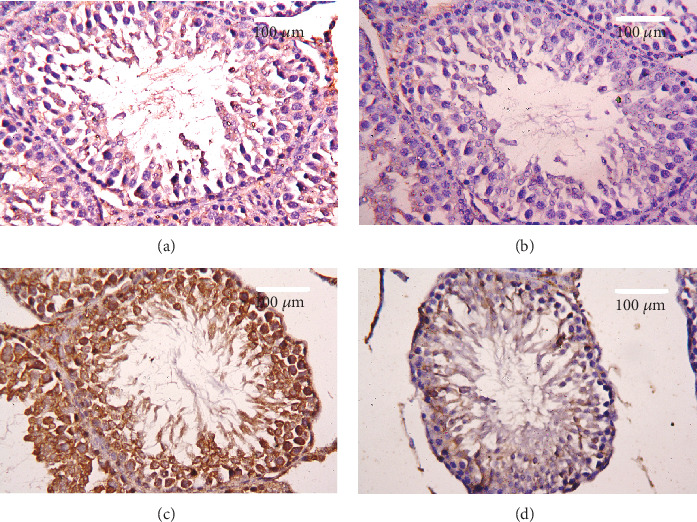
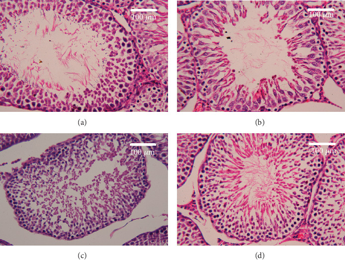
Exposure to lead (Pb) causes multiorgan dysfunction including reproductive impairments. Here, we examined the protective effects of coenzyme Q10 (CoQ10) administration on testicular injury induced by lead acetate (PbAc) exposure in rats. This study employed four experimental groups (n=7) that underwent seven days of treatment as follows: control group intraperitoneally (i.p.) treated with 0.1 ml of 0.9% NaCl containing 1% Tween 80 (v:v), CoQ10 group that was i.p. injected with 10 mg/kg CoQ10, PbAc group that was i.p. treated with PbAc (20 mg/kg), and PbAc+CoQ10 group that was i.p. injected with CoQ10 2 h after PbAc. PbAc injection resulted in increasing residual Pb levels in the testis and reducing testosterone, luteinizing hormone, and follicle-stimulating hormone levels. Additionally, PbAc exposure resulted in significant oxidative damage to the tissues on the testes. PbAc raised the levels of prooxidants (malondialdehyde and nitric oxide) and reduced the amount of endogenous antioxidative proteins (glutathione and its derivative enzymes, catalase, and superoxide dismutase) available in the cell. Moreover, PbAc induced the inflammatory response as evidenced by the upregulation of inflammatory mediators (tumor necrosis factor-alpha and interleukin-1 beta). Further, PbAc treatment induced apoptosis in the testicular cells, as indicated by an increase in Bax and caspase 3 expression, and reduced Bcl2 expression. CoQ10 supplementation improved testicular function by inhibiting Pb accumulation, oxidative stress, inflammation, cell death, and histopathological changes following PbAc exposure. Our findings suggest that CoQ10 can act as a natural therapeutic agent to protect against the reproductive impairments associated with PbAc exposure.
Overview
The claims and statistics of this paper haven’t been checked yet.
Run the check — free, no account needed. It reads the open-access text and checks the sources it cites.
Integrity signals
Registry
Cites
Statistics
Figures
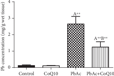
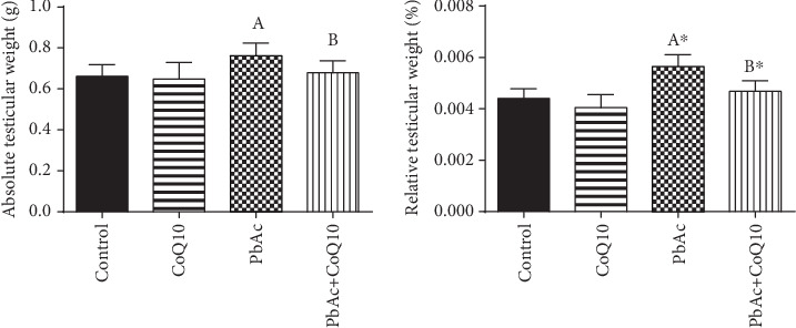
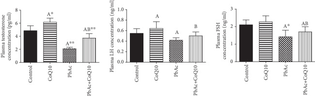
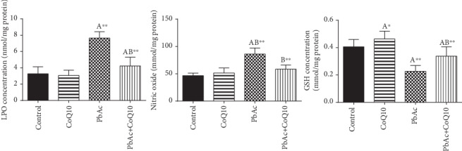
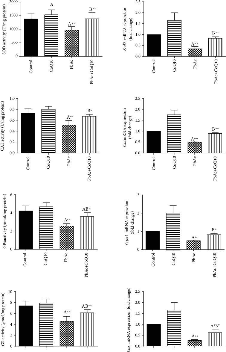
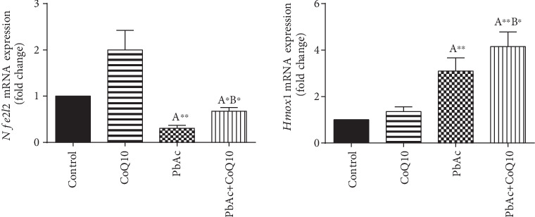
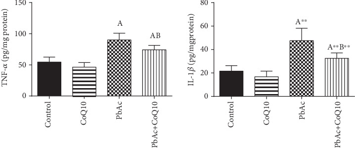
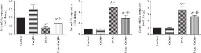
Verification
Claims the paper makes
The claims and statistics of this paper haven’t been checked yet.
Run the check — free, no account needed. It reads the open-access text and checks the sources it cites.
Claims other papers make about this one
Each is checked against this paper’s own text: does it say what the citing paper says it does?
The source backs part of the claim, not all of it — or backs it for a different population, condition or comparison.
In the citing paper
“This is in line with Sumi et al. (2018) and Fatima et al. (2021) who stated that CoQ10 inhibited the activation of caspase-3 and the release of cytochrome c from mitochondria via stabilizing the mitochondrial membrane, thus preventing the release of apoptotic mediators.”
From Can coenzyme Q10 alleviate the toxic effect of fenofibrate on skeletal muscle? (2023) · cites this paper as El-Khadragy et al. 2020
Experimental — accuracy not yet measured
Self-check
Percentages, sums and reported differences re-computed from the paper’s own numbers.
The self-check needs the paper’s full text.
Citations
Cites
1 retracted
Hamadouche, N. A., Sadi, N., Kharoubı, O., et al. (2013) · Archives of Biological Sciences · doi:10.2298/abs1304435h · Cited by 22
RetractedAssay for lipid peroxides in animal tissues by thiobarbituric acid reaction
Ohkawa, H., Ohishi, N., & Yagi, K. (1979) · Analytical Biochemistry · doi:10.1016/0003-2697(79)90738-3 · Cited by 29,746
Ellman, G. L. (1959) · Archives of Biochemistry and Biophysics · doi:10.1016/0003-9861(59)90090-6 · Cited by 26,824
Analysis of nitrate, nitrite, and [15N]nitrate in biological fluids
Green, L. C., Wagner, D. A., Glogowski, J. A., Skipper, P. L., Wishnok, J. S., & Tannenbaum, S. R. (1982) · Analytical Biochemistry · doi:10.1016/0003-2697(82)90118-x · Cited by 12,061
Studies on the quantitative and qualitative characterization of erythrocyte glutathione peroxidase.
Paglia, D. E., & Valentine, W. N. (1967) · PubMed · Cited by 10,776
A simple method for clinical assay of superoxide dismutase.
Sun, Y., Oberley, L. W., & Li, Y. (1988) · Clinical Chemistry · doi:10.1093/clinchem/34.3.497 · Cited by 4,537
Łoboda, A., Damulewicz, M., Pyza, E., Józkowicz, A., & Dulak, J. (2016) · Cellular and Molecular Life Sciences · doi:10.1007/s00018-016-2223-0 · Cited by 2,709
Metabolism and function of coenzyme Q
Turunen, M., Olsson, J., & Dallner, G. (2004) · Biochimica et Biophysica Acta (BBA) - Biomembranes · doi:10.1016/j.bbamem.2003.11.012 · Cited by 1,026
The chemical biology of nitric oxide: Implications in cellular signaling
Thomas, D. D., Ridnour, L. A., Isenberg, J. S., Flores-Santana, W., Switzer, C. H., Donzelli, S., Hussain, P., Vecoli, C., Paolocci, N., Ambs, S., et al. (2008) · Free Radical Biology and Medicine · doi:10.1016/j.freeradbiomed.2008.03.020 · Cited by 975
Heme oxygenase: colors of defense against cellular stress
Otterbein, L. E., & Choi, A. M. K. (2000) · American Journal of Physiology-Lung Cellular and Molecular Physiology · doi:10.1152/ajplung.2000.279.6.l1029 · Cited by 870
Oxidative Stress in Lead and Cadmium Toxicity and Its Amelioration
Patra, R. C., Rautray, A. K., & Swarup, D. (2011) · Veterinary Medicine International · doi:10.4061/2011/457327 · Cited by 579
Lück, H. (1965) · Elsevier eBooks · doi:10.1016/b978-0-12-395630-9.50158-4 · Cited by 473
Heme oxygenase-1-derived bilirubin ameliorates postischemic myocardial dysfunction
Clark, J., Foresti, R., Sarathchandra, P., Kaur, H., Green, C. J., & Motterlini, R. (2000) · American Journal of Physiology-Heart and Circulatory Physiology · doi:10.1152/ajpheart.2000.278.2.h643 · Cited by 392
Papucci, L., Schiavone, N., Witort, E. J., Donnini, M., Lapucci, A., Tempestini, A., Formigli, L., Zecchi‐Orlandini, S., Orlandini, G., Carella, G., et al. (2003) · Journal of Biological Chemistry · doi:10.1074/jbc.m302297200 · Cited by 308
Blood Lead, Blood Pressure, and Hypertension in Perimenopausal and Postmenopausal Women
Nash, D., Magder, L. S., Lustberg, M. E., Sherwin, R. W., Rubin, R. J., Kaufmann, R. B., & Silbergeld, E. K. (2003) · JAMA · doi:10.1001/jama.289.12.1523 · Cited by 248
Safarinejad, M. R. (2009) · The Journal of Urology · doi:10.1016/j.juro.2009.02.121 · Cited by 231
Olayan, E. M. A., Aloufi, A. S., Alamri, O., et al. (2020) · The Science of The Total Environment · doi:10.1016/j.scitotenv.2020.137969 · Cited by 198
Factor, V. M., Kiss, A., Woitach, J. T., Wirth, P. J., & Thorgeirsson, S. S. (1998) · Journal of Biological Chemistry · doi:10.1074/jbc.273.25.15846 · Cited by 173
Balercia, G., Mosca, F., Mantero, F., Boscaro, M., Mancini, A., Lamonica, G. R., & Littarru, G. P. (2004) · Fertility and Sterility · doi:10.1016/j.fertnstert.2003.05.009 · Cited by 139
So, H., Kim, H., Kim, Y., Kim, E., Pae, H., Chung, H., Kim, H. J., Kwon, K., Lee, K., Lee, H.-Y., et al. (2008) · Journal of the Association for Research in Otolaryngology · doi:10.1007/s10162-008-0126-y · Cited by 130
The effect of lead intoxication on endocrine functions
Doumouchtsis, K., Doumouchtsis, S. K., Doumouchtsis, E. K., & Perrea, D. (2009) · Journal of Endocrinological Investigation · doi:10.1007/bf03345710 · Cited by 128
Primary and secondary coenzyme Q10 deficiency: the role of therapeutic supplementation
Potgieter, M., Pretorius, E., & Pepper, M. S. (2013) · Nutrition Reviews · doi:10.1111/nure.12011 · Cited by 124
Gandhi, J., Hernandez, R. J., Chen, A. W., et al. (2017) · Zygote · doi:10.1017/s0967199417000028 · Cited by 123
Hepatoprotective effect of coenzyme Q10 in rats with acetaminophen toxicity
Fouad, A. A., & Jresat, I. (2011) · Environmental Toxicology and Pharmacology · doi:10.1016/j.etap.2011.12.011 · Cited by 121
Effect of Coenzyme-Q10 on Doxorubicin-Induced Nephrotoxicity in Rats
El‐Sheikh, A. A. K., Morsy, M. A., Refaie, M. M. M., et al. (2012) · Advances in Pharmacological Sciences · doi:10.1155/2012/981461 · Cited by 120
Genetic Activation of Nrf2 Protects against Fasting-Induced Oxidative Stress in Livers of Mice
Zhang, Y.-K. J., Wu, K. C., & Klaassen, C. D. (2013) · PLoS ONE · doi:10.1371/journal.pone.0059122 · Cited by 94
Oxidative Stress, Nrf2, and Epigenetic Modification Contribute to Anticancer Drug Resistance
Kang, K. A., & Hyun, J. W. (2017) · Toxicological Research · doi:10.5487/tr.2017.33.1.001 · Cited by 94
Tarry‐Adkins, J. L., Fernandez‐Twinn, D. S., Hargreaves, I. P., Neergheen, V., Aiken, C., Martin‐Gronert, M. S., McConnell, J. M. L., & Ozanne, S. E. (2015) · American Journal of Clinical Nutrition · doi:10.3945/ajcn.115.119834 · Cited by 89
Lead exposure reduces sperm quality and DNA integrity in mice
Li, C., Zhao, K., Zhang, H., et al. (2018) · Environmental Toxicology · doi:10.1002/tox.22545 · Cited by 83
Ramadan, L. A. A., Abd‐Allah, A. R. A., Aly, H. A. A., et al. (2002) · Pharmacological Research · doi:10.1016/s1043661802001718 · Cited by 81
Mirmalek, S. A., Boushehrinejad, A. G., Yavari, H., et al. (2016) · Oxidative Medicine and Cellular Longevity · doi:10.1155/2016/5818479 · Cited by 80
Furland, N. E., Zanetti, S. R., Oresti, G. M., Maldonado, E. N., & Aveldaño, M. I. (2007) · Journal of Biological Chemistry · doi:10.1074/jbc.m700708200 · Cited by 79
Palmeira, C. M., Santos, D. L., Seiça, R. M. F., Moreno, A. J., & Santos, M. S. (2001) · American Journal of Physiology-Cell Physiology · doi:10.1152/ajpcell.2001.281.3.c1023 · Cited by 75
Coenzyme Q(10) in male infertility: physiopathology and therapy.
Mancini, A., & Balercia, G. (2011) · Biofactors · doi:10.1002/biof.164 · Cited by 68
Hassan, E. M., El-Neweshy, M. S., Hassan, M. M., & Noreldin, A. E. (2019) · Life Sciences · doi:10.1016/j.lfs.2019.05.067 · Cited by 68
Coenzyme Q10 counteracts testicular injury induced by sodium arsenite in rats
Fouad, A. A., Al‐Sultan, A. I., & Yacoubi, M. T. (2011) · European Journal of Pharmacology · doi:10.1016/j.ejphar.2010.12.045 · Cited by 61
Erol, B., Bozlu, M., Hancı, V., Tokgöz, H., Bektaş, S., & Mungan, G. (2009) · Fertility and Sterility · doi:10.1016/j.fertnstert.2009.07.981 · Cited by 56
Interactions of lifetime lead exposure and stress: Behavioral, neurochemical and HPA axis effects
Rossi‐George, A., Virgolini, M. B., Weston, D., Thiruchelvam, M. J., & Cory‐Slechta, D. A. (2010) · NeuroToxicology · doi:10.1016/j.neuro.2010.09.004 · Cited by 55
Coenzyme Q10 Supplementation and Exercise in Healthy Humans: A Systematic Review
Ramírez, Á. S., Díaz‐Castro, J., Pulido‐Moran, M., Kajarabille, N., Guisado, R., & Ochoa, J. J. (2016) · Current Drug Metabolism · doi:10.2174/1389200216666151103115654 · Cited by 55
Khodir, A. E., Atef, H., Said, E., El‐Kashef, H. A., & Salem, H. A. (2017) · Inflammopharmacology · doi:10.1007/s10787-016-0305-0 · Cited by 55
Soleimanzadeh, A., Kian, M., Moradi, S., et al. (2019) · PubMed · doi:10.22038/ajp.2019.13513 · Cited by 54
Alahmar, A. T. (2019) · Daehan saengsik uihak hoeji/Clinical and experimental reproductive medicine · doi:10.5653/cerm.2019.00136 · Cited by 54
Li, N., Hou, Y.-H., Ma, D., Jing, W., Dahms, H.-U., & Wang, L. (2015) · Ecotoxicology and Environmental Safety · doi:10.1016/j.ecoenv.2015.03.019 · Cited by 53
Moneim, A. E. A. (2012) · Biological Trace Element Research · doi:10.1007/s12011-012-9370-4 · Cited by 51
Moniem, A. E. A., Dkhil, M. A., & Al‐Quraishy, S. (2010) · AFRICAN JOURNAL OF BIOTECHNOLOGY · Cited by 50
Salama, S. A., Arab, H. H., Maghrabi, I. A., et al. (2016) · Biological Trace Element Research · doi:10.1007/s12011-016-0624-4 · Cited by 49
Impact of lead toxicity on male rat reproduction at “hormonal and histopathological levels”
El-Sayed, Y. S., & El-Neweshy, M. S. (2010) · Toxicological & Environmental Chemistry Reviews · doi:10.1080/02772240902984453 · Cited by 48
Lead Toxicity on Male Reproductive System and its Mechanism: A Review
Kumar, S. R., & Devi, S. A. (2018) · Research Journal of Pharmacy and Technology · doi:10.5958/0974-360x.2018.00228.7 · Cited by 38
Exposure of women in general populations to lead via food and air in East and Southeast Asia
Ikeda, M., Zhang, Z.-W., Shimbo, S., Watanabe, T., Nakatsuka, H., Moon, C., Matsuda‐Inoguchi, N., & Higashikawa, K. (2000) · American Journal of Industrial Medicine · doi:10.1002/1097-0274(200009)38:3 · Cited by 36
Critical aspects of the antioxidant function of coenzyme Q in biomembranes
Nohl, H., Gille, L., & Kozlov, A. V. (1999) · BioFactors · doi:10.1002/biof.5520090210 · Cited by 35
He, Y., Zou, Q., Chen, H., et al. (2016) · The Tohoku Journal of Experimental Medicine · doi:10.1620/tjem.238.295 · Cited by 34
Indigofera oblongifolia mitigates lead-acetate-induced kidney damage and apoptosis in a rat model
Moneim, A. E. A., Dkhil, M. A., Al-Khalifa, M. S., Al-Quraishy, S., & Zrieq, R. M. (2016) · Drug Design Development and Therapy · doi:10.2147/dddt.s105511 · Cited by 34
Li, H., Chen, G., Ma, W., et al. (2014) · International Journal of Molecular Sciences · doi:10.3390/ijms150813388 · Cited by 32
Zuscik, M. J., Ma, L., Buckley, T., Puzas, J. E., Drissi, H., Schwarz, E. M., & O’Keefe, R. J. (2007) · Environmental Health Perspectives · doi:10.1289/ehp.10028 · Cited by 27
INHIBITORY ACTIONS OF LEAD ON STEROIDOGENESIS IN MA-10 MOUSE LEYDIG TUMOR CELLS
Huang, B.-M., & Liu, M. (2004) · Archives of Andrology · doi:10.1080/01485010490250434 · Cited by 23
Toward a new definition of essential nutrients: is it now time for a third ‘vitamin’ paradigm?
Challem, J. (1999) · Medical Hypotheses · doi:10.1054/mehy.1997.0685 · Cited by 21
Kabel, A. M., & Elkhoely, A. A. (2017) · Europe PMC (PubMed Central) · doi:10.22034/apjcp.2017.18.6.1629 · Cited by 17
Research works printed but not linked: 4
- 2010, African Journal of Biotechnology, 9, 7216
- 2005, Bulletin of the Veterinary Institute in Pulawy, 49, 89
- 1967, The Journal of Laboratory and Clinical Medicine, 70, 158
- 2019, Avicenna journal of phytomedicine
Cited by
Sorted by citations
Can coenzyme Q10 alleviate the toxic effect of fenofibrate on skeletal muscle?
El-Bassouny, D. R., Mansour, A. A., Ellakkany, A. S., Ayuob, N. N., & AbdElfattah, A. A. (2023) · Histochemistry and Cell Biology · doi:10.1007/s00418-023-02205-5
Metadata
- Authors
- El-khadragy, M., Al-Megrin, W. A., AlSadhan, N. A., Metwally, D. M., El-Hennamy, R. E., Salem, F. E. H., Kassab, R. B., & Abdel Moneim, A. E.
- Affiliations
- Biology Department, Faculty of Science, Princess Nourah Bint Abdulrahman University, Riyadh 84428, Saudi Arabia
- Almaarefa University (SA)
- Zagazig University (EG)
- Zoology and Entomology Department, Faculty of Science, Helwan University, Cairo 11795, Egypt
- Taibah University (SA)
- Al Baha University (SA)
- ORCID
- Venue
- Oxidative Medicine and Cellular Longevity
- Publisher
- Wiley
- Published
- 2020-06-03
- Volume
- 2020
- Pages
- 1-12
- ISSN
- 1942-0994 (ISSN-L)
- 1942-0900
- PMC
- PMC7290879
- OpenAlex
- W3033636297
- Cited by
- 56
- References
- 57
- Citations by year
- 2026: 4 · 2025: 9 · 2024: 9 · 2023: 13 · 2022: 12 · 2021: 7 · 2020: 2
- FWCI
- 3.40
- Type
- journal article
- Language
- en
- Open access
- hybrid
- doi.org
- Locations
- Oxidative Medicine and Cellular Longevity (publishedVersion, cc-by)
- PubMed Central (publishedVersion, cc-by)
- PubMed (publishedVersion)
- DOAJ (DOAJ: Directory of Open Access Journals) (submittedVersion)
- Funders
- Princess Nourah Bint Abdulrahman University
- Topics
- Physical Sciences › Environmental Science › Health, Toxicology and Mutagenesis › Heavy Metal Exposure and Toxicity
- Health Sciences › Medicine › Reproductive Medicine › Sperm and Testicular Function
- Life Sciences › Biochemistry, Genetics and Molecular Biology › Molecular Biology › Coenzyme Q10 studies and effects
- Keywords
- coenzyme Q10, lead acetate, lead toxicity, testicular damage, oxidative stress, reproductive toxicity, antioxidant enzymes, inflammation, apoptosis, testosterone, luteinizing hormone, follicle-stimulating hormone, lead accumulation, neuroprotection
- MeSH terms
- Animals, Follicle Stimulating Hormone, Glutathione, Male, Nitric Oxide, Organometallic Compounds, RNA, Messenger, Testis, Testosterone, Tumor Necrosis Factor-alpha, Ubiquinone, Lipid Peroxidation, Rats, Wistar, bcl-2-Associated X Protein, Caspase 3, Interleukin-1beta
About these checks
Registry, retraction, Cites and statistics results are computed, not judged, with one exception: a printed reference that no index lists is read once by a model before it counts as a citation error. Claims are extracted by a model and kept only when their quote appears verbatim in the text; each cited source is then checked, with a model’s help, for whether it says what the claim relies on. Those verdicts can be wrong — read the claim’s quote and the cited source before relying on one.
Statistics and arithmetic are recomputed, not judged.
Confidence bands are shown only for fields calibrated on our annotated test set. None is calibrated yet, so no band is shown.
What counts as a claim ›How we measure confidence ›How we check this ›
Are you an author, or do you see a problem with this page? Request a review
