

The DOI matches its Crossref record; 36 of the 36 references it cites were found in a registry or an index.
Curcumin Administered in Combination with Glu-GNPs Induces Radiosensitivity in Transplanted Tumor MDA-MB-231-luc Cells in Nude Mice
Open accessAbstract
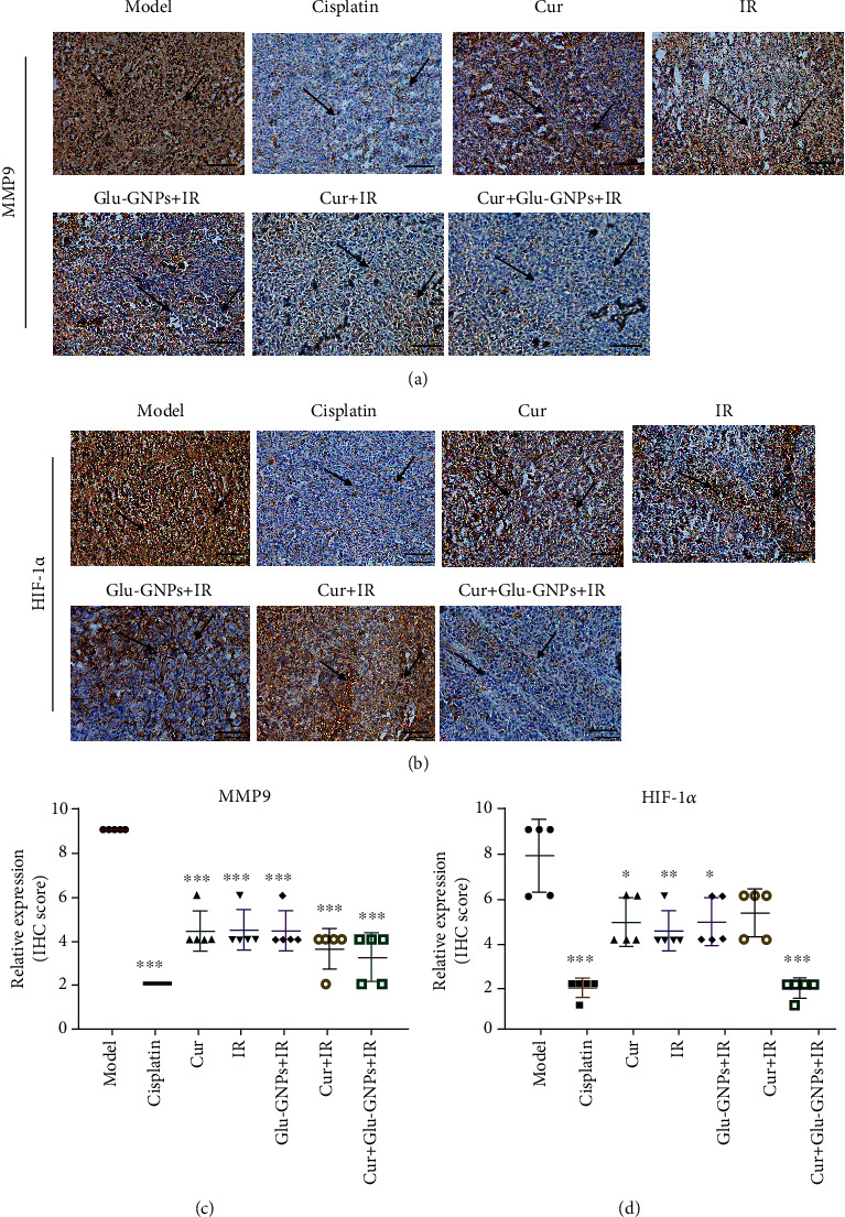
Curcumin is a type of plant polyphenol extracted from Curcuma longa L. rhizome, which demonstrates antitumor activity in breast cancer cells in vitro. To investigate the combined effect and possible mechanism of curcumin and glucose‐gold nanoparticles (Glu‐GNPs), the radiosensitivity of breast carcinoma xenografts was assessed in nude mice. MDA-MB-231 cells labeled with firefly luciferase were inoculated into the mammary fatty pads of nude mice to establish a transplantation tumor model of human breast cancer. The tumor‐bearing mice were treated with different drugs (curcumin, Glu‐GNPs, and cisplatin) for 3 weeks prior to radiotherapy. The body weights and tumor volumes of the mice were measured in regular intervals. Tumor bioluminescence intensity was determined in real‐time using an in vivo bioluminescence imaging system to monitor tumor growth. Transplanted tumor tissue samples were taken for hematoxylin and eosin (HE) staining, and the expression of VEGF, HSP90, HIF‐1α, and MMP9 was evaluated via reverse transcription‐quantitative PCR or immunohistochemistry. The results revealed that the breast tumor‐bearing nude mouse model was successfully established, as evidenced by a stable expression of luciferase. Curcumin inhibited the growth of tumors without causing significant weight loss in mice. Furthermore, additive inhibition was demonstrated when curcumin was administered in combination with Glu‐GNPs and irradiation. Tumor bioluminescence intensity was decreased in the model group following curcumin, Glu‐GNPs, and irradiation treatment. HE staining demonstrated that transplanted tumors were malignant, with necrotic tissue exhibited centrally. It was concluded that curcumin administered in combination with Glu‐GNPs and X‐ray irradiation could reduce the protein expression of VEGF, HSP90, HIF‐1α, and MMP9 in tumor tissue when compared with the model group. Curcumin and Glu‐GNPs administered with X‐ray irradiation significantly inhibited tumor growth and induced radiosensitivity, which may be associated with the inhibition of angiogenesis in tumor tissue.
Overview
The claims and statistics of this paper haven’t been checked yet.
Run the check — free, no account needed. It reads the open-access text and checks the sources it cites.
Integrity signals
Registry
Cites
Statistics
Figures
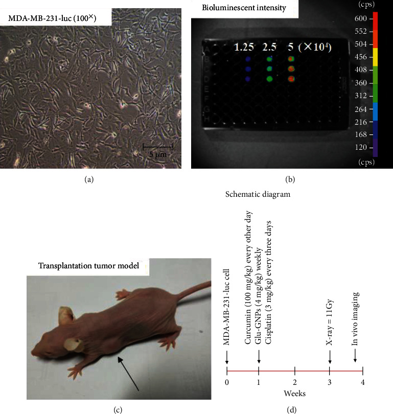
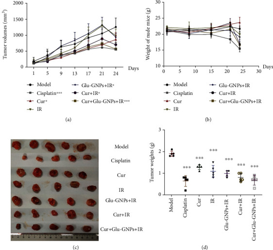
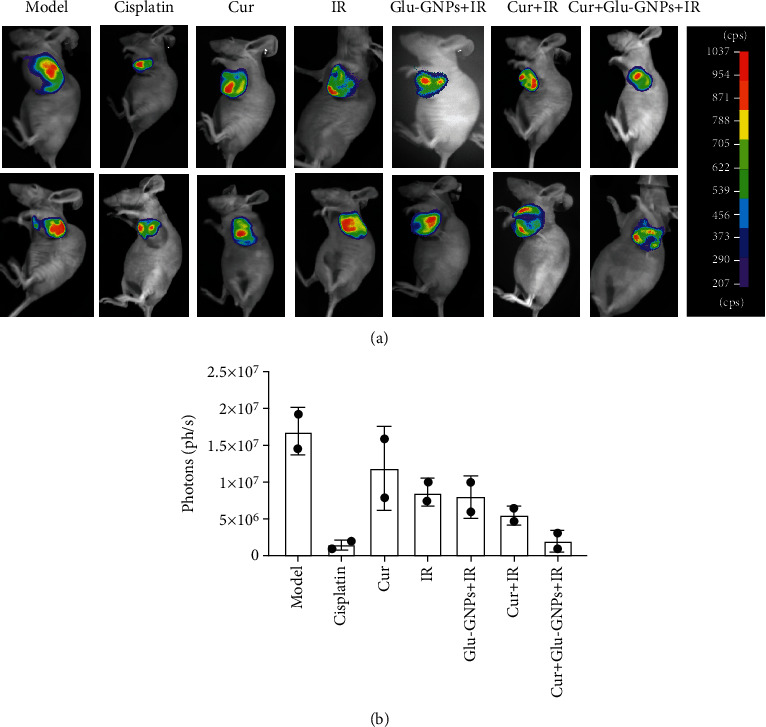
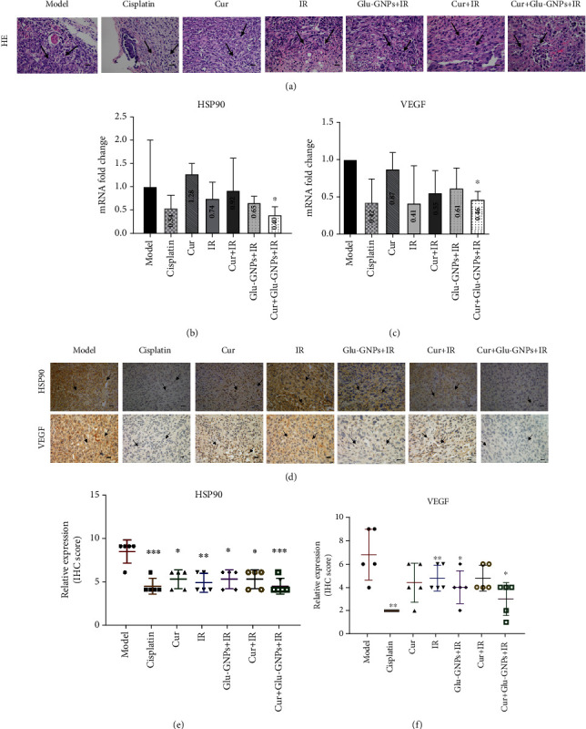
Verification
Claims the paper makes
The claims and statistics of this paper haven’t been checked yet.
Run the check — free, no account needed. It reads the open-access text and checks the sources it cites.
Claims other papers make about this one
Each is checked against this paper’s own text: does it say what the citing paper says it does?
The cited source says what the claim attributes to it.
In the citing paper
“Multimodal therapeutic approaches were also described, combining radiotherapy with chemotherapy, photothermal therapy, photodynamic therapy, or immunomodulatory strategies [39,43,49,50,67,68,69].”
From Nanoparticle-Mediated Radiosensitization in Breast Cancer: A Systematic Review of Preclinical Evidence and Translational Challenges (2026) · cites this paper as [69]
The check wasn’t confident either way, so it gives no verdict — never disputed.
In the citing paper
“Additional strategies included depletion of intracellular glutathione (GSH) and modulation of redox balance [17,19,45,47,68], as well as metabolic interventions targeting glycolysis or cellular energetics [49,69,75,76].”
From Nanoparticle-Mediated Radiosensitization in Breast Cancer: A Systematic Review of Preclinical Evidence and Translational Challenges (2026) · cites this paper as [69]
Experimental — accuracy not yet measured
Self-check
Percentages, sums and reported differences re-computed from the paper’s own numbers.
The self-check needs the paper’s full text.
Citations
Cites
1 retracted
Shen, Y., Han, Z., Liu, S., Jiao, Y., Li, Y., & Yuan, H. (2020) · Cancer Management and Research · doi:10.2147/cmar.s246691 · Cited by 14
RetractedBray, F., Ferlay, J., Soerjomataram, I., Siegel, R. L., Torre, L. A., & Jemal, A. (2018) · CA: A Cancer Journal for Clinicians · doi:10.3322/caac.21492 · Cited by 84,109
Nanoshell-mediated near-infrared thermal therapy of tumors under magnetic resonance guidance
Hirsch, L. R., Stafford, R. J., Bankson, J. A., Sershen, S. R., Rivera, B., Price, R. E., Hazle, J. D., Halas, N. J., & West, J. L. (2003) · Proceedings of the National Academy of Sciences · doi:10.1073/pnas.2232479100 · Cited by 3,961
The Tumour Microenvironment after Radiotherapy: Mechanisms of Resistance and Recurrence
Barker, H. E., Paget, J. T. E., Khan, A. A., & Harrington, K. J. (2015) · Nature Reviews Cancer · doi:10.1038/nrc3958 · Cited by 2,144 · cited 1×
“Hypoxic cells demonstrate high radiation tolerance and resist tumor stem cell DNA damage [29].”
BackgroundImmunotargeted Nanoshells for Integrated Cancer Imaging and Therapy
Loo, C. H., Lowery, A. R., Halas, N. J., West, J. L., & Drezek, R. A. (2005) · Nano Letters · doi:10.1021/nl050127s · Cited by 1,792
Kim, B. Y. S., Rutka, J. T., & Chan, W. C. W. (2010) · New England Journal of Medicine · doi:10.1056/nejmra0912273 · Cited by 1,153
Zhang, C., Samanta, D., Lu, H., Bullen, J. W., Zhang, H., Chen, I., He, X., & Semenza, G. L. (2016) · Proceedings of the National Academy of Sciences · doi:10.1073/pnas.1602883113 · Cited by 977
The HSP90 Family: Structure, Regulation, Function, and Implications in Health and Disease
Hoter, A., El-Sabban, M. E., & Naim, H. Y. (2018) · International Journal of Molecular Sciences · doi:10.3390/ijms19092560 · Cited by 693
Breast cancer in young women: an overview
Anastasiadi, Z., Lianos, G. D., Ignatiadou, E., Harissis, H. V., & Mitsis, M. C. (2017) · Updates in Surgery · doi:10.1007/s13304-017-0424-1 · Cited by 604
Breast Cancer in Young Women: Poor Survival Despite Intensive Treatment
Fredholm, H., Eaker, S., Frisell, J., Holmberg, L., Fredriksson, I., & Lindman, H. (2009) · PLoS ONE · doi:10.1371/journal.pone.0007695 · Cited by 494
Cited by
Sorted by citations
The Role of IL-6 in Cancer Cell Invasiveness and Metastasis—Overview and Therapeutic Opportunities
Rašková, M., Lacina, L., Kejík, Z., Venhauerova, A., Skaličková, M., Kolář, M., Jakubek, M., Rösel, D., Smetana, K., & Brábek, J. (2022) · Cells · doi:10.3390/cells11223698 · Cited by 241
Malekan, M., & Ebrahimzadeh, M. A. (2022) · Current Topics in Medicinal Chemistry · doi:10.2174/1568026622666220308161710 · Cited by 29
Guillén‐Meléndez, G. A., Pérez-Hernández, R. A., Chávez-Montes, A., Castillo-Velázquez, U., Loera‐Arias, M. D. J., Montes-de-Oca-Saucedo, C. R., Rodríguez‐Rocha, H., Contreras‐Torres, F. F., Cárdenas, O. S., & Soto‐Domínguez, A. (2024) · Toxicon · doi:10.1016/j.toxicon.2024.107753 · Cited by 10
Adjuvant Anti-tumor Therapy with Polyphenolic Compounds: A Review
Фанилевич, Г. И., Jiang, J., Beylerli, O., Beilerli, A., Ilyasova, T., Shumadalova, A. V., Bai, Y., Du, W., & Yang, B. (2024) · Current Medicinal Chemistry · doi:10.2174/0109298673284605240301035057 · Cited by 9
Li, M., Chen, L., Wang, M., Huang, X., Ke, Q., & Hu, C. (2024) · Biochemical Pharmacology · doi:10.1016/j.bcp.2024.116690 · Cited by 7
Self-citationGolden era of radiosensitizers
Čížková, J., Dolezal, O. J., Buchta, V., Pospichal, J., Blanař, V., Šinkorová, Z., & Lierová, A. (2024) · Frontiers in Veterinary Science · doi:10.3389/fvets.2024.1450776 · Cited by 6
Lunca, S., Morarasu, S., & Dimofte, G. M. (2026) · International Journal of Molecular Sciences · doi:10.3390/ijms27146522 · Cited by 2
Metadata
- Authors
- Li, M., Lin, L., Guo, T., Wu, Y., Lin, J., Liu, Y., Yang, K., & Hu, C.
- Affiliations
- Guangzhou University of Chinese Medicine (CN)
- Corresponding author
- Hu, Chenxia
- Venue
- BioMed Research International
- Publisher
- Wiley
- Published
- 2021-01
- Volume
- 2021
- Issue
- 1
- Pages
- 9262453-9262453
- ISSN
- 1110-7243 (ISSN-L)
- 2314-6133
- 2314-6141
- PMC
- PMC8610687
- OpenAlex
- W3211849558
- Cited by
- 7
- References
- 36
- Citations by year
- 2026: 1 · 2024: 4 · 2022: 2
- FWCI
- 0.48
- Type
- journal article
- Language
- en
- Open access
- hybrid
- doi.org
- Locations
- BioMed Research International (publishedVersion, cc-by)
- PubMed Central (publishedVersion, cc-by)
- PubMed (publishedVersion)
- DOAJ (DOAJ: Directory of Open Access Journals) (submittedVersion, cc-by-sa)
- Funders
- National Natural Science Foundation of China (81873052, 81503301)
- Topics
- Life Sciences › Biochemistry, Genetics and Molecular Biology › Molecular Medicine › Curcumin's Biomedical Applications
- Physical Sciences › Physics and Astronomy › Radiation › Radiation Therapy and Dosimetry
- Physical Sciences › Engineering › Biomedical Engineering › Nanoplatforms for cancer theranostics
- Keywords
- curcumin, radiosensitization, breast cancer, MDA-MB-231 cells, xenograft tumor model, angiogenesis inhibition, vascular endothelial growth factor, HIF-1α, HSP90, MMP-9, combination therapy, X-ray irradiation, cisplatin
- MeSH terms
- Animals, Antineoplastic Agents, Phytogenic, Breast Neoplasms, Combined Modality Therapy, Curcumin, Female, Glucose, Gold, Humans, Mice, Inbred BALB C, Mice, Nude, Neovascularization, Pathologic, Radiation Tolerance, Radiation-Sensitizing Agents, RNA, Messenger, HSP90 Heat-Shock Proteins, Matrix Metalloproteinase 9, Xenograft Model Antitumor Assays, Vascular Endothelial Growth Factor A, Cell Line, Tumor, Mice, Hypoxia-Inducible Factor 1, alpha Subunit, Metal Nanoparticles
About these checks
Registry, retraction, Cites and statistics results are computed, not judged, with one exception: a printed reference that no index lists is read once by a model before it counts as a citation error. Claims are extracted by a model and kept only when their quote appears verbatim in the text; each cited source is then checked, with a model’s help, for whether it says what the claim relies on. Those verdicts can be wrong — read the claim’s quote and the cited source before relying on one.
Statistics and arithmetic are recomputed, not judged.
Confidence bands are shown only for fields calibrated on our annotated test set. None is calibrated yet, so no band is shown.
What counts as a claim ›How we measure confidence ›How we check this ›
Are you an author, or do you see a problem with this page? Request a review
