Cited by 5Acute Myeloid Leukemia ResearchChronic Myeloid Leukemia TreatmentsLung Cancer Treatments and Mutations

The DOI matches its Crossref record; 4 of the 4 references it cites were found in a registry or an index.

Osimertinib Covalently Binds to CD34 and Eliminates Myeloid Leukemia Stem/Progenitor Cells

Open access
Xia, Li, Liu, Jie-Yang, Yang, Meng-Ying, Zhang, Xue-Hong, Jiang, Yue, Yin, Qian-Qian, Luo, Chen-Hui, Liu, Hong-Chen, Kang, Zhi-Jie, Zhang, Cheng-Tao, Gao, Bei-Bei, Zhou, Ai-Wu, Cai, Hai-Yan, Waller, Edmund K., Yan, Jin-Song, Lu, Ying·2024·Cancer Research

Published version of a preprint

Abstract

Osimertinib is a third-generation covalent EGFR inhibitor that is used in treating non–small cell lung cancer. First-generation EGFR inhibitors were found to elicit pro-differentiation effect on acute myeloid leukemia (AML) cells in preclinical studies, but clinical trials yielded mostly negative results. Here, we report that osimertinib selectively induced apoptosis of CD34+ leukemia stem/progenitor cells but not CD34− cells in EGFR-negative AML and chronic myeloid leukemia (CML). Covalent binding of osimertinib to CD34 at cysteines 199 and 177 and suppression of Src family kinases (SFK) and downstream STAT3 activation contributed to osimertinib-induced cell death. SFK and STAT3 inhibition induced synthetic lethality with osimertinib in primary CD34+ cells. CD34 expression was elevated in AML cells compared with their normal counterparts. Genomic, transcriptomic, and proteomic profiling identified mutation and gene expression signatures of patients with AML with high CD34 expression, and univariate and multivariate analyses indicated the adverse prognostic significance of high expression of CD34. Osimertinib treatment induced responses in AML patient-derived xenograft models that correlated with CD34 expression while sparing normal CD34+ cells. Clinical responses were observed in two patients with CD34high AML who were treated with osimertinib on a compassionate-use basis. These findings reveal the therapeutic potential of osimertinib for treating CD34high AML and CML and describe an EGFR-independent mechanism of osimertinib-induced cell death in myeloid leukemia. Significance: Osimertinib binds CD34 and selectively kills CD34+ leukemia cells to induce remission in preclinical models and patients with AML with a high percentage of CD34+ blasts, providing therapeutic options for myeloid leukemia patients.

Overview

The claims and statistics of this paper haven’t been checked yet.

Run the check — free, no account needed. It reads the open-access text and checks the sources it cites.

A few free checks a day · papers already verified open instantly · results are public

Integrity signals

Registry

Crossref · 10.1158/0008-5472.can-23-1632

Cites

4 of 4 references verified0 citation errors · 0 not checkable

Statistics

Figures

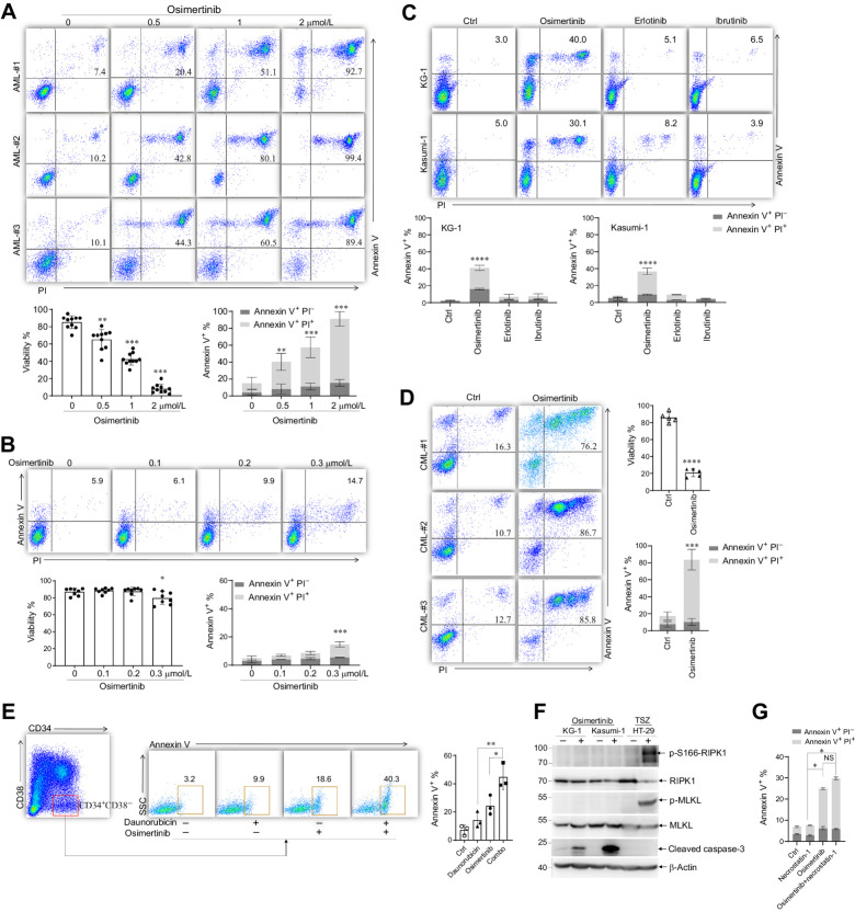
Figure 1. EGFR inhibitors induce apoptosis of CD34+ leukemia cells. A, CD34+ cells purified from BMMC of patients with AML were treated with indicated concentrations of osimertinib for 72 hours. Cell apoptosis was measured by Annexin V/PI staining. Representative flow cytometry plots of CD34+ cells from three AML individuals are shown. Bottom, cell viability measured by trypan blue (n = 10 patient samples; left) and quantified Annexin V+PI− and Annexin V+PI+ cells (n = 10 patient samples; right). B, CD34+ cells purified from BMMCs of patients with AML were treated with indicated concentrations of osimertinib for 72 hours. Cell apoptosis was quantified by Annexin V/PI staining. Flow cytometry plots are shown. Bottom, cell viability measured by trypan blue and quantified Annexin V+PI− and Annexin V+PI+ cells (n = 8). C, KG-1 and Kasumi-1 were treated with 2 μmol/L osimertinib, erlotinib, or ibrutinib for 72 hours. Representative flow cytometry plots and quantified Annexin V+PI− and Annexin V+PI+ cells are shown. D, Primary CML CD34+ cells were treated with 1 μmol/L osimertinib for 72 hours. Representative flow cytometry plots, cell viability measured by trypan blue, and quantified Annexin V+PI− and Annexin V+PI+ cells are shown. **, P < 0.005; ***, P < 0.001; ****, P < 0.0001, against control (ctrl) group. E, CD34+CD38− cells purified from patients with AML (n = 3) were treated with 2 μmol/L osimertinib, 2 μmol/L daunorubicin or osimertinib plus daunorubicin (combo), and cell apoptosis was measured by Annexin V staining. *, P < 0.05; **, P < 0.001, between the line-pointed group. F, KG-1 and Kasumi-1 cells were treated with osimertinib and the indicated proteins were detected by Western blot. G, KG-1 cells were treated with osimertinib, necrostatin-1, or osimertinib plus necrostatin-1, and cell apoptosis was measured by Annexin V/PI staining. *, P < 0.05, between the line-pointed group; NS, not significant.
Figure 1. EGFR inhibitors induce apoptosis of CD34+ leukemia cells. A, CD34+ cells purified from BMMC of patients with AML were treated with indicated concentrations of osimertinib for 72 hours. Cell apoptosis was measured by Annexin V/PI staining. Representative flow cytometry plots of CD34+ cells from three AML individuals are shown. Bottom, cell viability measured by trypan blue (n = 10 patient samples; left) and quantified Annexin V+PI− and Annexin V+PI+ cells (n = 10 patient samples; right). B, CD34+ cells purified from BMMCs of patients with AML were treated with indicated concentrations of osimertinib for 72 hours. Cell apoptosis was quantified by Annexin V/PI staining. Flow cytometry plots are shown. Bottom, cell viability measured by trypan blue and quantified Annexin V+PI− and Annexin V+PI+ cells (n = 8). C, KG-1 and Kasumi-1 were treated with 2 μmol/L osimertinib, erlotinib, or ibrutinib for 72 hours. Representative flow cytometry plots and quantified Annexin V+PI− and Annexin V+PI+ cells are shown. D, Primary CML CD34+ cells were treated with 1 μmol/L osimertinib for 72 hours. Representative flow cytometry plots, cell viability measured by trypan blue, and quantified Annexin V+PI− and Annexin V+PI+ cells are shown. **, P < 0.005; ***, P < 0.001; ****, P < 0.0001, against control (ctrl) group. E, CD34+CD38− cells purified from patients with AML (n = 3) were treated with 2 μmol/L osimertinib, 2 μmol/L daunorubicin or osimertinib plus daunorubicin (combo), and cell apoptosis was measured by Annexin V staining. *, P < 0.05; **, P < 0.001, between the line-pointed group. F, KG-1 and Kasumi-1 cells were treated with osimertinib and the indicated proteins were detected by Western blot. G, KG-1 cells were treated with osimertinib, necrostatin-1, or osimertinib plus necrostatin-1, and cell apoptosis was measured by Annexin V/PI staining. *, P < 0.05, between the line-pointed group; NS, not significant.CC BY-NC-ND · PubMed Central
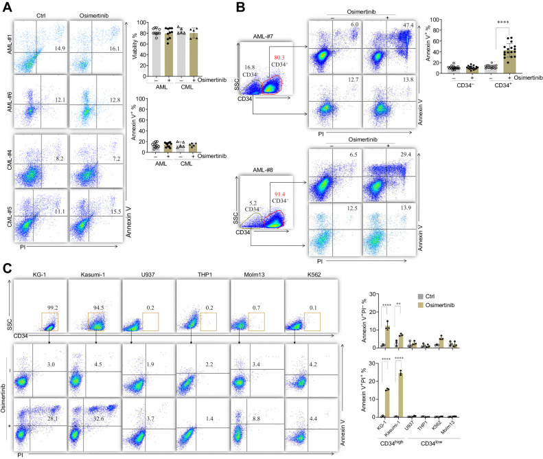
Figure 2. Osimertinib selectively kills CD34+ AML and CML cells. A, Primary CD34− cells were treated with 2 μmol/L osimertinib for 72 hours. Cell apoptosis was measured by Annexin V/PI staining. Representative flow cytometry plots, cell viability measured by trypan blue, and quantification of Annexin V+% are shown. B, BMMCs of patients with AML (n = 17) were treated with 2 μmol/L osimertinib for 48 hours and cell apoptosis was measured by Annexin V/PI assay co-staining with CD34-APC. Flow cytometry plots of two representative patients and quantification of Annexin V+% are shown. C, Leukemia cell lines KG-1, Kasumi-1, U937, THP1, Molm13, and K562 were treated with 2 μmol/L osimertinib for 72 hours and Annexin V+% was measured by Annexin V/PI assay co-staining with CD34-APC. Right, quantification of Annexin V+PI− and Annexin V+PI+ cells. *, P < 0.01; ****, P < 0.0001, between the line-pointed group.
Figure 2. Osimertinib selectively kills CD34+ AML and CML cells. A, Primary CD34− cells were treated with 2 μmol/L osimertinib for 72 hours. Cell apoptosis was measured by Annexin V/PI staining. Representative flow cytometry plots, cell viability measured by trypan blue, and quantification of Annexin V+% are shown. B, BMMCs of patients with AML (n = 17) were treated with 2 μmol/L osimertinib for 48 hours and cell apoptosis was measured by Annexin V/PI assay co-staining with CD34-APC. Flow cytometry plots of two representative patients and quantification of Annexin V+% are shown. C, Leukemia cell lines KG-1, Kasumi-1, U937, THP1, Molm13, and K562 were treated with 2 μmol/L osimertinib for 72 hours and Annexin V+% was measured by Annexin V/PI assay co-staining with CD34-APC. Right, quantification of Annexin V+PI− and Annexin V+PI+ cells. *, P < 0.01; ****, P < 0.0001, between the line-pointed group.CC BY-NC-ND · PubMed Central
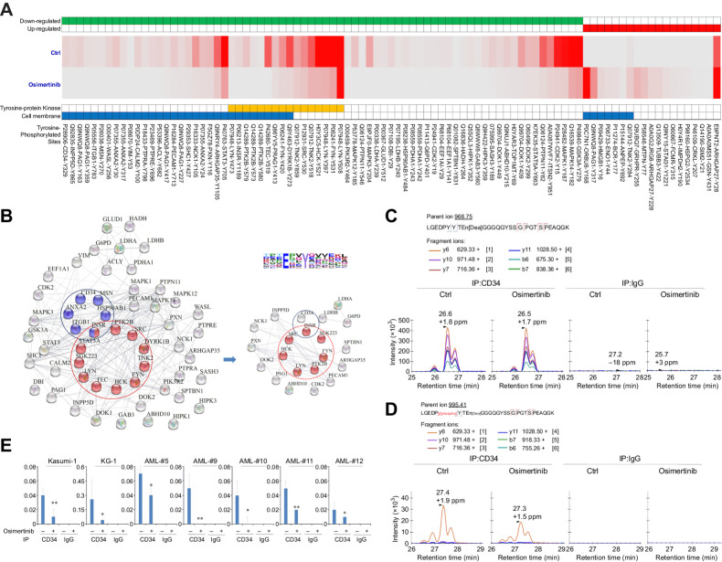
Figure 3. Osimertinib downregulates level of tyrosine phosphorylation of CD34. A, Heat map of levels of the indicated phosphorylated tyrosine sites in osimertinib-treated and untreated (ctrl) CD34+ cells. The CD34+ cells were purified from three AML individuals and pooled, followed by treatment with 2 μmol/L osimertinib for 48 hours. Tyrosine-phosphorylated peptides were enriched and analyzed by MS. The colors in the map represent the quantitative value (normalized total spectra) according to the Scaffold_4.3.3. B, Phosphoproteome changes observed in osimertinib-treated CD34+ cells analyzed by String database. The edge indicates known interaction between two proteins. Sequence logo represents the motif clustering from the tyrosine phosphorylation data set. C and D, The osimertinib treated or untreated CD34+ cells were collected and the cell lysates were immunoprecipitated with anti-CD34 or IgG antibodies, followed by PRM-MS analysis. Intensity histogram of nonphosphorylated (C) and phosphorylated Y329-containing peptide (D) of CD34 protein was quantified. E, Level of Y329-phosphorylation of CD34 quantified by PRM-MS assays in AML cell lines and 5 primary AML CD34+ samples. Data are representative of three independent experiments. *, P < 0.05; **, P < 0.01, against untreated group.
Figure 3. Osimertinib downregulates level of tyrosine phosphorylation of CD34. A, Heat map of levels of the indicated phosphorylated tyrosine sites in osimertinib-treated and untreated (ctrl) CD34+ cells. The CD34+ cells were purified from three AML individuals and pooled, followed by treatment with 2 μmol/L osimertinib for 48 hours. Tyrosine-phosphorylated peptides were enriched and analyzed by MS. The colors in the map represent the quantitative value (normalized total spectra) according to the Scaffold_4.3.3. B, Phosphoproteome changes observed in osimertinib-treated CD34+ cells analyzed by String database. The edge indicates known interaction between two proteins. Sequence logo represents the motif clustering from the tyrosine phosphorylation data set. C and D, The osimertinib treated or untreated CD34+ cells were collected and the cell lysates were immunoprecipitated with anti-CD34 or IgG antibodies, followed by PRM-MS analysis. Intensity histogram of nonphosphorylated (C) and phosphorylated Y329-containing peptide (D) of CD34 protein was quantified. E, Level of Y329-phosphorylation of CD34 quantified by PRM-MS assays in AML cell lines and 5 primary AML CD34+ samples. Data are representative of three independent experiments. *, P < 0.05; **, P < 0.01, against untreated group.CC BY-NC-ND · PubMed Central
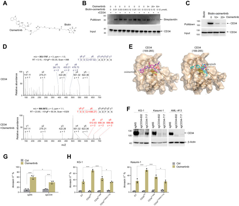
Figure 4. Osimertinib directly binds to CD34 protein. A, Chemical structure of biotin-tagged osimertinib. B, Pulldown assays were conducted using biotin-osimertinib against recombinant CD34 (rCD34) in the presence/absence of free osimertinib as a competitor. C, Pulldown assays were conducted using biotin-osimertinib against KG-1 cell lysates in the presence/absence of free osimertinib as a competitor. D, MS/MS analysis of the cysteine 199 (C199) tryptic peptide of rCD34 protein incubated with (bottom) or without (top) osimertinib for 30 minutes. E, Predicted conformations of compound osimertinib (magenta) and ibrutinib (cyan) in the binding pocket of CD34. The protein of CD34 is shown in wheat surface and cartoon. Compounds are shown as sticks. Cys177 and Cys199 of CD34 are shown as sticks. The dark dash lines show the distance between Cys199/Cys177 to the atom that is involved in Michael addition (in dash cycles). F, KG-1, Kasumi-1, and primary AML cells were infected with CD34-targeting CRISPR-Cas9 to deplete CD34 expression. Expression of CD34 was analyzed with immunoblot assay. G, CD34 was knocked out by CRISPR-Cas9 system in CD34+ cells purified from patients with AML (n = 3) and was exposed to 1 μmol/L osimertinib for 72 hours. Apoptosis was measured by flow cytometry. H, KG-1 and Kasumi-1 cells were infected with CD34-targeting CRISPR-Cas9 to deplete endogenous CD34 and then infected with plasmids encompassing WT, C199S, or C199S/C177S mutant CD34. These cells were treated with 2 μmol/L osimertinib for 72 hours and apoptosis was determined by flow cytometry. *, P < 0.05; ***, P < 0.001, between the line-pointed group. EV, empty vector; sgCD34, specific guide CD34; sgNS, specific guide nonspecific.
Figure 4. Osimertinib directly binds to CD34 protein. A, Chemical structure of biotin-tagged osimertinib. B, Pulldown assays were conducted using biotin-osimertinib against recombinant CD34 (rCD34) in the presence/absence of free osimertinib as a competitor. C, Pulldown assays were conducted using biotin-osimertinib against KG-1 cell lysates in the presence/absence of free osimertinib as a competitor. D, MS/MS analysis of the cysteine 199 (C199) tryptic peptide of rCD34 protein incubated with (bottom) or without (top) osimertinib for 30 minutes. E, Predicted conformations of compound osimertinib (magenta) and ibrutinib (cyan) in the binding pocket of CD34. The protein of CD34 is shown in wheat surface and cartoon. Compounds are shown as sticks. Cys177 and Cys199 of CD34 are shown as sticks. The dark dash lines show the distance between Cys199/Cys177 to the atom that is involved in Michael addition (in dash cycles). F, KG-1, Kasumi-1, and primary AML cells were infected with CD34-targeting CRISPR-Cas9 to deplete CD34 expression. Expression of CD34 was analyzed with immunoblot assay. G, CD34 was knocked out by CRISPR-Cas9 system in CD34+ cells purified from patients with AML (n = 3) and was exposed to 1 μmol/L osimertinib for 72 hours. Apoptosis was measured by flow cytometry. H, KG-1 and Kasumi-1 cells were infected with CD34-targeting CRISPR-Cas9 to deplete endogenous CD34 and then infected with plasmids encompassing WT, C199S, or C199S/C177S mutant CD34. These cells were treated with 2 μmol/L osimertinib for 72 hours and apoptosis was determined by flow cytometry. *, P < 0.05; ***, P < 0.001, between the line-pointed group. EV, empty vector; sgCD34, specific guide CD34; sgNS, specific guide nonspecific.CC BY-NC-ND · PubMed Central
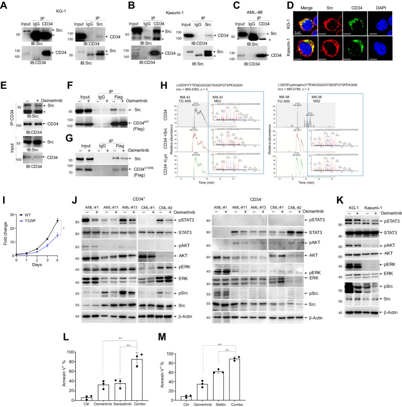
Figure 5. Osimertinib impairs interaction between CD34 and the Src protein kinase. A–C, IP assays were conducted using antibodies against Src or CD34 in KG-1, Kasumi-1, and primary CD34+ AML cells. *, the heavy chain of IgG. D, Immunofluorescent staining of Src and CD34 in KG-1 and Kasumi-1. E, IP assay was conducted using antibody against CD34 in primary CD34+ AML cells treated with/without osimertinib. F and G, Kasumi-1 cells were infected with flag-tagged WT (F) or C199S mutant (G) CD34 and then treated with/without osimertinib. IP assay was conducted using anti-Flag antibody. H, Recombinant CD34 peptide (aa324–352) was incubated with Src (middle) or Lyn (bottom), followed by MS/MS analysis. Total ion chromatogram (TIC, sum of MS1 intensities over time) and fragment-ion spectra (MS2, MS/MS) for the major peaks in the TIC-MS1 spectrum of nonphosphorylated peptides (left) and phosphorylated peptides (right) are shown. The b ions and y ions are labeled in blue and red, respectively. I, KG-1 cells were infected with CD34-targeting CRISPR-Cas9 to deplete CD34 expression, followed by overexpression with flag-tagged WT or Y329F mutant, and cell proliferation was evaluated. J and K, Immunoblot analysis showing activity of the STAT3, AKT, ERK, Src in response to osimertinib (1 μmol/L, 72 hours) in primary CD34+ or CD34− cells (J) or KG-1 and Kasumi-1 cells (2 μmol/L, 72 hours; K). L, Primary CD34+ cells purified from patients with AML (n = 3) were treated with 1 μmol/L osimertinib, 2 μmol/L saracatinib, or osimertinib plus saracatinib (combo), and apoptosis was measured by Annexin V/PI staining. M, Primary CD34+ AML cells were treated with 1 μmol/L osimertinib, 1 μmol/L stattic, or osimertinib plus stattic (combo), and apoptosis was measured by Annexin V/PI staining. Quantified apoptotic cells are shown. **, P < 0.005 between the line-pointed group.
Figure 5. Osimertinib impairs interaction between CD34 and the Src protein kinase. A–C, IP assays were conducted using antibodies against Src or CD34 in KG-1, Kasumi-1, and primary CD34+ AML cells. *, the heavy chain of IgG. D, Immunofluorescent staining of Src and CD34 in KG-1 and Kasumi-1. E, IP assay was conducted using antibody against CD34 in primary CD34+ AML cells treated with/without osimertinib. F and G, Kasumi-1 cells were infected with flag-tagged WT (F) or C199S mutant (G) CD34 and then treated with/without osimertinib. IP assay was conducted using anti-Flag antibody. H, Recombinant CD34 peptide (aa324–352) was incubated with Src (middle) or Lyn (bottom), followed by MS/MS analysis. Total ion chromatogram (TIC, sum of MS1 intensities over time) and fragment-ion spectra (MS2, MS/MS) for the major peaks in the TIC-MS1 spectrum of nonphosphorylated peptides (left) and phosphorylated peptides (right) are shown. The b ions and y ions are labeled in blue and red, respectively. I, KG-1 cells were infected with CD34-targeting CRISPR-Cas9 to deplete CD34 expression, followed by overexpression with flag-tagged WT or Y329F mutant, and cell proliferation was evaluated. J and K, Immunoblot analysis showing activity of the STAT3, AKT, ERK, Src in response to osimertinib (1 μmol/L, 72 hours) in primary CD34+ or CD34− cells (J) or KG-1 and Kasumi-1 cells (2 μmol/L, 72 hours; K). L, Primary CD34+ cells purified from patients with AML (n = 3) were treated with 1 μmol/L osimertinib, 2 μmol/L saracatinib, or osimertinib plus saracatinib (combo), and apoptosis was measured by Annexin V/PI staining. M, Primary CD34+ AML cells were treated with 1 μmol/L osimertinib, 1 μmol/L stattic, or osimertinib plus stattic (combo), and apoptosis was measured by Annexin V/PI staining. Quantified apoptotic cells are shown. **, P < 0.005 between the line-pointed group.CC BY-NC-ND · PubMed Central
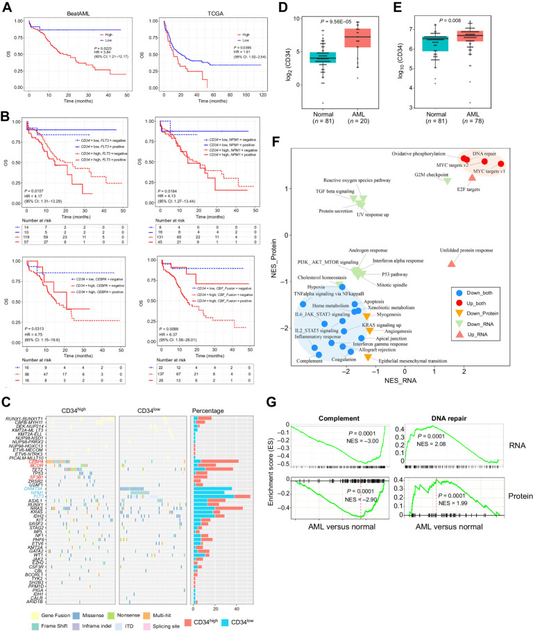
Figure 6. CD34 is an adverse prognosis marker in AML and associates with specific pathogenic events. A, Differences in OS in de novo patients with AML from the BeatAML dataset (n = 200; left) and TCGA dataset (n = 173; right) by expression of CD34. P values, HRs, and 95% confidence interval (CI) are shown from univariate Cox analysis. B, Differences in OS in de novo patients with AML from the BeatAML dataset (n = 200) by combination of CD34 expression with presence of FLT3, NPM1, CEBPA, and CBF fusion. P values, HRs, and 95% CI are for the CD34 high expression from the multivariate Cox analysis. CBF fusion, RUNX1-RUNX1T1 fusion or CBFB-MYH11 fusion. C, Landscape and percentage of mutations and fusions in patients with AML with high or low CD34 expression level. D and E, RNA-seq and quantitative proteomics analysis of sorted CD34+ cells samples collected from de novo patients with AML and healthy donors (Normal). The mRNA (D) and protein (E) expression level of CD34 are shown. F, HALLMARK genesets from the GSEA analysis of RNA-seq and quantitative proteomic data of CD34+ cells from patients with AML versus normal donors. G, GSEA for complement and DNA repair pathways in AML versus normal samples.
Figure 6. CD34 is an adverse prognosis marker in AML and associates with specific pathogenic events. A, Differences in OS in de novo patients with AML from the BeatAML dataset (n = 200; left) and TCGA dataset (n = 173; right) by expression of CD34. P values, HRs, and 95% confidence interval (CI) are shown from univariate Cox analysis. B, Differences in OS in de novo patients with AML from the BeatAML dataset (n = 200) by combination of CD34 expression with presence of FLT3, NPM1, CEBPA, and CBF fusion. P values, HRs, and 95% CI are for the CD34 high expression from the multivariate Cox analysis. CBF fusion, RUNX1-RUNX1T1 fusion or CBFB-MYH11 fusion. C, Landscape and percentage of mutations and fusions in patients with AML with high or low CD34 expression level. D and E, RNA-seq and quantitative proteomics analysis of sorted CD34+ cells samples collected from de novo patients with AML and healthy donors (Normal). The mRNA (D) and protein (E) expression level of CD34 are shown. F, HALLMARK genesets from the GSEA analysis of RNA-seq and quantitative proteomic data of CD34+ cells from patients with AML versus normal donors. G, GSEA for complement and DNA repair pathways in AML versus normal samples.CC BY-NC-ND · PubMed Central
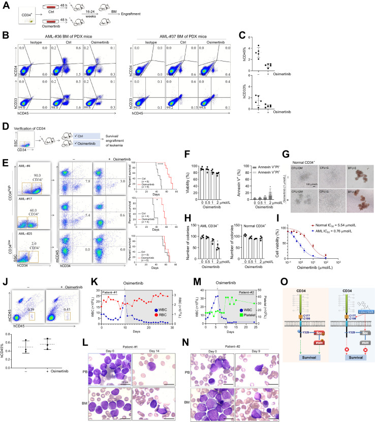
Figure 7. Effects of osimertinib in human AML and CML PDX models. A, Diagram of experimental design. B, Flow cytometry analyses of human CD45, CD33, and CD34 (hCD45, hCD33, and hCD34, respectively) expression in mouse bone marrow were used to assess engraftment of AML cells. The isotype controls were used to define the gating strategy. C, Percentages of hCD45+ and hCD33+ cells in the bone marrow are shown. D, Diagram of experimental design. E, Representative analyses of human CD34 and mouse CD45 (hCD34 and mCD45, respectively) expression in mouse bone marrow. Right, survival curves of PDX mice. F, CD34+ cells purified from BMMCs of healthy donors were treated with indicated concentrations of osimertinib for 72 hours. Cell apoptosis was quantified by Annexin V/PI staining. Cell viability measured by trypan blue and quantified Annexin V+PI− and Annexin V+PI+ cells (n = 5) are shown. G and H, Colony-forming unit assay of CD34+ AML and normal cells treated with indicated concentrations of osimertinib for 72 hours. Representative image of clones of normal CD34+ cells treated with 1 μmol/L osimertinib (G) and colony numbers (H) are shown. I, The AML and normal CD34+ cells were treated with osimertinib from 0.1 nmol/L to 100 μmol/L and the IC50 curve was constructed. J, Representative analyses of human and mouse CD45 (hCD45 and mCD45, respectively) expression in mouse bone marrow. Bottom, percentages of hCD45+ cells in the bone marrow at 16 weeks. K, White blood cell (WBC) and red blood cell (RBC) counts of AML patient-#1. L, Microscopic appearance of peripheral blood (PB) and bone marrow (BM) smear of AML patient-#1. Day 0 was defined as the day that osimertinib was started. M, White blood cell and platelet counts of AML patient-#2. N, Microscopic appearance of peripheral blood and bone marrow smear of AML patient-#2. O, A model proposing the mechanism by which osimertinib induces apoptosis through CD34-dependent and -independent ways.
Figure 7. Effects of osimertinib in human AML and CML PDX models. A, Diagram of experimental design. B, Flow cytometry analyses of human CD45, CD33, and CD34 (hCD45, hCD33, and hCD34, respectively) expression in mouse bone marrow were used to assess engraftment of AML cells. The isotype controls were used to define the gating strategy. C, Percentages of hCD45+ and hCD33+ cells in the bone marrow are shown. D, Diagram of experimental design. E, Representative analyses of human CD34 and mouse CD45 (hCD34 and mCD45, respectively) expression in mouse bone marrow. Right, survival curves of PDX mice. F, CD34+ cells purified from BMMCs of healthy donors were treated with indicated concentrations of osimertinib for 72 hours. Cell apoptosis was quantified by Annexin V/PI staining. Cell viability measured by trypan blue and quantified Annexin V+PI− and Annexin V+PI+ cells (n = 5) are shown. G and H, Colony-forming unit assay of CD34+ AML and normal cells treated with indicated concentrations of osimertinib for 72 hours. Representative image of clones of normal CD34+ cells treated with 1 μmol/L osimertinib (G) and colony numbers (H) are shown. I, The AML and normal CD34+ cells were treated with osimertinib from 0.1 nmol/L to 100 μmol/L and the IC50 curve was constructed. J, Representative analyses of human and mouse CD45 (hCD45 and mCD45, respectively) expression in mouse bone marrow. Bottom, percentages of hCD45+ cells in the bone marrow at 16 weeks. K, White blood cell (WBC) and red blood cell (RBC) counts of AML patient-#1. L, Microscopic appearance of peripheral blood (PB) and bone marrow (BM) smear of AML patient-#1. Day 0 was defined as the day that osimertinib was started. M, White blood cell and platelet counts of AML patient-#2. N, Microscopic appearance of peripheral blood and bone marrow smear of AML patient-#2. O, A model proposing the mechanism by which osimertinib induces apoptosis through CD34-dependent and -independent ways.CC BY-NC-ND · PubMed Central

Verification

Claims the paper makes

The claims and statistics of this paper haven’t been checked yet.

Run the check — free, no account needed. It reads the open-access text and checks the sources it cites.

A few free checks a day · papers already verified open instantly · results are public

Self-check

Percentages, sums and reported differences re-computed from the paper’s own numbers.

The self-check needs the paper’s full text.

Citations

Cites

Research works printed but not linked: 51

  1. Martin-Subero, 2018, Predicting leukemia relapse, Nat Med, 24, 385, 10.1038/nm.4529 · DOI
  2. Marchesi, 2013, Genetics: the AML mutational landscape, Nat Rev Clin Oncol, 10, 305
  3. Ciardiello, 2008, EGFR antagonists in cancer treatment, N Engl J Med, 358, 1160, 10.1056/NEJMra0707704 · DOI
  4. Cross, 2014, AZD9291, an irreversible EGFR TKI, overcomes T790M-mediated resistance to EGFR inhibitors in lung cancer, Cancer Discov, 4, 1046, 10.1158/2159-8290.CD-14-0337 · DOI · Paper page
  5. Janne, 2015, AZD9291 in EGFR inhibitor-resistant non, N Engl J Med, 372, 1689, 10.1056/NEJMoa1411817 · DOI · Paper page
  6. Chan, 2007, Complete remission in a patient with acute myelogenous leukemia treated with erlotinib for non–small cell lung cancer, Blood, 110, 1079, 10.1182/blood-2007-01-069856 · DOI
  7. Pitini, 2008, Erlotinib in a patient with acute myelogenous leukemia and concomitant non, J Clin Oncol, 26, 3645, 10.1200/JCO.2008.17.0357 · DOI
  8. Boehrer, 2008, Erlotinib exhibits antineoplastic off-target effects in AML and MDS: a preclinical study, Blood, 111, 2170, 10.1182/blood-2007-07-100362 · DOI
  9. Stegmaier, 2005, Gefitinib induces myeloid differentiation of acute myeloid leukemia, Blood, 106, 2841, 10.1182/blood-2005-02-0488 · DOI
  10. Hahn, 2009, Proteomic and genetic approaches identify Syk as an AML target, Cancer Cell, 16, 281, 10.1016/j.ccr.2009.08.018 · DOI
  11. Saleh, 2019, Gilteritinib changes AML landscape, Oncology, 33, 683729
  12. Boehrer, 2011, Erlotinib antagonizes constitutive activation of SRC family kinases and mTOR in acute myeloid leukemia, Cell Cycle, 10, 3168, 10.4161/cc.10.18.16599 · DOI
  13. Mahmud, 2016, Epidermal growth factor receptor is expressed and active in a subset of acute myeloid leukemia, J Hematol Oncol, 9, 64, 10.1186/s13045-016-0294-x · DOI
  14. Abou Dalle, 2018, A pilot Phase II study of erlotinib for the treatment of patients with relapsed/refractory acute myeloid leukemia, Acta Haematol, 140, 30, 10.1159/000490092 · DOI
  15. Deangelo, 2014, A Phase II study of the EGFR inhibitor gefitinib in patients with acute myeloid leukemia, Leuk Res, 38, 430, 10.1016/j.leukres.2013.10.026 · DOI
  16. Sayar, 2015, Pilot study of erlotinib in patients with acute myeloid leukemia, Leuk Res, 39, 170, 10.1016/j.leukres.2014.11.022 · DOI
  17. Thepot, 2014, A phase I/II trial of Erlotinib in higher risk myelodysplastic syndromes and acute myeloid leukemia after azacitidine failure, Leuk Res, 38, 1430, 10.1016/j.leukres.2014.09.014 · DOI
  18. Dobin, 2013, STAR: ultrafast universal RNA-seq aligner, Bioinformatics, 29, 15, 10.1093/bioinformatics/bts635 · DOI · Paper page
  19. Trapnell, 2010, Transcript assembly and quantification by RNA-seq reveals unannotated transcripts and isoform switching during cell differentiation, Nat Biotechnol, 28, 511, 10.1038/nbt.1621 · DOI · Paper page
  20. Li, 2009, Fast and accurate short read alignment with Burrows, Bioinformatics, 25, 1754, 10.1093/bioinformatics/btp324 · DOI · Paper page
  21. McKenna, 2010, The genome analysis toolkit: a MapReduce framework for analyzing next-generation DNA sequencing data, Genome Res, 20, 1297, 10.1101/gr.107524.110 · DOI · Paper page
  22. Wang, 2010, ANNOVAR: functional annotation of genetic variants from high-throughput sequencing data, Nucleic Acids Res, 38, e164, 10.1093/nar/gkq603 · DOI
  23. Ma, 2019, iProX: an integrated proteome resource, Nucleic Acids Res, 47, D1211, 10.1093/nar/gky869 · DOI
  24. Degterev, 2008, Identification of RIP1 kinase as a specific cellular target of necrostatins, Nat Chem Biol, 4, 313, 10.1038/nchembio.83 · DOI · Paper page
  25. Tao, 2020, A dominant autoinflammatory disease caused by non-cleavable variants of RIPK1, Nature, 577, 109, 10.1038/s41586-019-1830-y · DOI
  26. Weber, 2012, Dual phosphoproteomics and chemical proteomics analysis of erlotinib and gefitinib interference in acute myeloid leukemia cells, J Proteomics, 75, 1343, 10.1016/j.jprot.2011.11.004 · DOI
  27. Patel, 2019, Expression of myeloid Src-family kinases is associated with poor prognosis in AML and influences Flt3-ITD kinase inhibitor acquired resistance, PLoS One, 14, e0225887, 10.1371/journal.pone.0225887 · DOI
  28. Dos Santos, 2008, A critical role for Lyn in acute myeloid leukemia, Blood, 111, 2269, 10.1182/blood-2007-04-082099 · DOI
  29. Saito, 2017, Overcoming mutational complexity in acute myeloid leukemia by inhibition of critical pathways, Sci Transl Med, 9, eaao1214, 10.1126/scitranslmed.aao1214 · DOI
  30. Saito, 2013, A pyrrolo-pyrimidine derivative targets human primary AML stem cells in vivo, Sci Transl Med, 5, 181ra52, 10.1126/scitranslmed.3004387 · DOI
  31. Weir, 2018, Selective inhibition of the myeloid Src-family kinase Fgr potently suppresses AML cell growth in vitro and in vivo, ACS Chem Biol, 13, 1551, 10.1021/acschembio.8b00154 · DOI
  32. Krause, 1996, CD34: structure, biology, and clinical utility, Blood, 87, 1, 10.1182/blood.V87.1.1.1 · DOI
  33. Hu, 1998, The cytoplasmic domain of stem cell antigen CD34 is essential for cytoadhesion signaling but not sufficient for proliferation signaling, Blood, 91, 1152, 10.1182/blood.V91.4.1152 · DOI
  34. Varadi, 2022, AlphaFold protein structure database: massively expanding the structural coverage of protein-sequence space with high-accuracy models, Nucleic Acids Res, 50, D439, 10.1093/nar/gkab1061 · DOI · Paper page
  35. Zhou, 2010, A redox switch in angiotensinogen modulates angiotensin release, Nature, 468, 108, 10.1038/nature09505 · DOI
  36. Xia, 2013, Phosphoproteomics study on the activated PKCdelta-induced cell death, J Proteome Res, 12, 4280, 10.1021/pr400089v · DOI
  37. Johnson, 2018, Targeting the IL-6/JAK/STAT3 signaling axis in cancer, Nat Rev Clin Oncol, 15, 234, 10.1038/nrclinonc.2018.8 · DOI · Paper page
  38. Zeijlemaker, 2015, Absence of leukemic CD34(+) cells in acute myeloid leukemia is of high prognostic value: a longstanding controversy deciphered, Br J Haematol, 171, 227, 10.1111/bjh.13572 · DOI
  39. Zhu, 2013, CD34 expression on bone marrow blasts is a novel predictor of poor prognosis independent of FlT3-ITD in acute myeloid leukemia with the NPM1-mutation, Leuk Res, 37, 624, 10.1016/j.leukres.2013.02.007 · DOI
  40. Tyner, 2018, Functional genomic landscape of acute myeloid leukemia, Nature, 562, 526, 10.1038/s41586-018-0623-z · DOI
  41. Cancer Genome Atlas Research N, 2013, Genomic and epigenomic landscapes of adult de novo acute myeloid leukemia, N Engl J Med, 368, 2059, 10.1056/NEJMoa1301689 · DOI · Paper page
  42. Dohner, 2017, Diagnosis and management of AML in adults: 2017 ELN recommendations from an international expert panel, Blood, 129, 424, 10.1182/blood-2016-08-733196 · DOI
  43. Wu, 2020, Osimertinib in resected EGFR-Mutated non, N Engl J Med, 383, 1711, 10.1056/NEJMoa2027071 · DOI
  44. Sidney, 2014, Concise review: evidence for CD34 as a common marker for diverse progenitors, Stem Cells, 32, 1380, 10.1002/stem.1661 · DOI
  45. Baumheter, 1993, Binding of L-selectin to the vascular sialomucin CD34, Science, 262, 436, 10.1126/science.7692600 · DOI
  46. Healy, 1995, The stem cell antigen CD34 functions as a regulator of hemopoietic cell adhesion, Proc Natl Acad Sci USA, 92, 12240, 10.1073/pnas.92.26.12240 · DOI
  47. Annageldiyev, 2020, The novel Isatin analog KS99 targets stemness markers in acute myeloid leukemia, Haematologica, 105, 687, 10.3324/haematol.2018.212886 · DOI
  48. Drew, 2005, CD34 and CD43 inhibit mast cell adhesion and are required for optimal mast cell reconstitution, Immunity, 22, 43, 10.1016/j.immuni.2004.11.014 · DOI
  49. Felschow, 2001, The adapter protein CrkL associates with CD34, Blood, 97, 3768, 10.1182/blood.V97.12.3768 · DOI
  50. Wilson, 2015, Targeting B cell receptor signaling with ibrutinib in diffuse large B-cell lymphoma, Nat Med, 21, 922, 10.1038/nm.3884 · DOI
  51. Kuntz, 2017, Targeting mitochondrial oxidative phosphorylation eradicates therapy-resistant chronic myeloid leukemia stem cells, Nat Med, 23, 1234, 10.1038/nm.4399 · DOI

Metadata

Authors
Xia, L., Liu, J.-Y., Yang, M.-Y., Zhang, X.-H., Jiang, Y., Yin, Q.-Q., Luo, C.-H., Liu, H.-C., Kang, Z.-J., Zhang, C.-T., Gao, B.-B., Zhou, A.-W., Cai, H.-Y., Waller, E. K., Yan, J.-S., & Lu, Y.
Affiliations
  • 1Institute of Dermatology, Xinhua Hospital, School of Medicine, Shanghai Jiao Tong University, Shanghai, China.
  • Shanghai Jiao Tong University (CN)
  • 3Center of Genome and Personalized Medicine, Institute of Cancer Stem Cell, Dalian Medical University, Liaoning, China.
  • Dalian Medical University (CN)
  • 5Shanghai Institute for Advanced Immunochemical Studies, Shanghai Tech University, Shanghai, China.
  • 4Department of Hematology, Liaoning Medical Center for Hematopoietic Stem-Cell Transplantation, Liaoning Key Laboratory of Hematopoietic Stem-Cell Transplantation and Translational Medicine, Dalian Key Laboratory of Hematology, the Second Hospital of Dalian Medical University, Dalian, China.
  • 6Basic Medical Institute, Key Laboratory of Cell Differentiation and Apoptosis of the Chinese Ministry of Education, Shanghai Jiao Tong University School of Medicine, Shanghai, China.
  • Winship Cancer Institute (US)
Corresponding author
Lu, Ying
Venue
Cancer Research
Published
2023-12-14
Volume
84
Issue
3
Pages
479-492
ISSN
  • 0008-5472 (ISSN-L)
  • 1538-7445
OpenAlex
W4389732350
Cited by
5
References
4
Citations by year
2025: 3 · 2024: 2
FWCI
1.03
Type
journal article
Language
en
Open access
Locations
  • Cancer Research (publishedVersion, cc-by-nc-nd)
  • PubMed Central (publishedVersion, cc-by-nc-nd)
  • PubMed (publishedVersion)
Funders
  • National Natural Science Foundation of China (82170179, 81970131, 81370652, 81770146, 32171431)
  • Science and Technology Commission of Shanghai Municipality (22S11900400)
Topics
  • Health Sciences › Medicine › Hematology › Acute Myeloid Leukemia Research
  • Health Sciences › Medicine › Hematology › Chronic Myeloid Leukemia Treatments
  • Health Sciences › Medicine › Pulmonary and Respiratory Medicine › Lung Cancer Treatments and Mutations
Keywords
osimertinib, CD34, acute myeloid leukemia, chronic myeloid leukemia, covalent binding, apoptosis, Src family kinases, STAT3 signaling, synthetic lethality, prognostic factors, patient-derived xenografts, compassionate use
MeSH terms
Acrylamides, Aniline Compounds, Carcinoma, Non-Small-Cell Lung, Humans, Indoles, Lung Neoplasms, Pyrimidines, Neoplastic Stem Cells, Leukemia, Myeloid, Acute, Antigens, CD34, Myeloid Progenitor Cells, Proteomics, Cell Proliferation, ErbB Receptors

About these checks

Registry, retraction, Cites and statistics results are computed, not judged, with one exception: a printed reference that no index lists is read once by a model before it counts as a citation error. Claims are extracted by a model and kept only when their quote appears verbatim in the text; each cited source is then checked, with a model’s help, for whether it says what the claim relies on. Those verdicts can be wrong — read the claim’s quote and the cited source before relying on one.

Statistics and arithmetic are recomputed, not judged.

Confidence bands are shown only for fields calibrated on our annotated test set. None is calibrated yet, so no band is shown.

What counts as a claim ›How we measure confidence ›How we check this ›

Are you an author, or do you see a problem with this page? Request a review